






Abstract
Purpose
Dispersible hydroxyapatite (HAp) nanoparticles are very useful for applying a monolayer to implantable medical devices using the nano-coating technique. To improve tolerance to infection on implanted medical devices, silver-doped HAp (Ag-HAp) nanoparticles with dispersiblity and crystallinity were synthesized, avoiding calcination-induced sintering, and evaluated for antibacterial activity.
Methods
The Ca10-xAgx(PO4)6(OH)2 with x = 0 and 0.2 were prepared by wet chemical processing at 100°C. Before calcination at 700°C for 2 h, two kinds of anti-sintering agents, namely a Ca(NO3)2 (Ca salt) and a poly-acrylic acid/Ca salt mixture (PAA-Ca), were used. Escherichia coli was used to evaluate the antibacterial activity of the nanopowder.
Results
When PAA-Ca was used as an anti-sintering agent in calcination to prepare the dispersible nanoparticles, strong metallic Ag peaks were observed at 38.1° and 44.3° (20) in the X-ray diffraction (XRD) profile. However, the Ag peak was barely observed when Ca salt was used alone as the anti-sintering agent. Thus, using Ca salt alone was more effective for preparation of dispersible Ag-HAp than PAA-Ca. The particle average size of Ag-HAp with 0.5 mol% of Ag content was found to be 325 ± 70 nm when the formation of large particle-aggregations was prevented, as determined by dynamic light scattering instrument. The antibacterial activity of the Ag-HAp nanoparticles possessing 0.5 mol% against E. coli was greater than 90.0%.
Conclusions
Dispersible and crystalline nano Ag-HAp can be obtained by using Ca salt alone as an anti-sintering agent. The nanoparticles showed antibacterial activity.
Introduction
Hydroxyapatite [HAp, Ca10(PO4)6(OH)2], a synthetic material that is similar to the inorganic component of bone, enamel, and dentin, has been widely used for orthopedic applications due to its good biocompatibility, bioactivity, and osteoconductivity (1–3). Sintered HAp has also been reported to be compatible with soft tissues such as skin and vascular tissue (4–5). The biological functionalities of HAp are improved by incorporation of ions, such as Ag, Zn, Ti, Ni, Cu, Co, Cr, Al, La, Fe, F into the HAp lattice (6–11). For example, several in vitro studies have reported that small quantities of Cu and Zn ions are essential for various metabolic processes in most living organisms, whereas in higher amounts they are potentially toxic. Stanić et al reported that they could produce Cu- and Zn-doped nanosized HAp powders that contained no discernible crystalline impurities. They showed that Cu- and Zn-doped HAp could reduce the number of viable bacterial cells of all tested strains (12).
It has been well known since ancient times that Ag ions possess extraordinary inhibitory and bactericidal properties. Although it is relatively nontoxic to human cells (13, 14), Ag possesses antibacterial properties against a broad spectrum of bacterial strains found in industrial processes as well as in human bodies (15–16). Several reviews have described the mechanisms underlying the antibacterial activity of Ag-containing materials (17–22). Ag ions interact with membrane proteins and phosphorus-containing compounds, such as DNA, resulting in inhibition of bacterial functions (23). Ag ions also attack the respiratory chains in mitochondria, ultimately leading to cell death (24). In addition to these mechanisms, the formation of reactive oxygen species (ROS) plays a large role in the antibacterial activity of Ag (25). Ag catalyzes various reactions with oxygen, leading to excess production of free radicals (26), i.e., generation of ROS, which cause damage to cell membranes, membrane proteins, and DNA, ultimately resulting in cell death (27, 28). Because of these characteristics, Ag is widely used in medical devices and applications, such as wound dressings, scaffolds, skin donation, recipient sites, sterilization of materials in hospitals, medical catheters, contraceptive devices, surgical instruments, bone prostheses, artificial teeth, and bone coatings (29–32).
Several products that combine Ag and HAp have been fabricated via various synthetic routes: Ag nanoparticle/HAp hybrids via a wet process (33), Ag nanoparticles/HAp composite via a sonochemical approach (34), an Ag-containing HAp coating via a sol-gel process (35), an Ag-containing HAp coating via co-precipitation or thermal spraying (6, 36). In addition, metallic ion-substituted HAp has been reported, in the form of HAp nanoparticles doped with Ag ions, prepared via microwave processing (37) or a wet method at 100°C (38). However, the dispersibility of the calcinated HAp produced using these synthetic processes has not been investigated.
Recently, our research group developed a novel synthetic method for producing highly dispersed and crystalline HAp nanoparticles, by using anti-sintering agents for calcination (39, 40). The function of the anti-sintering agent is to penetrate individual crystals during calcination before it is subsequently removed by washing with water. Using this method, dispersible and crystalline nanoparticles were obtained. Surface nano-coating with a monolayer of the dispersed HAp nanoparticles through chemical bonding was shown to provide improved biocompatibility and bioactivity for medical devices such as percutaneous devices (4, 41) and artificial vascular grafts (5).
The present study focused on the effect of anti-sintering agents on calcination of Ag-doped HAp (Ag-HAp) to improve dispersibility of particles. Wet chemical processing was selected because it allows easy control of the chemical composition of the final product by ion doping. Ag-HAp was prepared using wet chemical processing at 100°C before calcination at 700°C. To avoid calcination-induced Ag-HAp particle-agglomeration, two kinds of anti-sintering agents were examined. A preliminary antibacterial examination using Escheria coli for Ag-HAp was also conducted. The synthesis of dispersible and crystalline HAp nanoparticles doped with metal ions by using anti-sintering agents during calcination has not yet been reported.
Materials and Methods
Materials
Analytical grade calcium nitrate tetrahydrate [Ca(NO3)2. 4H2O], silver nitrate [Ag(NO3)], and diammonium hydrogen phosphate [(NH4)2H(PO4)] were purchased from Wako Pure Chemical Industries Ltd., Osaka, Japan and were used for the preparation of Ag-doped hydroxyapatite without further purification. Polyacrylic acid (PAA, Mw = 6,000–8,000) used as an anti-sintering agent was purchased from Toagosei Co., Ltd., Tokyo, Japan.
Synthesis of Ag-HAp with anti-sintering agents
HAp with an Ag content of x = 0, 0.2 in Ca10-xAgx(PO4)6(OH)2 was prepared to verify the extent of Ag inclusion (37, 38). The entire reaction was performed at 100°C (38), and the amount of reactants required was calculated based on a Ca2+ + Ag+: PO43-molar ratio of 10:6. A mixture of 80 mmol of Ca(NO3)24H2O and 20 mmol of AgNO3 in 300 mL of deionized water was prepared to obtain a 0.3 M solution, which was placed in an oil bath and stirred at 100°C for 30 min. A solution of 0.2 M (NH4)2H(PO4) was prepared in addition to 300 mL of deionized water. The pH of the Ca(NO3)2 and AgNO3 mixture solution was adjusted to 10.0 by adding 28% of NH4OH, after which stirring was continued for 30 min. The (NH4)2H(PO4) solution was then added to the Ca(NO3)2 and AgNO3 mixture solution, and the reaction solution was stirred for 2 h at 100°C. The solution was stored for 12 h and then washed with deionized water 3 times using a centrifugal method to remove impurities. Then, the mixture was filtered using a fine filter paper to produce a wet cake, which was dried under reduced pressure and finally placed overnight in an oven at 60°C.
The 2 types of anti-sintering agents, a Ca(NO3)2 (Ca salt) and a PAA/Ca salt mixture (PAA-Ca), were used to prevent particle-aggregation during calcination. The anti-sintering agents (twice the amount of the dried sample cake) were added to the low-crystalline Ag-HAp nanoparticle dispersion in an aqueous solution. After the Ag-HAp powders with/without the anti-sintering agents were filtered and dried, the products were calcinated at 700°C for 2 h. The final products were obtained by washing with NH4(NO3) solution and deionized water and drying.
Measurements
Samples of the as-prepared Ag-HAp and the calcinated Ag-HAp (with/without the anti-sintering agent) were characterized using X-ray diffraction (XRD), Fourier transform infrared (FT-IR) spectroscopy, scanning electron microscopy (SEM) and inductively coupled plasma atomic emission spectroscopy (ICP-AES). The XRD was performed on a RAD-X Rigaku diffractometer, with Cu Ka (λ = 1.5418 A) radiation. The diffraction patterns were collected in the 20 range 10° to 90°, with a step size of 0.01° and a measuring time of 0.6 s per step. The cell parameters were calculated using the free software named “unit cell” with the value of 20 obtained from the measured XRD peaks (42). For the FTIR measurements, 1% Ag-doped powder was mixed with 99% KBr. The samples for FTIR measurements were prepared by pressing the powder mixture, and the spectra were captured in the range of 400 cm1 to 4000 cm1, with a 16x scanning resolution. The structure and morphology of the samples were studied using a SEM (JSM-6301F, JEOL, Akishima, Japan), operating at 5 kV with an emission current of 8 mA. The composition of the Ag, Ca, and P ions in the Ag-doped HAp was determined using ICP-AES (Optima, 2000DV; Perkin-Elmer, Yokohama, Japan). The ICP measurements were obtained by using 100 ppm of Ag-HAp solutions diluted in 1 M nitric acid solution. The size distribution of the HAp nanoparticles was measured by dynamic light scattering (DLS; ELS-8000, Otsuka Electronics, Kyoto, Japan). The measurements were carried out by using a 10 ppm solution prepared in an ethanol medium.
Anti-microbial assay
Evaluation of the antimicrobial activity of the Ag-HAp was performed based on ISO 22196 (43). Briefly, 10 mg of dried Ag-HAp was milled between 2 glass plates to disperse aggregates. After washing the powder twice with 70% ethanol and twice with 140 mM NaCl, the Ag-HAp nanoparticles were re-suspended in 1 mL of 140 mM NaCl. Wild-type E. coli K-12 W3110 was cultivated in 2 mL of LB medium (1% tryptone, 1% NaCl and 0.5% yeast extract) at 37°C for 20 h. The culture was diluted with 140 mM NaCl to adjust the OD600 to 0.1. Equal amounts of the diluted culture and the washed Ag-HAp were mixed and incubated at 25°C for 1 h with gentle agitation. For the agar spot method, a 10-fold dilution series of the samples were prepared with 140 mM NaCl, and 5 μL of each sample was dropped onto a plate containing LB agar medium (LB medium with 1.5% agar) and incubated at 37°C for 20 h. The survival rates of E. coli were measured on the basis of colonies formed in the spots and the dilution rates. This work was fully approved by the Kinki University Bio-safety Committee and was conducted in accordance with the regulations for use of recombinant pathogens.
Results and Discussion
XRD measurement
In our previous report, normal HAp nanoparticles prepared using PAA-Ca as an anti-sintering agent showed greater dispersibility than those prepared with Ca salt alone (44). The COO-groups of the PAA show strong minus-electrolytes and effectively penetrate surrounding HAp nanoparticles by interacting with the Ca sites in HAp in aqueous media before calcination. As a result, calcination-induced sintering between the nanoparticles is effectively prevented (See Figure 1S available online as supplementary material at www.artificial-organs.com).

XRD profiles of the samples: (
The HAp sample doped with Ag at 20 mol% in feed was calcinated at 700°C for 2 h with/without Ca salt alone or PAA-Ca as the anti-sintering agents. Figure 1 shows the XRD profiles of (a) as-prepared HAp, (b) HAp prepared without an anti-sintering agent, calcinated at 700°C for 2 h, (c) HAp prepared with Ca salt as the anti-sintering agent, calcinated at 700°C for 2 h, and (d) HAp prepared with PAA-Ca as the anti-sintering agent, calcinated at 700°C for 2 h. The identification of phases was achieved by comparing the diffraction patterns of HAp obtained in the laboratory with ICCD-PDF (The International Center for Diffraction Data-Powder Diffraction File) standards. The major phase, as expected, was HAp, which was confirmed by comparing the data obtained here with the ICCD-PDF card: 00-009-0432. Metallic Ag with XRD peaks at 38.1° and 44.3 (2θ) (JCPDS 4–783) was neither observed in the as-prepared or calcinated without anti-sintering agent at 700°C for 2 h (Figs. 1a and b). Even in samples calcinated with Ca salt at 700°C for 2 h, the Ag peak was barely observable (Fig. 1c). Thus, using this method for preparing Ag-HAp, the XRD analysis did not detect crystalline contamination by Ag in the nanoparticles after calcination, and it was confirmed that samples calcinated at 700°C became highly crystalline.
Moreover, to increase the dispersibility of the nanoparticles, PAA-Ca as an anti-sintering agent was used in the synthetic procedure (44). As a result, strong peaks for metallic Ag were observed at 38.1° and 44.3° (2θ) (Fig. 1d). The Ag peak arises from the (111) and (200) plane of a face-centered cubic Ag crystal. PAA, which contains COO” moieties, and which is a strong electrolyte, may have interacted with Ag ions in the HAp lattice, thereby evacuating the lattice when it was mixed with PAA-Ca as an anti-sintering agent in aqueous media. The Ag ions present around the HAp particles were ultimately converted to metallic Ag. Thus, when Ca salt alone was used as an anti-sintering agent, a dispersible calcinated Ag-HAp was successfully synthesized without metallic Ag contamination.
For the as-prepared sample (Fig. 1a), the XRD pattern revealed the presence of a relatively poor crystalline phase. The crystalline phase of the sample powder calcinated at 700°C (shown in Figs, 1b to d), resulted in a sharper XRD pattern than that of the as-prepared sample. The values of the full-width half-maximum (FWHM) for a peak at (211) in the XRD sample profiles are also shown in Table I. The FWHM value of a spectrum is known to decrease with increasing annealing temperature, as materials become more perfectly crystalline. In the present study, the FWHM value was observed to decrease when the samples were calcinated at 700°C, i.e., Ag-doped HAp became more perfectly crystalline when calcination was performed. The FWHM of the sample calcinated using PAA-Ca at 700°C for 2 h was lower than those of the other samples; as shown in Table I, it changed from 0.276–0.261, in (b) and (c), to 0.190, in (d). Thus, it appears that there is a relationship between the FWHM and crystal size. The cell parameters, i.e., the a-axis and c-axis of the Ag-doped HAp samples were compared with those of standard HAp and are shown in Table II. The cell parameters were observed to increase for the Ag-HAp samples (b) and (c), which may be due to the substitution of the larger Ag+ ion (with an ionic radius of 1.28 A) for Ca2+ (0.99 Å) (37, 45). However, the a- and c-axis values for sample (d), with PAA-Ca, were almost identical to the values of the standard HAp, because the Ag ions were forced out of the HAp lattice and simultaneously replaced with Ca ions from the anti-sintering agent. The unit cell parameters of the samples shown in (b) and (c) also shrunk following calcination, but not as much as for the sample in (d) compared to that of the sample in (a), which was prepared without calcination. This might be because the calcination, rather than the Ag segregation, was the dominating effect responsible for increasing the crystallinity.
The values of the full width half maximum (FWHM) for a peak at (211) in the Ag-HAp XRD profiles
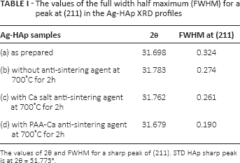
The values of 2θ and FWHM for a sharp peak of (211). STD HAp sharp peak is at 2θ = 31.773°.
Unit cell parameters of Ag-HAp
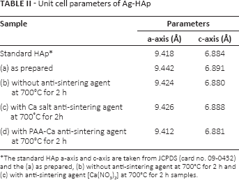
The standard HAp a-axis and c-axis are taken from JCPDS (card no. 09-0432) and the (a) as prepared, (b) without anti-sintering agent at 700°C for 2 h and (c) with anti-sintering agent [Ca(NO3)2] at 700°C for 2 h samples.
Fourier Transform Infrared (FT-IR) spectroscopy
FT-IR spectroscopy was performed to investigate the functional groups present in the HAp, Ca10-xAgx (PO4)6(OH)2, feeding with x = 0.2. The functional groups present in the prepared powder and in the powders calcinated at 700°C for 2 h were identified by FT-IR. These data clearly revealed the presence of the various vibrational modes corresponding to the phosphate and hydroxyl groups. Figure 2 shows the spectra of the samples as-prepared (Fig. 2a), without an anti-sintering agent calcinated at 700°C for 2 h (Fig. 2b), with a Ca-salt anti-sintering agent calcinated at 700°C for 2 h (Fig. 2c), and with a PAA-Ca anti-sintering agent calcinated at 700°C for 2 h (Fig. 2d). The bands at 604/575 and 474 cm−1 were assigned to v4(PO43-) and v2(PO43-) in the lattice. Peaks at 963 cm−1 reflected v1(PO43-) and 1091/1052 cm−1 indicated v3(PO43-). The presence of hydroxyl ions in the lattice was confirmed by absorption bands at 3573 cm−1 and 635 cm1, attributed to the stretching and bending modes of the OH group. The 2 OH bands observed in the samples calcinated at 700°C (Figs. 2b-d) were more intense than those observed in the as-prepared sample (Fig. 2a). In addition, a broad band was observed at approximately 3400 cm1, corresponding to absorbed H2O molecules, which decreased drastically with calcination at 700°C for 2 h, because the Ag-HAp calcinated at 700°C was highly crystalline. These results were concordant with the XRD results, shown in Figure 1. The bands at 874 cm−1 and 1420 cm−1 to 1470 cm−1 indicated the presence of B-type carbonate CO32- ions, which were substituted for PO43- in the apatite lattice (46). The carbonate ions may have originated from a reaction between carbon dioxide and a high-pH solution. In particular, the carbonate bands of the PAA-Ca-treated samples (Fig. 2d), were the strongest of all spectra, because the organic carbon in PAA heated at 700°C was converted to carbonate by reaction with atmospheric oxygen. These main adsorption bands were almost identical to those produced by pure HAp nanocrystals (47). Ciobanu et al reported that the broad bands observed at 1600 cm−1 to 1700 cm1 and 3200 cm−1 to 3600 cm1, which were attributed to adsorbed H2O, were weaker for the as-prepared Ag-HAp than for pure HAp (38). They also reported that the changes provided evidence for the substitution of Ag ions for Ca ions in the lattice. In the present study, no sign of interaction between Ag and the lattice was observed, except for a decrease in water molecules by calcination at 700°C.

FT-IR spectra of the samples: (

SEM images of Ag-Hap: (
Elemental analysis by ICP-AES
Rameshbabu et al (37) and Ciobanu et al (38) have successfully prepared Ag-HAp. The Ag contents of the samples prepared by the former, calcinated at 900°C, were below x = 0.6 of the feed ratio in [Ca10-xAgx(PO4)6(OH)2] before sample preparation, and the samples prepared by the latter, heated at 100°C, were below x = 0.2 of the feed ratio. Unfortunately, neither of these sample products was characterized to determine the quantitative value of the Ag contents in the powder by elemental analyses such as ICP-AES. In the present study, the composition of the HAp crystals and the amount of Ag in the Ag-HAp were determined using ICP-AES, as shown in Table III. The data of the samples calcinated with PAA-Ca at 700°C for 2 h are not shown in the table, because the product was a mixture of HAp and metallic Ag, as shown on the XRD profiles in Figure 1d. Initially, Ag-HAp was prepared by 20 mol% Ag content in the starting materials to verify the extent of Ag inclusion in the HAp. However, the doped Ag content in the HAp lattice was below approximately 1/13 (Tab. III). The fraction value refers to the ratio between 1.5 mol% of actual Ag content in the product determined by ICP and 20 mol% of Ag content in the starting materials. The reduction in Ag seems to be due to the washing of the Ag-HAp during the synthetic procedure, because the substitution of the larger Ag+ (1.28 Å) ions for Ca2+ (0.99 Å) ions in the lattice would be difficult because of the difference in their ionic radii. Furthermore, the Ag content of the Ag-HAp calcinated with Ca salt diminished to approximately 1/40 as shown in Table III. This is because the Ca ions in the lattice were removed during the washing process of CaO that converted from the anti-sintering agent by calcination as shown in Fig. 1S (48), as well as Ag ions. Actually, the (Ca+Ag)/P ratio of the Ag-HAp that was prepared with Ca salt as an anti-sintering agent decreased from 1.69 to 1.65, compared to the product that was prepared without the anti-sintering agent.
Ag content and (Ca+Ag)/P of Ag-HAp
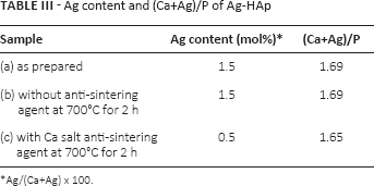
Ag/(Ca+Ag) × 100.
SEM observation and DLS measurement
SEM observations were performed on the powder samples. Figure 3 shows SEM microphotographs of (a, a′) as-prepared, (b, b′) without Ca salt, calcinated at 700°C for 2 h, and (c, c′) with the anti-sintering agent, calcinated at 700°C for 2 h samples, respectively. Most of the Ag-HAp in all 3 reaction systems showed an almost rod-like morphology. This is because the reaction temperature used in the wet method was 100°C, which had the effect of elongating the c-axis. The edges of the particles in the as-prepared sample (a, a′) showed an irregular morphology due to their poorer crystalline phase compared to that of the other calcinated particles. The size of the calcinated Ag-HAp particles did not appear to change, regardless of the presence or absence of anti-sintering agent.
In the as-prepared samples (a, a′), the particles were observed to be close together. Calcination was required to prepare highly crystalline HAp. Calcination in the absence of an anti-sintering agent resulted in considerable agglomeration (Figs. 3b and b′). This was suppressed by adding the Ca salt as anti-sintering agent to the HAp powders before calcination. After adding the anti-sintering agent, the Ag-HAp powders were calcinated at 700°C for 2 h and then cleaned with an NH4(NO3) aqueous solution to remove the anti-sintering agent. Figures 3c and c′ show the SEM images of Ag-HAp samples calcinated at 700°C, when the Ag level was 0.5 mol%. From Figure 3c and c′, it can be observed that the presence of the anti-sintering agent together with the use of calcination resulted in sufficient separation between the Ag-HAp particles.
As shown in Figure 4, the dispersed average particle sizes were measured for the as-prepared (Fig. 4a), without Ca salt, calcinated at 700°C for 2 h (Fig. 4b), and with an anti-sintering agent, calcinated at 700°C for 2 h (Fig. 4c) samples using DLS. The anti-sintering agent performs as an inhibitor of particular aggregation by calcination. The average size of the as-prepared samples (Fig. 4a) containing of secondly particles was 428 ± 85 nm, but when the Ag-HAp was calcinated at 700°C for 2 h without an anti-sintering agent (Fig. 1b), the average size of the particles, which consisted of large agglomerated particle clusters, was 1540 ± 994 nm. This is because calcination causes particle agglomeration. On the other hand, when the Ag-HAp nanoparticles were calcinated in the presence of the anti-sintering agent, the average particle size and distribution of sample (c) was reduced to 325 ± 70 nm (Fig. 4c), which is almost coincidental with the SEM images in Figures 3c and c′. Using Ca salt as an anti-sintering agent inhibited the particle-agglomeration and hence, increased the dispersibility of the Ag-HAp. Therefore, dispersible and crystalline Ag-HAp nanoparticles could be fabricated even with Ca salt alone as an anti-sintering agent, which is expected to be sufficient for achieving a nano-coated monolayer on implantable medical devices.

Size distributions of Ag-HAp particles: (
Antibacterial test
To evaluate the antimicrobial effects of Ag-HAp prepared using different methods with/without an anti-sintering agent, Ag-HAp powders were added to E. coli wild-type cells, and the number of surviving cells was calculated (Tab. IV). Typical data showing the antibacterial effect of Ag-HAp against E. coli, using the agar spot method, are shown in Figure 2S, available online as supplementary material at www.artificial-organs.com. Based on data from 3 independent experiments using 4 types of Ag-HAp, the mean survival rate of E. coli with any Ag-doped HAp nanoparticles was less than 10.0%, and these rates differed significantly from those observed for the addition of pure HAp. In this preliminary assay, no significant differences were observed between the 3 types of Ag-HAp particles, possessing 0.5 to 1.5 mol% Ag content, and even HAp nanoparticles with only 0.5 mol% Ag content showed high antimicrobial activity. The dispersible carboxyl group-donated Ag nanoparticles showed even higher antibacterial activity (49). The antibacterial activity of Ag nanoparticles is well known to be high, because the nanoparticles penetrate the cytoplasm through the cell membrane, damaging proteins and DNA, etc., ultimately causing death (17–22). The antibacterial mechanism of nano-dispersible Ag-HAp nanoparticles may be similar to that of Ag nanoparticles.
Antimicrobial effects of Ag-HAp prepared using different processes
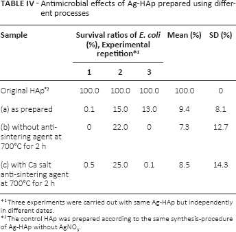
Three experiments were carried out with same Ag-HAp but independently in different dates.
The control HAp was prepared according to the same synthesis-procedure of Ag-HAp without AgNO3.
Conclusions
Dispersible Ag-doped HAp nanoparticles, Ag-HAp, were prepared in the presence and absence of anti-sintering agents in a wet-chemical reaction method and this was followed by calcination. When an anti-sintering agent was added before calcination, HAp-particle agglomeration was prevented. Although metallic Ag was barely detectable in the Ag-HAp that was calcinated with Ca salt as an anti-sintering agent, the XRD analysis clearly showed existence of metallic Ag in the powder samples calcinated using PAA-Ca as the anti-sintering agent. Thus, Ca salt as the anti-sintering agent was more suitable for synthesis of dispersible Ag-HAp. The 3 types of Ag-HAp powders were also assayed for their anti-bacterial activity. The mean sterilization rate of E. coli was greater than 90.0%. The dispersible Ag-HAp nanoparticles containing as little as 0.5 mol% Ag showed an antibacterial effect against E. coli. Vascular-accessing catheters and other medical implant devices coated with the Ag-HAp nanoparticles are now being developed. This is accompanied by a primary safety evaluation using human epithelial and smooth muscle cells.
Footnotes
Financial support: The study was supported in a part by JSPS Grant-in-Aid for Scientific Research (C) (KAKENHI), grant number 25350557, and a grant from the MEXT-Supported Program for the Strategic Research Foundation at Private Universities, grant number S1311045.
Conflict of Interest: T. Furuzono serves as a consultant to SofSera Corporation.