






Abstract
Introduction
We employed a nanosilver-collagen scaffold and tested its effects on inhibiting bacteria and facilitating nerve regeneration.
Methods
Based on our previous research, we prepared bionic scaffolds with different concentrations of nanosilver and examined their internal structures by scanning electron microscopy and energy dispersive spectroscopy. We implanted these scaffolds or autologous nerve grafts into rats to repair a 10-mm injury of the sciatic nerve.
Results
The 2 mg/ml group showed a >10 mm bacterial inhibition zone in all 3 types of bacterial culture dishes. At day 60 postsurgery, the 2 mg/ml group also showed the highest amplitude of evoked potential (AMP) and nerve conduction velocity (NCV). The regenerating nerves in the 2 mg/ml group were denser and more mature, and with thicker and well-arrayed myelin sheath.
Conclusions
These results demonstrate that nanosilver scaffolds (2 mg/ml group) were effective in inhibiting bacteria both in vitro and in vivo, and reduced the contamination-caused immune responses, which in turn promoted nerve regeneration and functional recovery.
Introduction
The repair and replacement of injured peripheral nerve segments have been major issues in clinical practice. In recent years, the use of tissue engineering in constructing peripheral nerve scaffolds has made great progress (1–3). Tissue- engineered artificial nerve scaffolds are mostly made of an extracellular matrix (ECM) that has good cell affinity and a basic structure of a hollow, nonuniform, axial, multichannel catheter. At present, collagen is the ideal material for ECM. Particularly, if the material is modified using a suitable cross-linking method (4), the limitations in the mechanical strength and degradation rate of the material can be overcome while the original tissue and cell affinity is unaffected. A single-channel nerve conduit made of collagen produced by Integra and Synovis is commercially available and has been used clinically.
Peripheral nerve injury often results from open trauma and is accompanied by severe wound contamination. Extensive and thorough wound cleaning should be carried out to prevent infection, followed by continuous drainage. Furthermore, any medical equipment that can easily cause contamination to spread or bacterial parasitism should not be placed into the wound area (5). In this event, if conventional tissue-engineering peripheral nerve scaffold is used, then subsequently early nerve regeneration cannot occur (6). Moreover, the contamination that causes immigration and aggregation of inflammatory cells such as leukocytes can induce a secondary inflammation and peroxide damage to the injured nerves, which in turn can result in severe neuronal apoptosis (7). As a result, some axonal terminals can form neuromas in the early neuronal injury stage, which subsequently seriously affects nerve regeneration (8). In addition, the immigration of a large number of inflammatory cells into the internal microtubules of the scaffold can block the path of growth cones and baffle nerve regeneration (9).
The construction of scaffolds with natural ECM components has become the new direction of tissue engineering. Collagens and gelatin are the main components of ECM. Due to its intrinsic or acquired nature, collagen has been widely used in medical implants, such as artificial skins and wound dressings (10). Furthermore, collagens are nonimmunogenic and their degradation products are not cytotoxic, which is useful to induce and improve the regeneration of peripheral nerves (11). In our previous study, we developed a nanosilver-collagen scaffold that has a highly biomimetic structure by using the improved freeze-dry method to simulate the directional guiding and regeneration promoting effects of the Bungner's band of Schwann cells on neuronal axons. The positively charged nanosilver particles can adhere to laminin (LN) and fibronectin (FN) through electrostatic attraction to modify the scaffold surface. The results of an in vivo experiment indicated that the nanosilver scaffold was an ideal tool to repair the 10-mm defect in the rat sciatic nerve (6, 12). Further physicochemical property testing revealed that when the final concentration of nanosilver within the scaffold was 1 mg/ml to 2 mg/ml, the scaffold had the greatest tensile strength and also showed an ideal internal structure and degradation rate (13, 14). Particularly, due to the strong antibacterial property of nanosilver, the release of silver particles in the body can provide a continuous antibacterial effect at the wound area. Therefore, in the present study, we aimed to compare the antibacterial effects of tissue engineering scaffolds that contain different concentrations of nanosilver to morphologically and functionally assess the efficiency of the nerve regeneration.
Materials and methods
Animals
A total of 36 Sprague Dawley (250–300 g) rats with normal leukocyte number were provided by the Experimental Animal Center of the Fourth Military Medical University. The experiments were approved by the Institutional Animal Care and Use Committee of the Fourth Military Medical University (Xi'an, China). All animal handling and procedures met the ethical guidelines, which determined that we appropriately minimized the number of animals used, and limited their suffering in this investigation.
Preparation of the nanosilver scaffolds
As described in our previous studies (6, 12), 150 mg collagen I (C9879; Sigma, St Louis, MO, USA) and 75 mg gelatin (G9382; Sigma) were dissolved in 0.05 M acetic acid (pH 3.2), stirred at 18 000 rpm at 4°C or 90 min in overhead blender (RW20, IKA Works, USA), and stocked at 4°C overnight after negative pressure vacuum. The liquid nanosilver particles (Shanghai Tinaph Nano-Tech, Shanghai, China) were added into the collagen-gelatin suspension at final concentrations of 1 mg/ml and 2 mg/ml, respectively. Each mixture was injected into a separate silicone tube (internal diameter, 3 mm; length, 10 mm). After sealing the 2 ends, the tube was immersed in the coolant along the axial direction at a speed of 2 × 10⁻⁵ m/s using a homemade micro-speed controller (Shanghai Precision Instruments, Shanghai, China) (6). The suspension and frozen tube were placed into a precooled aluminum plate. The 2 ends of the tube were opened, freeze-dried at -40°C and 100 mtorr by the Alpha 2–4 LDPlus freeze dryer (Martin Christ, Osterode am Harz, Germany) for 48 h to obtain the nanosilver-collagen material. The scaffolds were then cross-linked with a 1 mg/ml solution of genipin (Challenge Bio-products, Taiwan, China) for 48 h. Next, the cross-linked scaffolds were rinsed 3 times with distilled water, dehydrated for 30 min with 95% of ethanol, and air-dried for 1 week. The scaffolds were disinfected using Co60 and sealed for use.
Observation of internal scaffolds using scanning electron microscopy and energy dispersive spectroscopy
The scaffolds containing 1 mg/ml or 2 mg/ml nanosilver were cut open along the transverse and longitudinal cross sections to spray gold coating. Scanning electron microscopy (SEM) (S-3400; Hitachi Medical, Hitachi, Japan) was performed to observe the transverse and longitudinal cross sections and to measure the diameter of the micropore. Energy dispersive spectroscopy was performed to observe the distribution of the nanosilver particles in the material by using energy dispersive spectrometer and EMAX ENERGY software (EMAX-7021-H, HORIBA, Ltd., Japan).
In vitro antibacterial test of the nanosilver scaffold
The antibacterial test was performed according to the 2007 criteria of the Clinical and Laboratory Standards Institute (CLSI). Staphylococcus aureus, Pseudomonas aeruginosa and Escherichia coli colonies were inoculated into the screening plate at a dose of 0.5 McFarland units. Each bacteria was inoculated into 3 plates for the Autologous group, the 1 mg/ml scaffold nanosilver group, and the 2 mg/ml nanosilver group, respectively. The scaffold and 1 segment of normal rat sciatic nerve were attached to the center of the colonies, and the plate was incubated at 37°C for 24 h. If the scaffold or the sciatic nerve generated a bacterial inhibiting zone, the scaffold was determined to have an antibacterial effect on the corresponding bacteria. Furthermore, the diameters of inhibiting zone were measured. These in vitro antibacterial tests were repeated 2 more times for the 3 species of bacteria.
Preparation of S. aureus suspension
Under sterile conditions, 5. aureus was inoculated into the slant culture medium using the line method, cultured at 37°C for 24 h, and activated. Two ring bacteria moss were selected from the activated bacteria and diluted with sterile water to obtain the 10 7 ∼10 8 CFU/ml S. aureus suspension.
Bridging surgery
Animals were randomly divided into 3 groups. The rats received anesthesia through intraperitoneal injection of 3% sodium pentobarbital. Routine skin preparation, disinfection, and draping were performed at the surgical area of the left thigh. An incision was made at the middle of the left thigh to cut open the skin and fascia. The sciatic nerve was isolated along the intermuscular cleft, and a 10-mm nerve stem was cut off. The nanosilver scaffold (1 and 2 mg/ml) was conjugated to the proximal and distal cutting ends of the sciatic nerve. For the Autologous group, the removed nerve segment was rotated 180° and sutured to both the proximal and distal nerve stumps with nondamaging thread. The epineurium was fixed with the superficial layer of scaffold with nondamaging 10/0 sutures, which reduce the injury to the nerve bundle. For each group, 2 mL S. aureus suspension was injected into the injury site and the surrounding deep fascia. The incision was closed layer by layer. After surgery, the rats were fed and monitored regularly without antibiotic treatment.
Blood collecting and leukocyte counting
From 1 day before surgery to 60 days post-surgery, 20 μL blood was collected from the tail vein of each rat every other day, and the number of rat peripheral blood leukocytes was counted using the Mek-5216k Hematology Analyzer (Nihon Kohden, Tokyo, Japan), until the number was stabilized at the level of the operation day for 3 consecutive days.
Sciatic functional index
Footprint analysis was performed on all rats at days 20, 40, and 60 after surgery and the sciatic functional index (SFI) was used to evaluate the recovery of hind limb function (15). Pre-Operatively, the rats were trained to walk down a wooden track (50 × 7 cm) into a darkened goal box. Postoperatively, the rats’ hind paws were painted with nontoxic finger paint, and any changes in their paw prints that resulted from nerve injury and denervation were recorded. The recordings continued until 5 measurable footprints were collected. The SFI was calculated as follows:
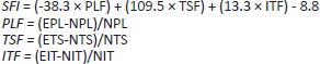
where print length (PL) is the distance from the heel to the top of the third toe, toe spread (TS) is the distance between the first and the fifth toe, and intermediary toe spread (IT) is the distance from the second to the fourth toe. NPL, NTS, and NIT represent the PL, TS, and IT, respectively, recorded from the nonoperated foot. EPL, ETS, EIT represent the PL, TS and IT, respectively, recorded from the operated, experimental foot. The calculations for print length factor (PLF), toe spread factor (TSF), and intermediary toe spread factor (ITF) are shown above. Changes in the SFI correlate with changes in the rat paw print and are indicative of nerve injury and regeneration.
Although the SFI is an indirect measurement, it has been well established as an indicator of functional nerve restoration. SFI values that are close to zero indicate better recovery. An SFI value of -100 indicates complete loss of function.
Electrophysiology
After determination of SFI, the rats were anesthetized with 350 mg/kg ketamine, the sciatic nerve on the operative side was exposed, and 2 electrodes were positioned on the nerve 1.5 cm from the end of the scaffold. Viking IV EEG Unit evoked potential equipment (Nicolet, Middleton, WI, USA) was used to record the evoked potential; the distance between the 2 electrodes was 3.0 cm; the wave width was 0.1 ms; the frequency was 5.0 Hz; the intensity was 25 mV to 50 mV; electric resistance was less than 15 MO.; the stimulation could induce the lower limb to tremble and the signal was amplified more than 100 times by a preamplifier. The amplitude of the evoked potential (AMP) and the nerve conduction velocity (NCV) were then recorded.
Transmission electron microscope examination and gastrocnemius muscle weighed
The 12 rats in each group were sacrificed by cervical dislocation and the scaffolds were removed 60 days after surgery. The middle portion of the scaffold (5 mm) was taken and fixed in 2.5% glutaraldehyde for 24 h, washed 3 × 10 min in 0.01 mol/l phosphate-buffered saline, postfixed in 1% OsO4 overnight, dehydrated in acetone series, embedded in Epon812, and trimmed and cut into ultra-thin transverse sections (0.95 μm). The sections were then stained in lead citrate and the thickness of the myelin sheath was examined under a transmission electron microscope (TEM). At the same time, the gastrocnemius muscles were carefully cleaned and dissected out, dividing their tendinous origin and insertion from the bone from intact and injured sides. Bilateral gastrocnemius muscles were weighed while still wet, using an electronic balance for determination of the wet weight ratio of the gastrocnemius muscle. In each rat, the gastrocnemius muscle wet weight ratio was calculated based on the weight of the gastrocnemius on the experimental leg versus that on the normal leg. All measurements were made by 2 independent observers unaware of the analyzed group.
Statistics
All data are expressed as mean ± SD. One-way ANOVA followed by Bonferroni's test for comparisons among multiple groups were used to assess statistical significance. In all cases, a value of P<.05 was considered to be significantly different.
Results
Internal structures of the nanosilver scaffolds and spectroscopy results
The constructed scaffold with nanosilver was in a cylindrical shape (Fig. 1A); scaffolds containing different concentrations of nanosilver had identical internal structures of parallel microtubules along the axial direction (Figs. 1B and C). The microtubules showed the uniform internal diameter of 20 urn to 80 urn, which was similar to the sciatic nerve and thus can provide a temporal scaffold and channel for nerve regeneration. The energy spectroscopy results indicated that the silver particles existed (Fig. 2A) and were evenly distributed in both concentrations of nanosilver scaffolds (Figs. 2B and C).

Structure of tissue-engineered scaffold containing nanosilver: (A) a picture of nanosilver-collagen type I scaffolds appeared in cylindrical shape; SEM showed the internal structure of the stent, indicating that both the axial (B) and cross (C) sections of the nanosilver scaffold are arranged as axially paralleled microtubules. Scale bar = 1 cm.

(A) energy spectrometry of a nanosilver-embedded scaffold showing C, 0, and silver elements. Energy spectrometry results showed evenly-distributed nanosilver particles in both 1 (B) and 2 (C) mg/ml groups, and the content of nanosilver particles in the latter was significantly higher than the former. Scale bars = 800 nm in B-C.
In vitro antibacterial test
As shown in Figure 3, the 2 mg/ml scaffold group showed a >10 mm bacterial inhibiting zone in all 3 types of culture dishes including 5. aureus, P. aeruginosa, and E. coli, indicating that the scaffold containing 2 mg/ml nanosilver can take an obvious antibacterial effect on all these 3 types of bacteria. However, neither the 1 mg/ml group nor the Autologous group showed a bacterial inhibiting zone, indicating the absence of an antibacterial effect in these 2 groups. To investigate the effects of nanosilver scaffolds in vivo, S. aureus was chosen to be injected into the injury site. First, 5. aureus is one of the most frequent causes of traumatic wound contamination and the infection is difficult to treat (16, 17). Second, given the results of our in vitro investigations, the inhibiting zone of S. aureus is larger than the others. Finally, single bacterial infection is far more common than the compound infections of these 3 bacteria.

Scaffolds of 2 mg/ml group showed evident antibacterial effects in all three culture plates with Staphylococcus aureus (A), Pseudomonas aeruginosa (B) and E. coli (C), respectively. Scale bars = 2 cm.
Post-surgery observation
The procedures for animal surgery are shown in Figure 4. The sciatic nerve was intact before surgery (Fig. 4A) and the auto sciatic nerve and scaffolds were sutured with the residual nerve (Figs. 4B and C). After surgery, the wound healing, food intake, and activity of the rats were monitored daily. At first, the lower limb activity on the operative side was limited, and the posture could only be maintained by the residual tensile strength. With the increase of time, the lower limb activity on the operative side was obviously improved. Starting from day 3 postsurgery, some rats in the 1 mg/ml and the Autologous groups showed wound swelling and excessive exudates, less food and water intake, and emotional irritability. The rats did not receive any treatment, and the symptoms were relieved after 1 week. The 2 mg/ml group rats showed ideal wound healing without wound swelling or excessive exudates.

Pictures for animal surgery. The sciatic nerve was exposed in (A). For the autologous group, the removed nerve segment was rotated 180° and sutured to both the proximal and distal nerve stumps (B). The nanosilver scaffold (1 mg/ml and 2 mg/ml) was conjugated to the proximal and distal cutting ends of the sciatic nerve (C). Scale bar = 1 cm in B (applies in A and C).
Leukocyte counting
Starting from 1 day before the surgery, blood samples were collected from the tail vein every other day, and the number of leukocytes was counted (Fig. 5). The results indicated that the number of leukocytes showed a remarkable increase from the first day, and reached a peak about two times the base value on the third day. The peak number of leukocytes was lower in the 2 mg/ml group than in Autologous nerve grafting group (P<.001). Starting from day 7 postsurgery, the number of leukocytes started to decline, and at day 15, the number returned to the pre-operative level. For the 2 mg/ml scaffold group, the leukocyte number also showed a peak at day 3, which was 50% higher than before the surgery; the number started to decline from day 5, and returned to the pre-operative level at day 11.

Postoperative peripheral leukocyte counts showed that there were various degrees of increase in peripheral leukocyte counts after the surgery. The 2 mg/ml nanosilver group showed the lowest peak leukocyte count and the fastest recovery, indicating a mild infection state, followed by the 1 mg/ml nanosilver group; the infection in autologous nerve graft group was relatively severe.
SFI measurement
As shown in Figure 6, at day 20 postsurgery, the difference between the 3 groups did not show significance in the SFI (1-way ANOVA, F(2,33) = 0.52, P = 0.60). At day 40 (1-way ANOVA, F(2,33) = 37.19, P<0.0001) and day 60 (1-way ANOVA, F(2,33) = 62.20, P<0.0001) postsurgery, there was a significant difference between the 3 groups. Additionally, the 2 mg/ml scaffold group showed better functional recovery of the sciatic nerve than the 1 mg/ml scaffold group (P<.0001 at day 40; P<.05 at day 60) and the Autologous group (P<.0001 at day 40; P<.0001 at day 60). It is remarkable that the 1 mg/ml group had better functional recovery than the Autologous group (P<.0001).

The sciatic functional index (SFI) in each group. No significant difference in SFI was observed among groups at day 20 after the surgery, and all groups showed severe nerve damage. SFI at days 40 and 60 after the surgery exhibited significant difference among groups, i.e., due to a relatively severe infection, the Autologous group showed the slowest recovery, while the 2 mg/ml nanosilver group exhibited the best functional recovery under the infection condition.
Electrophysiological recordings and gastrocnemius muscle weighed
As shown in Table I, there was a significant difference between the 3 groups in AMP (1-way ANOVA, F(2,33) = 88.02, P<.0001) and NCV (1-way ANOVA, F(2,33) = 227.1, P<.0001) at 60 days postsurgery. The AMP and NCV of sciatic nerves in 2 mg/ml scaffold group were higher than in the 1 mg/ml scaffold group (P<.0001, P<.0001) and the Autologous group (P<.0001, P<.0001). Compared with the contralateral normal muscle, different degrees of gastrocnemius muscle atrophy in the operation side of the 3 groups were observed. The wet weight ratio of the gastrocnemius muscle on the operation side is shown in Figure 7. There was significant difference between the 3 groups (1-way ANOVA, F(2,33) = 34.62, P<.0001). Furthermore, the wet weight ration of gastrocnemius muscle in the 2 mg/ml group was significantly higher than those in the 1 mg/ml group (P<.0001) and Autologous group (P<.0001). The wet weight ration of gastrocnemius muscle in the 1 mg/ml group was also higher than in the Autologous group (P<.05).

The wet weight ratio of gastrocnemius muscle in each group. Gastrocnemius muscle wet weight ration measurement (gastrocnemius muscle weight of experimental leg/gastrocnemius muscle weight of normal leg) showed that at day 60 after the surgery, various degrees of atrophy were observed in the 3 groups. The muscle atrophy in the Autologous group was most serious, while the 2 mg/ml nanosilver group had the highest wet weight ratio of gastrocnemius muscle.
The electrophysiological assessments in each group
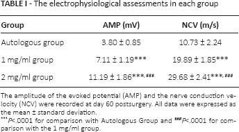
The amplitude of the evoked potential (AMP) and the nerve conduction velocity (NCV) were recorded at day 60 postsurgery. All data were expressed as the mean ± standard deviation.
P<.0001 for comparison with Autologous Group
P<.0001 for comparison with the 1 mg/ml group.
TEM examination
At day 60 after implantation into the injured nerve tissues in the rat, the newly formed nerve fibers were dissected and processed for TEM. The observations showed results: the regenerating nerves from the 2 mg/ml scaffold group were denser and more mature, with thicker and well-arrayed myelin sheath, similar to nerves in the native group (Fig. 8A). The mean thickness of the myelin sheath was 3.9 ± 0.4 mm in the native group, while the mean thickness was 3.2 ± 1.1 mm in the 2 mg/ml scaffold group (Figs. 8A and B). In the 1 mg/ml scaffold group, the regenerating nerves were growing along the microtubules and bypassing the defective area, but the nerve fibers were thin and sparse and not well arrayed; the newly grown myelin sheath was thin and with small cells and rich vessels. The mean thickness of the myelin sheath was 2.2 ± 0.5 mm (Fig. 8C), and the result of Autologous group was 1.7 ± 1.6 mm (Fig. 8D).

Transmission electron microscope (TEM) examination in each group. The nerve fibers in the native group were shown in (A). At day 60 after the bridging operation, TEM showed that the 2 mg/ml nanosilver group (B) exhibited the largest number of regenerated nerve fibers, which were arranged neatly, with significantly superior myelin thickness, compared to the 1 mg/ml nanosilver group (C) and the Autologous group (D). However, the nerve fibers in the native group (A) were better than those in the 2 mg/ml nanosilver group. Scale bars = 2 μm in A-D.
Discussion
In the present study, we investigated the efficacy of nanosilver-collagen scaffolds for enhancement of axonal regeneration after peripheral nerve injury under contamination. Our in vitro study indicates that 2 mg/ml nanosilver scaffolds possess wide-spectrum antibacterial activity for inhibiting all 3 bacteria. In addition, our study also showed that both 1 mg/ml and 2 mg/ml nanosilver scaffolds significantly reduced the immune response and led to better axonal regeneration and functional recovery than the Autologous group. These findings indicate that the nanosilver scaffolds are capable of inhibiting bacteria and facilitating nerve regeneration after peripheral nerve injury under contamination.
Biodegradable scaffolds were produced by using collagen type I and gelatin as the main materials in combination with nanosilver particles evenly distributed at the internal surface of the microtubules. They had the structure of axially arrayed microtubules and are highly biomimetic in both composition and structure, which is favorable to the induction of directional growth in the axons. The fine, dense surface structure can prevent the inward growth of fibrous connective tissue of the body. Furthermore, the scaffolds use ECM components as the main constituents and can be used for bionics in tissue engineering, efficiently stimulating the growth of axons and guiding the extension of regenerating axons toward the longitudinal direction along the microtubules.
Previous studies mostly suggested that the efficacy of the nerve autograft is better than any known tissue engineering scaffold, and thus it has been used as the gold standard for the treatment of peripheral nerve injury (9, 10). However, in the present study, using the most common 5. aureus to simulate surface contamination, we found that the 2 mg/ml scaffold group showed the best results in wound healing, axonal regeneration, and functional recovery. For the 1 mg/ml scaffold group, although we did not observe an obvious bacterial inhibiting zone in the in vitro experiment, the in vivo bridging experiment still showed a better repair effect compared to the autologous graft group. These observations indicated that wound contamination and inflammatory response can suppress the regeneration of peripheral nerves; the nanosilver peripheral nerve scaffold can not only directly kill bacteria at the wound site, it can also reduce the neuronal peroxide damage that induces inflammatory cells.
Nanosilver is an antibacterial product developed based on nanotechnology. Compared to Ag+, nanosilver shows more stable physicochemical properties, and thus has excellent electrical, optical, and catalytic characteristics. Due to the quantum effect, the small size effect, and the large specific surface area, nanosilver can generate a superb, safe, and longlasting antibacterial effect compared to the traditional inorganic antibacterial agents (18, 19). Dibrov et al (20) suggest that nanosilver employs the same antibacterial mechanism as silver, but at different effective concentrations. Usually, the diameter of the nanosilver particles is as low as 10 nm to 100 nm, which enables them to easily enter the pathogens. As a result, the nanosilver particles can rapidly bind to the thiol base (-SH) of the enzymes within the pathogens to inhibit the -SH dependent enzymes, resulting in bacterial death by disturbing their metabolism. By this means, nanosilver particles can exert an antibacterial effect and promote wound healing (21). Feng et al (22) applied silver to wounds and found that nanosilver could bind the DNA bases of the pathogens to form cross-linking to replace the hydrogen bonds between the adjacent nitrogen in the purine and pyrimidine. As a result, the bacterial DNA cannot replicate and the proteins are inactivated, resulting in the apoptosis of the pathogens. Lok et al (23) studied the antibacterial mechanisms of nanosilver using the proteomics approach and found that a short-term exposure of E.coli to nanosilver can lead to the accumulation of envelope protein precursors; additionally, the plasma membrane was damaged, resulting in decreased membrane potential and intracellular ATP concentration. Bhol et al (7) reported that nanosilver can also have an anti-inflammatory effect by suppressing the expression of IL-12 to induce the apoptosis of inflammatory cells. Demling et al (24) also suggest that silver can have an anti-inflammatory effect to inhibit DNA replication, and has strong permeability, which allows the silver material to penetrate into the subcutaneous tissue to continuously release nanosilver. The nanosilver particles at the wound surface rapidly diminish the pathogens at the wound and suture.
Due to its antibacterial mechanism, nanosilver can have a wide-spectrum, antibacterial effect. In addition, after the collapse of the bacteria, the nanosilver particles can enter new bacteria to further function without self loss. Therefore, in clinical practice, a low-concentration, nanosilver-embedded, peripheral nerve scaffold can continuously take effect at the wound site. In addition to reducing the local inflammatory response at the wound, the nanosilver particles can also reduce the time and dose of the systemic prophylactic antibiotics. This may be an important reason for the antibacterial differences between in vitro and in vivo investigations of the 1 mg/ml nanosilver scaffold group. The antibacterial effects might be more powerful with a longer observation time.
Bacterial biofilm (BF) is an extracellular, macromoleculewrapped, bacterial cluster that is attached to the surface of living or inanimate surfaces. BF is a highly organized multicell structure formed on the surface, and it is a very important environmental adaptation mechanism of bacteria (25). According to the U.S. Centers for Disease Control and Prevention (CDC), infection caused by built-in medical equipment is the fifth largest cause of death of hospitalized patients (26). Sixty-five percent of cases of human bacterial infection are related to the formation of BF (27). This protecting “shell” enables the bacteria to evade the host's immune surveillance. Even if the bacteria on the membrane surface are eliminated by drugs or other antibacterial products, it is difficult to affect the bacteria inside the membrane, and thus the bacteria can exist in the body for a long time. To effectively prevent the growth and proliferation of bacteria, it is necessary to prohibit the formation of BF on the surface of medical equipment. Hence, it is critical to prevent BF formation. In clinical practice, peripheral nerve injury is always combined with severe open trauma, and subsequently the wound harbors a high risk of surface contamination. During the application of the tissue engineering scaffold in the repair of an injured nerve, the bacteria in the wound can easily accumulate on the scaffold to form BF and subsequently cause continuous systemic infection. However, without the tissue engineering scaffold, the proximal part of the injured nerve degenerates and permanently loses its function. Hence, it has been difficult for surgeons to make a choice. The nanosilver scaffold not only has a simulative internal structure and biological mechanics, it can also continuously exert a stable antibacterial effect throughout the whole process from implantation to degradation. Therefore, the nanosilver scaffolds can be an appropriate choice used for repairing peripheral nerve defects under contamination conditions in clinical practice.
There are several limitations in our investigation. First, we only tested 1 kind of bacteria in vivo although we have investigated the antibacterial activity of nanosilver scaffolds on 3 kinds of bacteria in vitro. Second, the time points we chose to observe are limited and we should examine the functional recovery and axonal regeneration of the sciatic nerve on more time points, both in the early and late phases. Third, the suitable concentration of nanosilver in the scaffolds is unclear. The antibacterial activities of different concentrations of nanosilver scaffolds should be investigated both in vivo and in vitro.
Conclusions
Our study showed that the nanosilver-collagen scaffolds (especially the 2 mg/ml group) inhibit bacteria both in vitro and in vivo, with evenly distributed nanosilver particles in the internal surface of the microtubules. These nanosilver scaffolds efficiently repaired a 10-mm injury of the sciatic nerve in adult rat significantly better than the autologous group, under contamination. Nanosilver-embedded scaffolds may promote nerve regeneration and functional recovery by reducing the immune responses caused by contamination. It is tempting to speculate that these new scaffolds will have a promising application for nerve tissue engineering, especially for situations under contamination.
Footnotes
Financial support: This work was supported by grants from the National Natural Science Foundation of China (No. 81100900, No.31010103909, No.81371239) and Shaanxi Provincial Program for Science and Technology Development (No. 2011K12–59).
Conflict of interest: None.