






Abstract
Purpose
Implanting the largest valved conduit possible – oversizing – to reconstruct an absent connection from the right ventricle to the pulmonary artery in certain types of congenital heart defects has been suggested as a compensating measure for somatic outgrowth of the patient. However, one effect that has not been investigated yet is the hemodynamic consequence. For this purpose, virtual implantation and flow simulations were conducted in this study.
Methods
To isolate the effects of conduit oversizing on the hemodynamics observed after conduit implantation and outgrowth, calculated wall shear stresses (WSS) of image-based computational fluid dynamic (CFD) simulations were used as indicators. Three different sizes of valved conduits (20 mm, 22 mm, and 24 mm), including the largest possible conduit size, virtually implanted in a child-sized healthy pulmonary artery and the corresponding adult-sized model were investigated.
Results
The child and adult models show a decrease of the mean WSS (approx. 26%) in the whole domain with an increase of the conduit size. When looking at the mean WSS at the anastomosis, for the child model the WSS is significantly increased (approx. 40%) when oversizing (Z-score +3.21). In contrast, the stresses are decreased for the adult model (34%) when using the largest conduit (Z-score +0.25).
Conclusions
Based on the results of this study, it must be considered that choosing a prosthesis size that will lead to high WSS and an associated intimal reaction, possibly leading to stenosis, can defeat the benefit of having a nominally larger orifice area directly after implantation.
Keywords
Introduction
A wide variety of congenital heart defects associated with an absent or deficient right ventricular to pulmonary artery connection require surgical reconstruction, which may include placement of a conduit from the valved right ventricle (RV) to the pulmonary artery (PA). These conduits are plagued with relatively high failure rates: approximately only half of the patients are free from reoperation 10 years after the initial placement of the conduit (1). Commonly reported modes of failure for valved RV-PA conduits are stenoses at the site of distal anastomoses, valvular stenoses, calcification of the biological conduits and valves, proximal anastomosis stenoses (2, 3), and insufficiencies of the valve or infections. In small children whose body size increases very rapidly, conduit failure is also associated with the somatic outgrowth. The insertion of the largest conduit possible in children, known as oversizing (1, 4), is one method of compensating for the fact that today no RV to PA conduit possesses potential for growth.
The Z-score is usually used to define oversizing. It is given by

where x is the observed dimension in the patient with a known body surface area, μ is the mean normal dimension and σ the standard deviation around the mean normal dimension in normal individuals of the same body surface area as the patient.
Recent clinical studies in this field have shown that oversizing in children under the age of 2 years is beneficial (1, 5, 6) and oversizing of the RV-PA conduit even over Z>3 is recommended (1). There is still some controversy regarding oversizing in bigger children. In a study by Karamlou et al (7) from 2005, the conclusion was that there is no significant benefit to placing an oversized RV-PA homograft conduit in children aged >10 years. Askovich et al (4) even reported decreased durability in children (median age of 5.6 years) when oversizing excessively (Z>2.7).
With the advent of transcatheter pulmonary valves, a new possibility for treating patients with failing RV to PA conduits has emerged that offers a less traumatic option for reestablishing valvular function. When sizing an RV to PA conduit one must therefore also consider the option of a future transcatheter valve in valve implantation. With the smallest available transcatheter valve having a diameter of 18 mm, since such prostheses are currently not suitable for implantation in small conduits, implantation of oversized conduits may be favored to allow for later treatment via a catheter-based approach.
Taking all these factors into account, it seems that a large majority of surgeons would be inclined to oversize when implanting an RV-PA conduit. However, one effect that has not been investigated yet is the hemodynamic consequence of implanting a larger sized, orthotopically implanted conduit in a child's pulmonary artery position. Analyses isolating the effects of oversizing on altered flow conditions are difficult in a clinical setting, since the isolation of this factor is nearly impossible due to the anatomical and physiological variations of treated patients. For this reason, a computational fluid dynamics (CFD) simulation study was conducted. In vivo and in vitro studies have shown that altered stress exerted on a vessel wall leads to fibrin deposition and intimal overgrowth at the site of the disturbance (8, 9). This is why altered wall shear stress (WSS) was chosen as the indicator for possible intimal overgrowth, a cause for conduit or allograft failure (10). The hemodynamic effects of 3 differently sized valved conduits virtually implanted in a child-sized, healthy PA and in the corresponding adult-sized models were computationally investigated. The conduit sizes were selected to simulate a body surface adjusted conduit size and oversizing up to the 20 highest possible size that would fit the anatomy of the chest. The size of the child's was chosen to correspond to the age (10 years) and body surface of a child who's body growth has become more stable (11), so the oversizing would theoretically exert its hemodynamic effects for a longer period of time.
Methods
Anatomical Models
To create a generic 3D model of a pulmonary trunk, CT scan images of an adult patient with a healthy RV and pulmonary trunk and arteries were used. Imaging was helical and performed using a CT whole body scanner with a 0.7-mm slice increment and a 211-mm field of view. Mimics 14.11 (Materialise, Leuven, Belgium) was used for the segmentation and construction of a 3D model consisting of the infundibulum, main pulmonary artery (MPA), right pulmonary artery (RPA), and left pulmonary artery (LPA). Since the CT images are of an adult patient, the model was scaled in order to obtain a pulmonary artery of a 10-year-old child. The mean diameter of the MPA in adults is 25.1 ± 2.8 mm according to the literature (12), corresponding well to the presented case (27.4 mm). Knobel et al (13) investigated 69 children with a median age of 10 ± 4.9 years. They measured a MPA mean diameter of 17.6 ± 5.1 mm. Considering these measured values, a scaling factor of 0.7 (17.6/25.1) was chosen. The same ratio is calculated when using autopsy data from literature (14). Child and adult models are labeled with C and A in this study, respectively.
Three different sizes (diameters of 20 mm, 22 mm, and 24 mm) of a computer-aided design (CAD) model of a trileaflet valve inside a conduit with 3 sinuses were used. For each size, 2 states for the leaflet position (fully open and half open) were modeled based on structural finite element method (FEM) simulations. These models are labeled F and H, respectively.
To attach the conduit to the PA, both 3D models were imported in 3-Matic 7.0 (Materialise, Leuven, Belgium). The conduit models were cut and attached to the pulmonary arteries of the adult model as well as the scaled child model according to surgical procedures (15) under the guidance of an experienced pediatric heart surgeon. The resulting Z-scores are shown in Table I. This was performed for all conduit sizes and both leaflet positions, resulting in 12 CAD models.
Z-Scores for the different models and conduit sizes
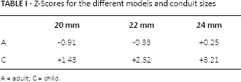
A = adult; C = child.
To minimize the effect of outlet boundary conditions, straight extensions were added at the outlets of all geometries investigated this study.
Numerical Setup
A hexahedral core mesh was generated using ICEM CFD 14.5 (Ansys Europe Ltd., Otterfing, Germany). To accurately resolve boundary layers of the flow, prism layers were added (Fig. 1). A mesh independence study was performed by comparing the pressures (average and profile), velocites (peak and profile), and averaged WSS values of steadily increasing mesh densities. A difference below 1% was aimed for. This resulted in 4 prism layers and a total mesh number between 4.3 million (20 mm conduit) and 5.3 million (24 mm conduit) for the child model, and between 5.6 million (20 mm conduit) and 6.6 million (24 mm conduit) for the adult model. The resulting y+ value (the nondimensional wall distance, defined as the wall distance times the shear velocity divided by the kinematic viscosity) was below 1 for all simulations, therefore the mesh was able to resolve the boundary layer appropriately.
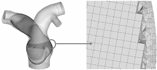
Final 3D model and used mesh (shown in 1 plane) of the CFD simulation.
The finite volume CFD solver ANSYS Fluent 14.5 (Ansys Europe, Otterfing, Germany) was used to solve the governing Navier-Stokes equations for the blood flow. To isolate the addressed problem from unknowns and uncertainties like material properties, anastomosis type, etc., a steady flow condition without wall motion was used. Motions of the heart valve leaflets and vessel wall were not considered. The Reynolds averaged Navier-Stokes (RANS) Shear Stress Transport (SST) k-ω turbulence model was used, which is well suited for the internal, low-Reynolds number flow (between 900 and 4000) and commonly used for arterial flow simulations (16).
According to Cheng et al (17), the average blood flow rate through the MPA at rest is 2.9 l/min per m2 for children and 2.2 l/min per m2 for adults. Considering a mean body surface area for children and adults of 1 m2 and 1.9 m2, respectively, this leads to average flow rates of 2.9 l/min for the child model and 4.18 l/min for the adult model. These values were used at the inlet for the half-open cases (early systole). For the fully open cases (peak systole), 3 times these flow rate values were applied. A no-slip condition (fluid has zero velocity relative to the boundary) was assumed on the walls and the gravitational effects on the blood flow were ignored.
The third-order Monotonic Upstream-Centered Scheme for Conservation Laws (MUSCL) (18) was used for spatial discretization to minimize numerical dissipation. The Semi-Implicit Method for Pressure-Linked Equations (SIMPLE) (19) algorithm was used for coupling between the pressure and the velocity field. To model the shear rate-dependent viscosity of blood, the non-Newtonian Carreau model (20) was applied (equation 1):

where η is the fluid dynamic viscosity, n = 0.3568 the flow index,
Pulmonary Resistance
To achieve the physiological flow split ratio between the RPA and LPA, the vascular resistances at the outlets have to be considered. Sun et al (22) used a porous model to simulate the pulmonary resistance in patient-specific CFD models of an extracardiac conduit Fontan connection. They concluded that a porous portion can be used to realistically simulate specific pulmonary resistances and pressures at the outlets, also during flow rate changes in the pulmonary arteries.
In this study, a fixed relative pressure of Prelative = 5 mmHg was combined with vessel-specific, flow-dependent resistances at the outlets to achieve the desired flow split between the RPA (∼55%) and the LPA (∼45%) according to Cheng et al (17). The resistance was calculated by using the dimensionless loss coefficient L, which is expressed according to the Darcy-Weisbach equation (2):

where ΔP is the pressure loss in the pulmonary arteries,


Using

where α
n
is the split ratio of the flow rate Q for each PA with the cross-sectional area of A
n
and velocity (

Therefore, LLPA can be calculated when LRPA is set:

In order to achieve the desired flow split ratio of RPA/LPA = 55%/45%, the value of LRPA must be estimated iteratively. For this purpose, the resulting flow split with values for LRPA varying between 0 and 256 was analyzed. Three different models of healthy adult human pulmonary anatomies (Figs. 2a, b, and c) were created using the segmentation and postprocessing methods previously described for the models with implanted conduits. These models were used to study the effects of changes in the LRPA value on the flow split. A flow rate of 4.18 l/min was applied at the inlets for this study.
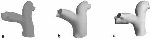
Three different models (
Wall Shear Stress as an Indicator of Intimal Overgrowth
Altered wall shear stress (WSS) patterns in the region of the anastomoses appear to be one major factor in the development of fibrin deposition and intimal growth leading to conduit or allograft failure (8–10). Therefore, altered WSS was chosen as the indicator for conduit dysfunction in this study. WSS, τ W , is given by
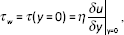
where η is the dynamic viscosity, u is the flow velocity parallel to the wall, and y is the distance to the wall. Previously Tang et al (23) performed a CFD study to compare the time-averaged central pulmonary arterial WSS in rest condition with that at exercise condition. They observed a significant increase in WSS during exercise. Using the same approach, Tang et al (24) compared pulmonary arterial hypertension (PAH) patients with normal subjects. In their study, WSS in the proximal pulmonary arteries of PAH patients was found to be significantly lower than in the control group.
Results
Anatomical Study
Figure 3 shows the virtually implanted conduits in a child PA (C) and the corresponding adult PA (A), respectively. Using a 20-mm conduit leads to a visible narrowing after somatic growth.
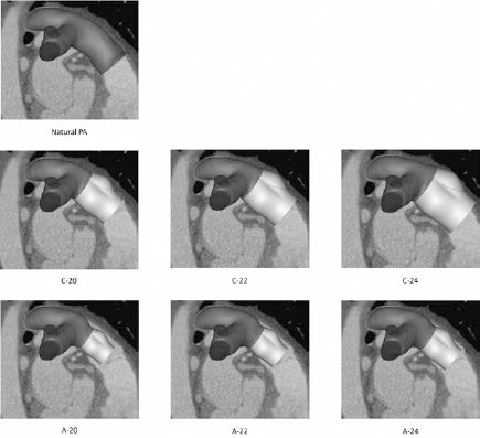
Implantation of different conduit sizes (20, 22, and 24 mm) on a child PA (
Pulmonary Resistance
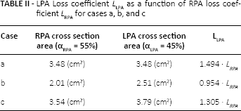
Table II shows LLPA as a function of LRPA for cases a, b, and c according to equation (6). Based on these ratios, starting from the RPA loss coefficient LRPA of 0, the simulation was performed with increasing values of LRPA. The resulting RPA flow rates and pressure differences between RPA and LPA are shown in Figures 4A and B, respectively.
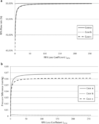
(
LPA Loss coefficient LLPA as a function of RPA loss coefficient LRPA for cases a, b, and c
Using an RPA loss coefficient of 0, which equals no pulmonary resistance, RPA flow rates of 48.81% and 39.90% are calculated for cases a and c, respectively. For case b, the desired flow rate is almost achieved when no resistance is used (54.73%). The results show that for an RPA loss coefficient above 100, the changes in split ratio and pressure difference between RPA and LPA are very small and negligible. Therefore, this value was used for all further simulations.
Wall Shear Stress Distribution
The WSS distributions are depicted in Figures 5, 6, 7, and 8 for the child model (fully opened valve and half-opened valve) and the adult model (fully opened valve and half-opened valve), respectively.
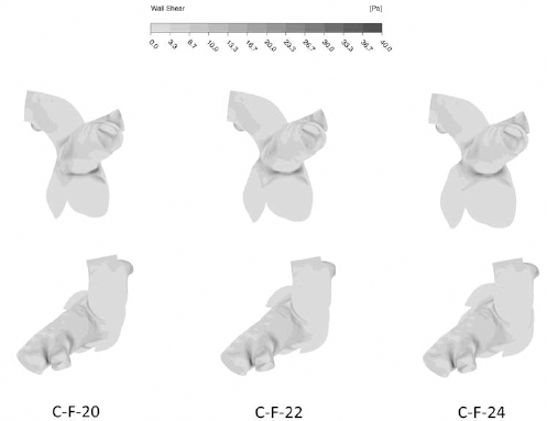
Wall shear stress contours on the conduit and arteries in different views (fully opened leaflets) in the child model (WSS is scaled between 0 and 40 Pa).
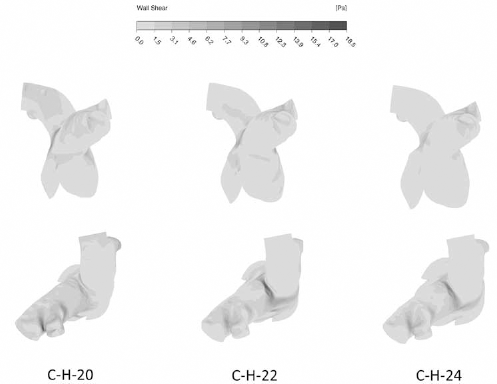
Wall shear stress contours on the conduit and arteries in different views (half-opened leaflets) in the child model (WSS is scaled between 0 and 18.5 Pa)
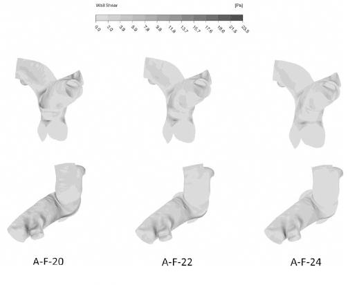
Wall shear stress contours on the conduit and arteries in different views (fully opened leaflets) in the adult model (WSS is scaled between 0 and 23.5 Pa).
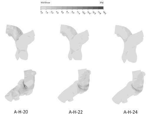
Wall shear stress contours on the conduit and arteries in different views (half-opened leaflets) in the adult model (WSS is scaled between 0 and 19.5 Pa).
Table III shows the mean WSS for each model and conduit size. All cases show a decrease of the mean WSS (about 26%) with an increase of the conduit size. Table IV depicts the mean WSS at the anastomosis. For the child model, the WSS is significantly increased (about 40%) for the fully open case when oversizing. In contrast, the stresses are decreased for the adult model (34%) when using a larger conduit.
Mean WSS (Pa) for the different models and conduit sizes
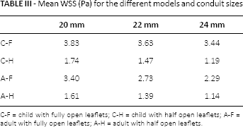
C-F = child with fully open leaflets; C-H = child with half open leaflets; A-F = adult with fully open leaflets; A-H = adult with half open leaflets.
Discussion
In a clinical setting, the implanting surgeon has a choice of different conduit or allograft sizes for replacement of diseased and dysfunctional pulmonary valves and right ventricular outflow tracts. Especially in a growing patient, this choice will potentially impact the mid- or long-term treatment strategy. Recent clinical studies (1, 5, 6) have shown that oversizing in small, rapidly growing children (under the age of 2) is beneficial and they recommend oversizing. Other studies (7, 4) that have included older children have not shown such an obvious benefit of oversizing and have actually shown decreased durability of these conduits with significant (Z +2.7) oversizing. No obvious reason for this discrepancy is readily available. One might speculate that the fast somatic growth seen in very small children demands implantation of as large as possible a conduit to counteract this effect, but when the somatic growth becomes more stable and linear, other effects become more important for the conduit longevity. The patient base, different prosthesis types, operational skill and technique all have a significant influence on conduit longevity (25). One of the factors that has a definite influence on native blood vessels is altered hemodynamics and flow conditions (8). Still the mechanism is currently not understood in detail. No previous study has considered the altered flow conditions when inserting a larger conduit.
To determine the impact of conduit oversizing on the hemodynamics, calculated WSSs of image-based CFD simulations were used in this study as an indicator. To calculate WSS in various conditions of oversizing, the anatomical and hemodynamic conditions had to be defined.
Anatomical Study
To create appropriate anatomical conditions, 3D models of pulmonary arteries (Fig. 1) were created using CT scan images of a healthy adult human. The model was then scaled to pediatric size (scaling factor 0.7) corresponding to a child with a BSA of 1 m2 or approximately 10 years of age. This size was chosen to simulate a more stable linear growth of a child, since somatic growth in very small children is more exponential. Taking into account a more steady rate of growth, the hemodynamic effects exerted with oversizing would also last longer.
In the next step, three different sizes (diameters of 20, 22, and 24 mm) of CAD models of valved conduits were virtually implanted into the child-sized model (Fig. 3). To simulate growth, the same-sized conduit models were implanted into the adult model as well (Fig. 3). The resulting Z-scores were +1.43, +2.32, and +3.21 for the child model and −0.91, −0.33, and +0.25 for the adult model. The sizes of conduits were chosen to simulate a clinical situation as closely as possible. The smallest conduit was chosen as best fit with regards to the size of the native vessel; some small oversizing was allowed. The next step includes the implantation of the next conduit by size usually commercially available, with the last one stepping up to the maximal possible size that would fit into the patient. Sizes larger than 24 mm were deemed not implantable due to anatomical constraints (Fig. 3). As can be seen in the anatomical study (Fig. 3), a conduit size of 20 mm corresponds best to the native size of the child's pulmonary trunk (Z-sore of +1.43). However, the same conduit produces a visible narrowing in the adult-sized 3D model (Z-score of −0.91). In this case, the most appropriate size of the conduit is a 24-mm conduit (Z-score of +0,25). Implanting this size of the conduit also corresponds best to the concept of “growing into” the conduit.
Pulmonary Resistances
To achieve a physiological flow split ratio of RPA/LPA = 55%/45% (17), appropriate pulmonary resistances were determined and incorporated. Without the use of resistances, unrealistic flow conditions were observed. We were able to establish a relation between RPA and LPA resistance depending solely on the anatomy and flow split ratio (equation 6). The resulting hemodynamic conditions with varying RPA resistances were analyzed for 3 different models of healthy adult human pulmonary anatomies (Figs. 2a, b, and c). The analysis shows that for an RPA loss coefficient above 100, the changes in split ratio and the pressure difference between the RPA and the LPA is not significant (Fig. 4). Therefore, this loss coefficient is used in all further simulations. This value is also in accordance with previous studies of flow conditions in the aorta (16).
In two investigated cases (Figs. 2a and c), a pressure difference between the RPA and LPA of less than 0.07 mmHg led to a change in the RPA flow ratio from approximately 43% to 55% and from 40% to 55%, respectively (Fig. 4a). Therefore, minor changes in pressures in the LPA and RPA result in significantly different flow splits. This indicates that effects on the pressure difference, such as PA branch stenosis, have a major impact on the flow field. Anastomoses, stent implantation, or vessel banding influence the ability of the vessels to react to pathological flow splits. This consideration is of particular interest in operations where ideal pulmonary flow conditions are required, for example Glenn and Fontan-type physiologies or hybrid Norwood procedures.
Evaluation of the Wall Shear Stress
Finally, after defining the flow resistances, the WSS for both the child-sized and adult-sized models were calculated. The WSS was calculated for conditions of fully opened and half-opened valves (Tab. II).
For both ages, the WSS is at least twice as high in the fully opened condition compared to the half-opened condition. This is mainly due to the higher inlet velocities and the shape of the leaflets. All cases show a decrease in the mean WSS with an increase of the conduit size (Tab. III). One reason for this is that the highest stresses are observed at the bifurcation and smaller conduits result in higher velocities of the jet flow hitting the bifurcation. However, to study the effect of oversizing on conduit failure, one needs to closely analyze the region of the distal anastomosis. For this purpose, the mean WSS at the anastomosis was calculated (Tab. IV). For the child model, the WSS is significantly increased (approx. 40%) for the fully open case when oversizing (Fig. 5). What this means practically is that the more oversizing is performed, the greater the WSS at the site of the anastomosis. In contrast, the stresses are decreased for the adult model (34% in total, 41% for the fully open case) when using a larger conduit (Figs. 7 and 8).
Mean WSS (Pa) at the anastomosis for the different models and conduit sizes
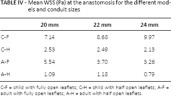
C-F = child with fully open leaflets; C-H = child with half open leaflets; A-F = adult with fully open leaflets; A-H = adult with half open leaflets.
As has been shown in previous studies, the altered WSS has a significant influence on the intima of the native vessels and was identified as a cause of intimal proliferation and stenosis formation (8) in different conditions. Shear stress is the hydrodynamic force exerted by the blood flow on the endothelial surface. The presence of disturbed blood flow, such as substantially high or low shear stress, induces a complex series of biochemical reactions in endothelium that eventually stimulates intimal hyperplasia (26–28). Since no studies exist that would identify the amount of shear stress increase or even the absolute forces needed to produce the biological responses of the endothelium in pulmonary circulation, it is not possible to quantify our results. However, a 40% increase of WSS in the child and 41% decrease of WSS in the adult when aggressively oversizing is significant. This could explain clinically observed problems of implanting certain types of conduits with well described problems of supravalvular/distal anastomotic stenosis in this patient group (10).
This would suggest that choosing the largest possible conduit could have a negative effect on the longevity of the conduits. This agrees with the findings of Karamlou et al (7) and Askovich et al (4), in which no positive effect of oversizing was seen; furthermore, aggressive oversizing had a negative impact on conduit longevity.
Study Limitations
The results of this study indicate that oversizing could have negative effects on the longevity of the conduit in certain clinical settings. The choice of conduit size, however, remains a decision to be made on a case-by-case basis to best benefit the patient. No specific estimations can be given about the value of altered WSS in inducing an intimal response and its clinical significance. The aim of this study was to isolate the effects of conduit oversizing on the WSS observed after conduit implantation and somatic growth in order to gain insight and a better understanding of the underlying mechanisms. Factors potentially influencing intimal overgrowth, such as material type, anastomosis type, anatomic constraints, and microstructure, are unknown and cannot be included in such a study. Therefore, only a steady flow condition without wall motion and healthy RVs and PAs were used to isolate the addressed problem. Furthermore, this model does not account for the widely adopted practice of patch enlargement of the PA bifurcation to prevent distal stenosis or interval procedures to prolong conduit durability.
A calculated scaling factor was used to scale the model of the PAs from an adult to the corresponding child size. Other factors involved during growth are not considered using this approach. This was nevertheless the method of choice for our model creation, since no CT data of the same patient as a child and adult was available to us.
In conclusion, based on the results of this study, it must be considered that choosing a prosthesis size that will lead to altered WSS and associated intimal ingrowth, leading to stenosis, will defeat the benefit of having a nominally larger orifice area directly after implantation. The long-term durability of such a surgical solution could also be affected.
Footnotes
Conflict of interest: There is no financial or personal conflict of interest in relation to this article.
Meeting presentation: This work was presented at the XLII ESAO Congress 2015, Leuven, Belgium, September 2015.