
Abstract

Editor,
A 48-year-old woman underwent dialysis for two years because of diabetic nephropathy. Her extremities had been too exhausted to construct vascular accesses (VA). Because of her vision loss due to vitreous hemorrhage, she could not manage peritoneal dialysis. We placed an arteriovenous graft (AVG) of the left axillary artery to the left axillary vein in her left upper arm in August 2010. We repeated percutaneous angioplasty because of her stenotic axillary vein (Fig. 1) and we had managed the patency of the VA for approximately two years. That VA occluded in June 2012 and finally we performed an anterior chest wall graft (ACWG). We used modified polyurethane (Vectra®, 5 mm, Thoratec®, Inc.) for the graft. We verified the location of stenosis in the left axillary vein on venography and chose to locate the anastomosis in the part proximal to the stenosis (Fig. 2).
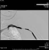
Venography shows the stenosis in the left axillary vein.
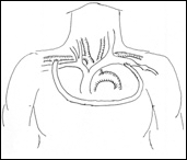
An anterior chest wall AVG of the right axillary artery to the left axillary vein.
We consider that ACWGs are indicated in the following patients; 1. Steal syndrome. The diameter of axillary artery is larger than that of the distal brachial artery, resulting in increased distal flow. 2. Patients whose veins are stenotic. 3. Patients whose patent central veins are difficult to be detected for inserting catheters.
ACWGs have some advantages. There are less possibilities of kinking and stenosis because no joints are crossed. It is possible to fix needles tightly on the chest and there are few problems even when the patient moves their body. Another advantage is the low infection rate. Generally, AVGs have a disadvantage compared to autogenous AVF. However, McCann reported that the incidence of infection of ACWGs was comparable with that of conventional AVFs (1). Maybe this is because the anterior chest wall skin is thick and resistant to infections. Moreover, the patency rate is possibly good. McCann reported a 77% three-year cumulative patency rate in over 50 patients (1). In other hands, there is a possible disadvantage of congestive heart failure. Ono reported four cases of that kind of graft and only one of them developed this 42 days after the operation (2). The patient was treated by narrowing the diameter of the graft by a suture and, subsequently, she recovered. This time we used a 5 mm tube and so far she does not have congestive heart failure or thrombosis in the graft.
In conclusion, the ACWG is a reasonable vascular access. For this type of AVG there is less possibility of steal syndrome and infections. Nowadays, the number of severe atherosclerosis patients is increasing, and this may be more helpful than autogenous AVF when they require dialysis.