
Abstract
Editor,
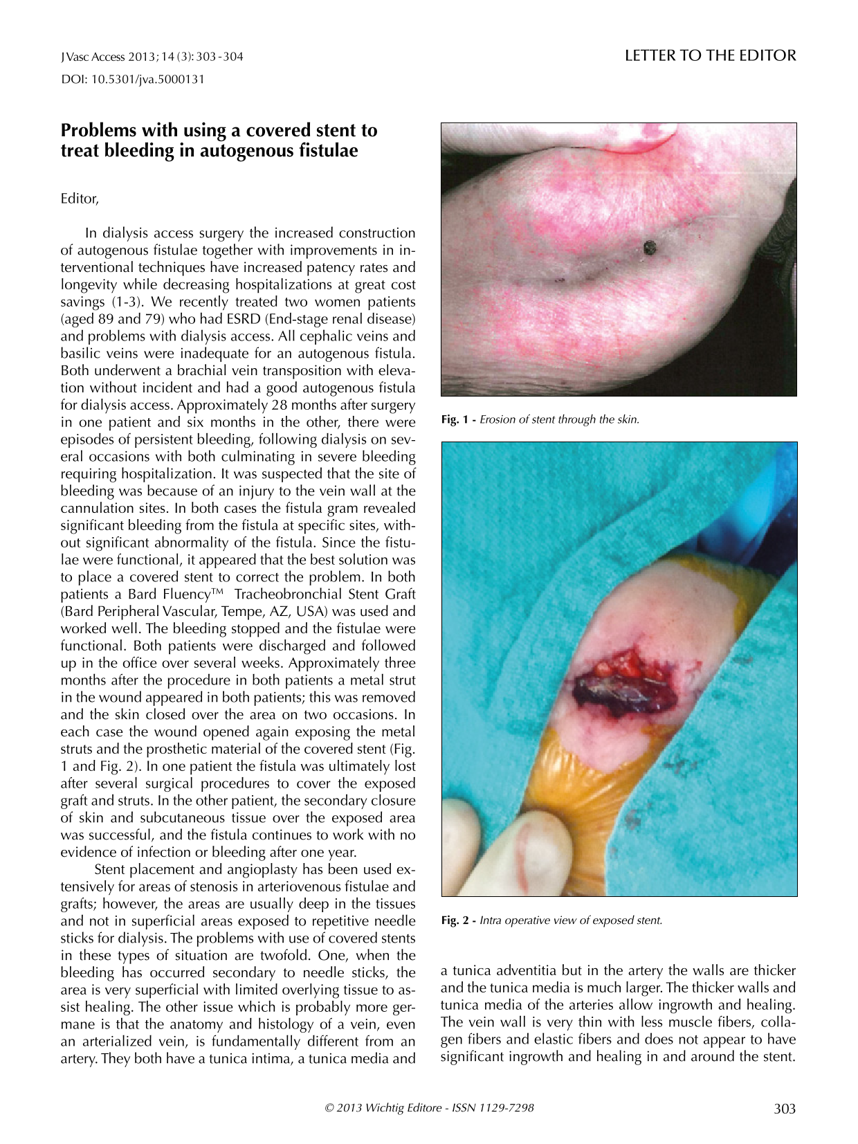
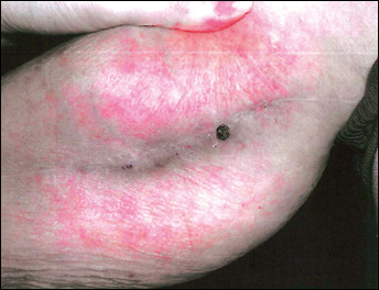
In dialysis access surgery the increased construction of autogenous fistulae together with improvements in interventional techniques have increased patency rates and longevity while decreasing hospitalizations at great cost savings (1-3). We recently treated two women patients (aged 89 and 79) who had ESRD (End-stage renal disease) and problems with dialysis access. All cephalic veins and basilic veins were inadequate for an autogenous fistula. Both underwent a brachial vein transposition with elevation without incident and had a good autogenous fistula for dialysis access. Approximately 28 months after surgery in one patient and six months in the other, there were episodes of persistent bleeding, following dialysis on several occasions with both culminating in severe bleeding requiring hospitalization. It was suspected that the site of bleeding was because of an injury to the vein wall at the cannulation sites. In both cases the fistula gram revealed significant bleeding from the fistula at specific sites, without significant abnormality of the fistula. Since the fistulae were functional, it appeared that the best solution was to place a covered stent to correct the problem. In both patients a Bard Fluency™ Tracheobronchial Stent Graft (Bard Peripheral Vascular, Tempe, AZ, USA) was used and worked well. The bleeding stopped and the fistulae were functional. Both patients were discharged and followed up in the office over several weeks. Approximately three months after the procedure in both patients a metal strut in the wound appeared in both patients; this was removed and the skin closed over the area on two occasions. In each case the wound opened again exposing the metal struts and the prosthetic material of the covered stent (Fig. 1 and Fig. 2). In one patient the fistula was ultimately lost after several surgical procedures to cover the exposed graft and struts. In the other patient, the secondary closure of skin and subcutaneous tissue over the exposed area was successful, and the fistula continues to work with no evidence of infection or bleeding after one year.
Erosion of stent through the skin.
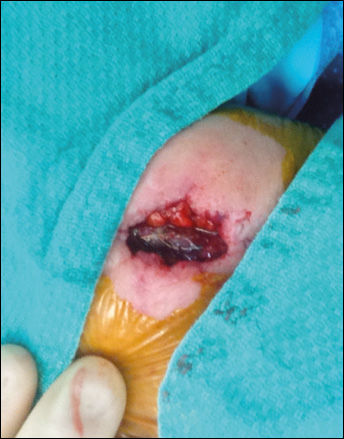
Intra operative view of exposed stent.
Stent placement and angioplasty has been used extensively for areas of stenosis in arteriovenous fistulae and grafts; however, the areas are usually deep in the tissues and not in superficial areas exposed to repetitive needle sticks for dialysis. The problems with use of covered stents in these types of situation are twofold. One, when the bleeding has occurred secondary to needle sticks, the area is very superficial with limited overlying tissue to assist healing. The other issue which is probably more germane is that the anatomy and histology of a vein, even an arterialized vein, is fundamentally different from an artery. They both have a tunica intima, a tunica media and a tunica adventitia but in the artery the walls are thicker and the tunica media is much larger. The thicker walls and tunica media of the arteries allow ingrowth and healing. The vein wall is very thin with less muscle fibers, collagen fibers and elastic fibers and does not appear to have significant ingrowth and healing in and around the stent. Furthermore, if the stented area is close to the skin, as in both these instances, the covered stent can erode through the injured thin walled vein and skin. It has been suggested that other stent grafts such as a VIABAHN (W. L. Gore and Associates, Flagstaff, Arizona, USA) may be more successful in these areas since they do not have metal struts outside the prosthetic graft. In theory, at least, there would be no metal stents present to erode through the vein wall and the skin.
This interesting and difficult problem just represents a new situation which must be recognized in an attempt to prolong the use of dialysis access in the dialysis dependent population. This is not to condemn the use of covered stents but instead to urge a thoughtful and careful approach to their use in order to avoid problems of infection and loss of the access. Endovascular techniques are invaluable in the treatment of dialysis access problems but as with each new technique there are unexpected problems and complications which must be recognized and responded to appropriately. It is this type of response to new difficulties which allows progress and advancement of the field of interventional radiology and dialysis access surgery.