






Abstract
Purpose
The United Kingdom Renal Association recommends duplex ultrasound to monitor arteriovenous fistula (AVF) flow rates during surveillance. Significant flow rate changes should prompt further investigation or treatment to avoid a failing fistula. Hemodialysis is known to alter the hemorrheologic and physiologic factors with a potential impact on measured flow rates. The aim of this study was to determine the difference in flow rate measured with duplex ultrasound before and after a single hemodialysis session in patients with brachial-cephalic fistulae.
Methods
Patients with brachial-cephalic AVFs in our dialysis populations who were undergoing regular hemodialysis without recent intervention (less than six weeks) were invited to participate. Flow measurements were made pre-and post-hemodialysis using a Zonare ultrasound machine. The vascular scientist was always blinded to the pre-hemodialysis flow.
Results
A total of 157 patients were identified with brachial-cephalic fistulae. Following exclusions, 119 patients were eligible. However, a further 58 were excluded because they declined or did not attend leaving 61 patients in the study. Paired t test showed a statistically significant reduction in flow rate of 105 mL/min (P=0.026) post-hemodialysis which equates to a −6.9% change in flow (95% C.I. −12.7 to −0.8%). Bland-Altman analysis showed limits of flow rate agreement between −599 mL/min and +810 mL/min (+/-1.96 s.d.).
Conclusions
Whilst we have shown a statistically significant change in flow rate post-hemodialysis, this is small and should be taken in the context of previously accepted interobserver variability. Therefore, the practical and financial considerations of implementing an AVF surveillance programme are likely to outweigh the minimal benefit of consistency that would be enabled by strict protocol of pre-hemodialysis flow measurements.
Keywords
Introduction
Following the implementation of the fistula first breakthrough initiative (FFBI) in the United States towards the end of the last century, native arteriovenous fistulae (AVF) have been identified internationally as the gold standard method of vascular access. The need for regular AVF monitoring to ensure prolonged patency and early detection of malfunction has been recognized for over a decade and was implemented by the European best practice guidelines (2002) (1). Prospective surveillance is an essential recommendation for the assessment of clinical parameters for functional adequacy and effective prevention of thromboocclusion, facilitating AVF longevity (2). Although in the past, static intra-access pressure ratios have been widely utilized for AVF surveillance, its diagnostic accuracy for assessment of access adequacy is questionable. Instead, it has been concluded that real-time volume flow (Qa) measurements are the best current option for access surveillance (3). Within the UK, current renal association guidelines (2011) recommend duplex ultrasound (DU) as one of the preferred monitoring modalities for access surveillance (4).
It is well established that dialysis results in various hemorrheologic changes. Many early studies have reported that whole blood viscosity increases within the immediate post-dialysis period (5, 6). The shear rates associated with normal flow in large arteries means blood can be regarded as a Newtonian fluid (7). Therefore, Poiseuille's law for fluid dynamics can be applied, which indicates the inverse relationship between blood viscosity and flow.

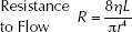
It can be postulated that volume flow rates may be affected by dialysis such that a pre-dialysis assessment differs significantly from a post-dialysis assessment. This has significant clinical relevance in terms of the timing of surveillance scans with respect to dialysis sessions. Because of the variability of dialysis sessions that can often begin early in the morning or end late in the evening, the practicality of duplex ultrasound surveillance scans has to be compromised to coincide with vascular scientist working hour constraints. Although it would be preferable to consistently perform the scan always before or after the session, this is not possible for logistic reasons. In practice, surveillance scans are performed before or after the hemodialysis session depending on the time of day.
The primary aim of this study was to test for a difference in duplex ultrasound acquired Qa measurements immediately before and after a single hemodialysis session for patients with brachial-cephalic fistulae. The primary outcome measure was the percentage difference in mean volume flow (mL/min) measured before and after the dialysis session.
Our conclusions may lead to adjustments to our vascular laboratory surveillance protocol to give more consistent and accurate Qa measurements of our dialysis patients.
Materials and Methods
A quasi-experimental design was adopted in the form of a before/after observational study. The Kings College Hospital, London (KCH) renal database was interrogated to identify all suitable patients and to extract the demographic and clinical data. Patients attending our main renal dialysis unit and our four satellite units were invited to the study via a patient information leaflet made available at each unit.
Patients eligible for inclusion were those aged over 18 years and diagnosed with end-stage renal failure (ESRF). Patients were required to have an adequately functioning native brachial-cephalic fistula (urea reduction ratio >65%).
Exclusion criteria were patients aged under 18 years of age and those unable to give informed consent because of a communication barrier or medical condition. In order to maintain consistency, all alternative types of AVF were excluded in this study other than the brachial-cephalic formation. Patients who had recent surgery or radiologic intervention (less than six weeks) on their brachial-cephalic fistula were excluded. This post-operative period is clinically recommended to re-establish stable AVF flow and often patients dialyze through a central venous catheter for this duration rather than their native fistula (3). Any patients that could be unacceptably delayed for emergency treatment or are critically unwell were also excluded.
Those patients who consented to participate had an additional scan limited to the specific measurements for this study as well as these measurements being taken during their normal surveillance scan. For the purpose of the study, a set of three consecutive Qa measurements was recorded from the same location during the pre-dialysis scan and again during the post-dialysis scan. This location was taken within a non-tortuous segment of the brachial artery 4-15 cm proximal to the anastomosis (with the exception of those with high brachial bifurcations where the axillary artery was used) to avoid inaccuracies associated with flow turbulence at the anastomotic site. During the pre-dialysis scan, this level was marked with a permanent marker to indicate the position of the transducer (avoiding the needling region), so as to provide a consistent position for the post-dialysis scan.
Seven trained vascular scientists participated in taking Qa measurements for this study. For every patient who consented to study participation, a different vascular scientist performed the post-dialysis scan and was blinded to the results of the pre-dialysis scan. All post-dialysis measurements were taken within ten minutes of bleeding cessation from the needling site.
The Zonare™ Z-one portable ultrasound machine and its linear array transducer (8-3 MHz) was utilized for every volume flow measurement made pre and post-hemodialysis and a strict protocol was followed to minimize instrumentation bias. Whilst our study was an observational one, this did require additional measurements and scanning time beyond the patient's normal clinical assessment. Therefore, we applied for and successfully received full ethical approval.
Duplex Ultrasound
Duplex ultrasound not only achieves the fundamental role of surveillance by measuring a significant reduction in access Qa, it also has the advantage of identifying the pathologic cause. The benefit of utilizing non-invasive imaging for surveillance enables the accurate grading of underlying stenotic lesions, visualization of any vascular abnormalities and regions of thrombosis. Thus, duplex ultrasound surveillance of dialysis access can also play a vital role in future endovascular and surgical interventional planning (8).
Duplex ultrasound acquired volumetric flow measurements are a function of time-averaged mean velocity (TAMV, the spatial mean velocity temporally averaged over a set number of cardiac cycles) and the arterial cross-sectional area (9). The volume flow (Qa) is calculated from the inflow axillary or brachial artery as opposed to the outflow vein as arterial flow is relatively uniform and unidirectional, permitting accurate velocity measurements (Fig. 1). Doppler frequencies from emitted pulsed echo waves are utilized to sample the time average mean velocity (TAMV) across the intraluminal flow (10). In addition, the cross-section tends to be more circular in geometry and, therefore, an intraluminal diameter can be measured to estimate cross-sectional area (Fig. 2).
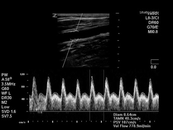
Image of volume flow calculation of brachial artery using duplex ultrasound.
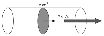
Calculation of volume flow from inflow artery.
Instantaneous flow rate (Qa) = Arterial cross-sectional area (A cm2) x Time-average mean velocity (V cm/s).
Volume flow measurements are measured against set criteria established by the UK Renal Association, whereby AVF flows between 300-600 mL/min are identified as a clinically critical AVF (11). A loss of more than 25% volume flow between surveillance scans within patients with <1000 mL/min is also considered a particular clinical concern for access patency and patients whose assessment fall within these criteria are considered for endovascular or surgical intervention to prolong patency (12).
Results
Using the KCH database, 157 patients were identified to have native brachial-cephalic fistulae across all dialysis units, of which 38 were excluded (Fig. 3).
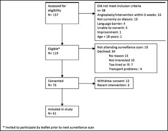
Reasons for exclusions from participation in the study.
For all study participants included in this analysis, 37 are women and 24 are men. Mean age was 62.3 years and the median duration participants had been dialyzing via their brachial-cephalic AVF at the time of the study was 16.3 months.
Pre and Post-Dialysis Flow Rate Measurements
There was an overall mean Qa reduction of 105 mL/min (95% CI 13 to 197) between pre and post-dialysis (P=0.026, paired t test).
Between pre- and post-hemodialysis Qa measurements, 39 patients (64%) had an overall reduction and 22 patients (36%) had an overall increase (Tab. I). The primary outcome measure of this study is the percentage mean difference in Qa measurement, which is illustrated within Table I.
Mean Differences in QA Measurement Between Pre and Post-Dialysis
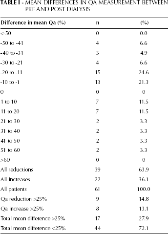
Most patients (69.8%) had Qa measurements that differed up to 20% within the post-dialysis period. Only nine of the 39 patients who showed an overall loss in duplex ultrasound acquired Qa measurement post-dialysis, had more than 25% loss in Qa, relative to their pre-hemodialysis measurement (Tab. I). Of 22 patients who showed an overall increase in duplex ultrasound acquired Qa measurement post-dialysis, only eight had a duplex ultrasound measurement increase in Qa of more than 25% relative to their pre-hemodialysis measurement (Tab. I).
When considering the absolute difference in flow rate (whether negative or positive), 44 patients (72.1%) therefore showed an absolute mean difference of <25% AVF Qa between the pre and post-dialysis measurements.
It should be remembered that changes in flow rate of >25% have particular clinical significance when the flow rate is <1000 mL/min. A total of nine patients had >25% reduction and eight patients showed >25% increase in duplex ultrasound acquired Qa between the pre and post-dialysis measures. This gives an incidence of 14.8% (CI 95%, 12.6–18.9) and 13.1% (CI 95%, 10.9–17.6) respectively. However, it should be noted that within those 17 patients, every one of these had a pre-dialysis Qa >1000 mL/min. No patients of the 61 observed had a reduction of 25% or more on a pre-procedure.
Therefore, no patients with pre-dialysis Qa <1000 mL/min (0%, CI 0% to 5.9%, by the exact binomial method) needed to be referred for intervention (Fig. 4). The average relative change is −6.9%, (95% CI −12.7% to −0.8%).
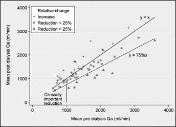
Scatterplot comparing mean pre and post-dialysis Qa measurements.
Bland-Altman analysis of the pre and post-dialysis Qa measurements reveals limits of Qa measurement agreement as −599 mL/min to +810 mL/min (+/-1.96 s.d.) (Fig. 5).
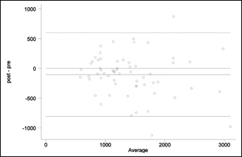
Bland-Altman analysis of pre and post-dialysis Qa measurements.
Discussion
We found a statistically significant mean reduction of 105 mL/min in Qa measurement between the pre and post-dialysis period. Most study participants (63.9%) showed an overall reduction in Qa within the post-dialysis period. This is coherent with the theoretical physics of Newtonian fluid dynamics as described by Poiseuille's law: an increase in blood viscosity reduces overall volume blood flow rate during hemodialysis. These results are consistent with that observed by Vaisman et al (13) who reports the persistence of ultrafiltration induced hyperviscosity into the immediate post-hemodialysis period. Resulting intravascular depletion can induce several hemorrheologic factors, namely hemoconcentration and hypovolemia, which can promote intravascular coaguability. Another factor that increases coaguability is the rise in overall hematocrit that occurs from blood entering the extra-corporeal circuit. Blood flow rates may also be affected by intra-dialytic hypotension, which can affect between 10% to 30% of patients having hemodialysis, another potential factor for changing access flow rates (14). These findings would suggest that as suspected, physiologic factors associated with ultrafiltration are likely to significantly lower duplex ultrasound flow measurements taken within the immediate post-hemodialysis period.
It is important to assess the post-dialysis Qa changes from a quantitative perspective in order to place this into a clinical context. A significant intersession difference in Qa measurement has been reported as >25% reduction in access flow, when Qa <1000 mL/min but before this reaches the critical range between 300-600 mL/min (11). This criteria is applied to native hemodialysis fistula within our local hospital protocol and has been used to define a significant change in Qa between the pre and post-hemodialysis period in this study.
Bland-Altman analysis of our study data revealed high variability of Qa measurements with limits of agreement from −599 mL/min to +810 mL/min between pre and post-hemodialysis Qa measurement. However, most study participants (69.8%) had an absolute difference within 20% of their Qa measurement post-dialysis and it can be argued that some of this variation may be attributed to errors in measurement from duplex ultrasound.
Previous studies have reported between 10% to 20% intra- and inter-observer variability in duplex ultrasound volume flow measurements because of errors associated with diameter and velocity measurement (15-17). Volume blood flow measurements are subject to inaccuracies from non-uniform insonation of the artery, incorrect angle correction, wall filters and errors in the calculation of intensity weighted mean velocity. However, recent in vitro tests reveal that the experienced operator using modern ultrasound machines can minimize these inaccuracies and achieve reproducible flow measurements and accuracy within 5% (18). This is particularly applicable in hemodialysis fistula surveillance, where the vessels lie relatively superficial, creating a higher resolution image, and Qa measurements are the recommended standard for monitoring.
The absolute incidence of Qa >25% was determined as 27.9% (CI 95%, 24.6–37.8). It is interesting that none of these 17 patients that had a significant loss in Qa >25% had a pre-hemodialysis Qa >1000 mL/min (0%, CI 0% to 5.9%, by the exact binomial method) with the average relative change noted as −6.9%, (95% CI −12.7% to −0.8%). No patients from this study were shown to have a brachial-cephalic fistula considered clinically significant. Thus, the study data suggests greater variability of Qa measurements with higher underlying flow rates. This may indicate that variation within duplex ultrasound Qa measurements taken pre and post-hemodialysis is unlikely to fall within the clinically significant referral criteria. However, it is not possible to determine whether this is because of a more influential physiologic impact within patients who have higher fistula flow rates.
In theory, the physiologic impact from hemodialysis on duplex ultrasound Qa measurements could be eliminated by ensuring the patient never has a surveillance scan within the post-dialysis period. However, in practice, this has several disadvantages. Firstly, this would require the vascular scientist to be present outside their working hours for patients using the morning dialysis session. This would have financial implications on the vascular laboratory as overtime pay would have to be given. Secondly, the hemodialysis patient would have to be within their unit at least half an hour before their session, which is likely to cause patient disruption and problems with hospital transportation. The alternative is to scan all morning patients on a non-dialysis day when another group of patients are being seen for surveillance. This would be efficient for the vascular scientist; however, this gives an unfair disadvantage to the morning dialysis patient. Many of these patients choose to dialyze at this time because of other commitments during the day.
Conclusions
Our study reveals a statistically significant reduction in duplex ultrasound Qa measurement between pre and post-dialysis measures taken relative to a single hemodialysis session. This suggests that there is a significant physiologic impact on fistula flow measurement from the ultrafiltration process. However, whether this is clinically significant is debatable as this is likely to be within the limits of interobserver variability.
The ideal strategy for surveillance would be to consistently scan patients before their hemodialysis session to eliminate errors that may be inflicted because of hemodialysis-induced hemorrheologic changes. However, there is a fine balance between the ideal strategy and the practical strategy, and in the absence of further evidence to the contrary, we shall continue with the latter pragmatic approach.
Further Work
This observational study has shown a significant physiologic impact of hemodialysis in duplex ultrasound volumetric flow analysis. This has not been investigated quantitatively within previous literature. Further analysis is required on a larger patient sample with Qa measurements repeated over several surveillance scans to determine whether the timing of the scan affects clinical diagnosis.
Footnotes
Acknowledgements
The authors are especially grateful to Fatima Defigiueiredo and to the renal access nursing team at Kings College Hospital and its dialysis satellite units. We would also like to express our gratitude to the other supporting vascular scientists of the Kings College Hospital Vascular Laboratory: Ben Freedman, Tim-Fernandez-Hart, Stuart Wind and Annette Quinn.