
Abstract
Purpose
To investigate the feasibility and safety of the interventional technique of retrieving the fractured peripherally inserted central catheter (PICC) segments within the vessels via the femoral vein.
Methods
From July 2007 to January 2012, we performed percutaneous retrieval of PICC fractures in six cancer patients who accepted chemotherapy via PICC. The fractures occurred during the traction of the catheter and were diagnosed with chest plain film radiography and/or computed tomography. The patients included four cases of ovarian cancer, one case of breast cancer and one case of cervical cancer. The fractures were retained in the vessels of the patients for 1 to 10 days. According to the location of the ends of the PICC fractures, three methods were employed using the most commonly used interventional devices in the digital subtraction angiography suite.
Results
The PICC fractures were located in the subclavian vein, superior vena cava, right atrium, right ventricle or pulmonary arteries. During the procedures, a goose neck snare, pigtail catheter and stone basket catheter were used individually or in combination. The PICC fractures were removed successfully in all six patients via unilateral or bilateral femoral vein access. No major complications occurred during the operation or the follow-up period of 7 to 10 days.
Conclusions
Via femoral vein access, PICC fractures could be removed with common interventional instruments such as a goose snare, basket catheter and pigtail catheter. The interventional retrieval is a safe, convenient and minimally invasive method for the removal of PICC fractures.
Introduction
In recent years, a peripherally inserted central catheter (PICC) has become widely used in patients for the administration of chemotherapy drugs, antibiotics and nutrients and is a cost-effective approach for central venous pathway access (1-3). The incidence of pain and vein injury due to repeated vein puncture has been reduced obviously with the use of PICCs. Compared with the subclavian venous catheter, the incidence of pneumothorax and hemopneumothorax has also obviously decreased (4). Although central catheter-related complications such as sepsis, effusion, phlebitis, rupture, blockage, migration and fracture still cannot be avoided completely, the development of new catheter materials, techniques and nurse training have contributed to decreased complications.
PICC has been commonly used in cancer patients for chemotherapy administration. However, the complication rates in cancer patients with PICCs have been shown to be greater than those in other patients (5). Catheter fracture is a relatively rare complication, and previously reported incidences of fracture range from 0.2% to 9.7% (6). Although the natural history of a PICC fracture retained in the vessels is unclear, it is necessary to remove the fracture to prevent from complications including pulmonary embolism, arrhythmia and vascular or cardiac perforation (7). Several interventional techniques and instruments have been used in the percutaneous retrieval of foreign bodies in the cardiovascular system (8-10). But, the goose neck snare and basket catheter with or without the assistance of a pigtail catheter are the most commonly used interventional devices (11). In this study, we successfully performed percutaneous retrieval of PICC fractures in six cancer patients using the most common interventional devices.
Patients and Methods
Patients
The patients in this study included four cases of ovarian cancer, one case of breast cancer and one case of cervical cancer. The patients received chemotherapy via PICC. The PICC catheters were silicone Groshong* NXT PICC and Groshong* NXT ClearVue PICC (Bard Access System, Salt Lake City, USA). All six patients were female. The average age of the patients was 51 years (range 37-71 years). Until the fracture, the catheters had been implanted in the patients for at least 26 days (range 26-105 days). The catheter fractures occurred during extraction of the PICC. In five patients, the fractured catheters were found promptly during the extractions and were identified with imaging. Then, the fractures were removed within 1 to 2 days. In the other patient, the fracture occurred abroad and the patient was treated in China. The proximal fragment of the fracture was removed with a venotomy in the Vascular Surgery Department of our hospital. The interventional retrieval of the retained fragment was performed 10 days after the fracture. Informed consent was obtained from all the patients before the procedures.
Diagnosis
Once the fracture occurred, X-ray plain film radiography was performed on the elbow, shoulder and chest to identify the location of the PICC fragments, especially the two ends of the fracture. The PICC fracture was identified in the body in the chest plain film. In one patient, the distal portion was identified in the lower right pulmonary artery and the proximal portion was identified in the left subclavian vein; in another patient, the distal portion of the fracture was identified at the junction of the subclavian vein and the superior vena cava and the proximal portion was identified in the right brachial vein; in two patients, the distal portion was identified in the lower right pulmonary artery and the proximal portion was identified in the right subclavian vein and in the last two patients, the distal portion was identified in the lower right pulmonary artery and the proximal portion was identified in the superior vena cava.
Interventional Technique
The procedures were performed via femoral vein access with the Seldinger technique under local anesthesia with 1% lidocaine. Before the interventional procedure, the locations of the fractures were estimated by X-ray fluorescence imaging and compared with the X-ray and computed tomography (CT) images. Three patterns were categorized according to the positions of the proximal and distal ends (relative to the puncture site in the arm) of the fractures. Pattern 1: The fracture was located across the subclavian vein (even to the brachial vein) and the right/left pulmonary arteries; briefly, the proximal end was located in the subclavian vein and the distal end was located in the pulmonary artery. Both ends were floating in the vessels. Pattern 2: The fracture had a free end floating in the superior vena cava, right atrium, right ventricle or pulmonary artery and the other end was located in the subclavian vein or pulmonary artery. Both ends were floating in the vessels. Pattern 3: The fracture had no free ends to snare, and the two ends were embedded in the vascular tissues.
The three methods were applied in the six patients, and the technique details are described as follows. Method 1 was used in the patients who had a fracture with no free end (with the distal end in the pulmonary artery and the proximal end in the subclavian vein), but the two ends were floating in the veins. Briefly, after introducing a 11F sheath to the unilateral femoral vein, a 11F guiding catheter was introduced and placed in the inferior vena cava. A goose snare and a 0.035-inch hydrophilic guide wire were placed on each side of the PICC line, respectively. After successfully snaring the soft head of the guide wire with the loop, the guide wire and the goose snare were together dragged back into the guiding catheter. Finally, the PICC fracture was dragged out with the guiding catheter. Method 2 was used in the patients who had fractures with a free end floating in the superior vena cava, right atrium or right ventricle. Briefly, a 6F sheath was introduced into the femoral vein. Then, a 5F pigtail catheter was introduced via the contralateral femoral vein to hook the fracture. After successfully hooking the fracture, the fracture was subsequently dragged carefully to the inferior vena cava. Then, the goose snare was introduced via the 6F sheath to the inferior vena cava to capture the fracture, and the fracture was dragged out via the femoral vein. Method 3 was used in the patients who had fractures with no free ends; the pigtail catheter failed to drag the catheter to the inferior vena cava because the fractures were embedded in the tissues. Briefly, a stone basket catheter was used to wring the floating part of the fracture and drag it out.
Results
The locations of the fractures were identified in the vein system using fluoroscopy and angiography, and they were consistent with the diagnosis provided by X-ray plain film radiography and/or CT. The procedures were finished within 50 min. Method 1 was used in three patients (Fig. 1), method 2 was used in two patients (Fig. 2) and method 3 was used in one patient (Fig. 3).
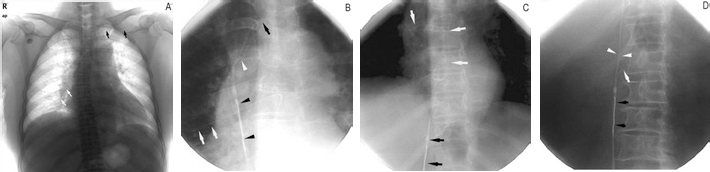
Female, 71 years old, ovarian cancer, the operation was performed 16 h after PICC breakage. A) Chest plain film radiography shows the distal portion of the fracture in the lower right pulmonary artery (white arrow) and the proximal portion in the left subclavian vein (black arrow). B) Overlap of the soft head of the 0 035-inch hydrophilic guide wire (white arrow head) to form a snare and drag the fracture with the loop to the guiding catheter. The proximal portion of the fracture was located in the superior cava vena (black arrow), and the distal portion was located in the right lower pulmonary artery (white arrow). C) The goose snare, guide wire, and guiding catheter were dragged to the inferior cava vena, and part of the PICC can be observed in the right heart and right pulmonary artery (white arrow). D) The fracture (white arrow) was dragged into the guiding catheter with the loop formed by the guidewire and goose snare.
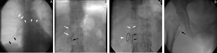
Female, 61 years old, cervical cancer, the operation was performed 18 h after the PICC fracture. A) The fracture was found by fluoroscopy. The distal portion of the fracture was located in the right lower pulmonary artery, the proximal portion was located in the superior cava vena, and the middle portion was curled in the right heart and right pulmonary artery. B) Via the left femoral vein access, the proximal portion was hooked with the fillet (black arrow) of the 5F pigtail catheter and dragged to the inferior cava vena (white arrow). C) After dragging the proximal portion to the inferior cava vena (black arrow), the fracture was overlapped with the goose snare loop (black arrow) via the right femoral vein access and dragged into the right femoral vein (white arrow). D) The fracture was removed from the right femoral vein with the goose snare (black arrow).
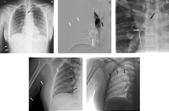
Female, 37 years old, ovarian cancer, the operation was performed 10 days after the PICC fracture. A) According to the chest plain film, the distal portion (black arrow) of the fracture and the proximal portion (white arrow) were located in the right elbow. B) The distal portion of the fracture was embedded at the junction of the superior cava vena and left subclavian vein. C) The fracture line was hooked with the fillet (white arrow) of the 5F pigtail catheter but failed to be dragged to the inferior cava vena because the distal portion was embedded in the left subclavian vein (black arrow). D) The fracture line was twisted into the basket by rotating the basket catheter and dragged to the guiding catheter (black arrow). The proximal portion was located in the right axillary vein (white arrow). E) The basket catheter was dragged to the guiding catheter (black arrow), and the proximal portion was moved to the right subclavian vein (black arrow).
It took 20 to 35 min to finish the procedures for the five patients treated with method 1 or method 2. The other patient with the distal portion of the fracture at the junction of the left subclavian vein and superior vena cava and the proximal portion in the right brachial vein required 50 min to remove the fracture with the basket catheter after failing to hook the fracture with a pigtail catheter because of embedded ends in the tissues. No severe complications occurred in the patients.
Discussion
PICC is a cost-effective and safe approach for central vein access in patients who need long-term intravenous infusion of anticancer drugs, antibiotics or nutrition (2, 4, 12). However, PICCs can cause multiple complications, predominately including leakage, infection/phlebitis, blockage, dislodgement, disruption, fracture, bleeding, embolism and allergic skin reactions. Although the incidence of complications has decreased with the professional training of personnel and even in the setting of professional PICC nurses, complications cannot be avoided completely.
Catheter fracture is one of the most severe PICC-related complications. Currently, the cause of the fracture is still unclear. It has been suggested that it is related to the tearing of the catheter during insertion or traction on the catheter–hub junction (2). With the development of new PICC designs and materials, this problem has been overcome somewhat. The fracture can also localize in the line of the catheter during the traction, and the breakage point may be located in any part of the catheter. The fracture can move with refluxed blood flow to the subclavian vein, right atrium, right ventricle and pulmonary artery (13, 14). Vein thrombosis due to PICC fracture is relatively rare, but it can lead to severe consequences such as pulmonary embolism (1, 15, 16). The occurrence of a PICC fracture has only been documented in a few case reports, and the fractures were removed with an interventional method or vascular incision. Here, we report six cases of PICC fracture treated with interventional therapy.
The patient tends to become nervous once the PICC is fractured. In that moment, the medical staff should reassure the patient with gentle speech and limit or completely stop the patient's movement, especially in the involved limb. Changing the upper extremity position, especially with abduction and adduction, can induce the movement of the PICC toward blood flow. Pressing the target vein may decrease the chance of movement of the fracture. Even if the PICC fracture has entered the vessel, the medical staff should keep the patient calm and prepare for further examination and therapy. An immediate imaging examination can show the fracture clearly. Chest plain film radiography is an easy and effective method to detect the PICC line. CT provides more details of the fracture. If permitted, the patient should be sent to the interventional department, and the fracture should be removed in time in order to prevent late complications such as embolism and infection. In this case series, the fractures were removed in five of the six patients within 1 to 2 days after the occurrence of the fracture. One patient received surgical therapy, but the fracture was not removed entirely. Therefore, the patient received another interventional therapy 10 days after the fracture. All fractures were removed integrally from the vein without any residue. All patients had no severe complications.
A retained catheter fragment in the cardiovascular system may lead to severe complications such as acute pulmonary embolism, cardiac perforation and cardiac arrest. The incidence of fatal complications and death due to catheter fragments was up to 71% in the early 1970s (7). In recent reports, catheter fragments have not caused fatal complications and the fracture could be retrieved by percutaneous interventional radiology methods (1, 13, 17, 18). Although improved biocompatibility and flexibility of the catheter have decreased the risk of fatal complications, Savage et al have advised the removal of intravascular foreign bodies to prevent potential complications even if the patient is asymptomatic (10). We categorized the PICC fractures of our patients into three patterns according to the position of the proximal and distal ends (relative to the puncture site in the arm) of the fractures. Although three methods were applied in the interventional retrieval procedures, both method 1 and method 2 can be used in pattern 1 and pattern 2 in clinical practice. A similar method as method 1 was first described in the interventional percutaneous retrieval of a bile duct catheter (19). The key point of this method is the formation of a snare with the guiding catheter, guidewire and loop snare. Compared with method 2, method 1 may save time during the operation; thus, the patient would receive less radiation. The capture operation in the right ventricle could lead to severe complications and should be avoided as much as possible (14). Therefore, the pigtail catheter was used to hook the free end of the fracture to the inferior vena cava in order to capture the fractures. In the event of failing to drag the end out of the tissues, besides the in situ formation of the snare, a basket catheter can also be used to retrieve the fracture by wringing the floating part of the catheter line. The head of the basket catheter consists of three interleaved wires. If the fracture can be wrung and trapped in the basket, the strength is enough to pull the fracture out of the vessels. Although many interventional devices have also been used in the clinic, the devices we used in this study are the most common retrieval devices and are affordable for most patients. Myocardial biopsy forceps or foreign body recovery pliers, which have crocodile-like fronts, can catch the fracture and are not limited by the fracture location (20). We successfully removed the ectopic embolic coils in the pulmonary artery and popliteal artery with myocardial biopsy forceps. However, these types of interventional instruments are not commonly used in most hospitals.
In summary, PICC fractures will occur more often with the widespread use of PICC. Once the PICC has fractured, the medical staff should reassure the patients and stabilize their emotions. Next, imaging examinations such as chest plain film radiography or CT should be performed. Interventional therapy should then be arranged to remove the fractures. Generally via femoral vein access, the fracture can be removed with common interventional instruments such as a goose snare, basket catheter and pigtail catheter. In conclusion, the interventional operation is a safe, convenient and minimally invasive method and should be considered as the first choice for the removal of PICC fractures.
Footnotes
Financial support: None.
Conflict of interest: None of the authors have identified a conflict of interest.