






Abstract
Purpose
Direct measurement of venous length is easy to cause contamination during bedside insertion of peripherally inserted central venous catheter (PICC). It is necessary to provide an equation for proper estimate of catheter length in case of bedside insertion of PICC in relation to patient height.
Methods
For 165 PICC cases through left arm vein in 151 adult patients (male: female = 72:79), the cubital crease to carina length (CCL) was calculated as follows: CCL = (distance from cubital crease to puncture point) + (length of PICC inside body) – (distance from carina to catheter tip on post-procedural chest radiograph). We analyzed the relationship between CCL and height with regression analysis and suggest a new equation of CCL based on height.
Results
The mean CCL through the left arm vein was 47.1 ± 2.6 cm in male and 44.0 ± 2.9 cm in female. CCL was significantly correlated with patient height. Equation of CCL (cm) based on height was as follows:
CCL = height* 0.19 + 14.
Conclusions
The equation of our study would provide a new equation for proper estimation of catheter length in case of bedside insertion of left arm PICC in relation to height and be helpful for optimal positioning of catheter tip of PICC.
Introduction
After the first trial of using upper extremity vein to access to the central venous system in 1912, usage of upper extremity veins is increasing for peripherally inserted central catheter (PICC) to provide reliable venous access routes for prolonged delivery of various medications, repeated laboratory testings, and hemodynamic monitoring especially for vulnerable patients requiring long-term clinical care (1-3).
Many reports are focusing on the importance of correct position of venous catheter tip (optimally lower superior vena cava, SVC) because improper catheter tip position is one of the most important factors to cause complications that can cause critical deterioration in these patients such as catheter malfunction resulting in repeated procedure, increased chance of infectious contamination, and venous thromboembolism (4-7).
As for central venous catheters, intracavitary ECG and/or echocardiography, with or without tracking devices, are safe and cost-effective for adequate catheter tip positioning for PICC, but in many institutions, bedside placement of PICC without such devices is common. And, individually incorporated methods of catheter length determination for each patient are used such as using tape ruler in the PICC sets, but measuring the length of catheter from the puncture point to central vein after venipuncture can be very cumbersome and vulnerable to infectious contamination. A ‘tailored fit formula’ to individual patient height based on the anatomic measurement results of humerus and clavicle has also been reported, which has a variable accuracy. In addition, incorrect estimation of the length of catheter is one of the causes of low rates of successful central tip positioning (44-99%) (6, 8-11). Although several reports about the length of insertion guidelines for the depth of a central venous catheter through internal jugular or subclavian veins in relation to the height are present, in our knowledge, for PICC, equation of venous length through left upper arm vein in relation to patient height is not present except our previous report about that of right side elbow crease to carina length (ECL) based on patient height (12-15). Because the left arm is non-dominant side than the right side and is more frequently selected during PICC procedure according to our policy, an equation for left-side PICC procedure is necessary.
This study was conducted to make a more convenient and accurate equation of PICC length determination for proper central position of PICC tip based on patient height through left upper arm vein, which is especially useful in cases of bedside insertion of PICC.
Materials and Methods
This retrospective study was approved by our institutional review board
Basic data of patients and the lengths of measurements in male and female groups

PICC Procedure and Measurement Methods
All PICC insertion procedures were done with the patient supine on the angiographic table. Before the procedure, all patients were asked which side would be more convenient for PICC and ultrasonography (US) examination was done to determine whether veins were usable, but catheter-to-vein ratio was not measured. The left elbow joint was fully extended and the arm was externally rotated as possible and abducted to about 40 degrees. Usable vein among basilic or cephalic veins was selected under US examination (basilic vein first) because of its superficial location and brachial vein was selected if no other veins were determined usable or prior venipunctures failed.
Venipuncture was done about 3-10 cm above the cubital crease with micropuncture needle in PICC set (5 Fr. Turbo-flo PICC Set. CookR, Bloomington, USA, or 6 Fr. dual lumen Pro-Picc CT Basic IR set, MedicompR, Harleysville, PA, USA). After insertion of guidewire and dilatation of the tract with dilater sheath, the catheter was trimmed. The catheter length was determined about 2-3 cm longer than expected to reduce the possibility of catheter shortness (the right side length by the formula of our previous report of ECL through right basilic vein based on patient height, plus 3.5 -4 cm considering the report that left innominate vein is 3.5 to 4 cm longer than the right one, and minus cubital crease to puncture point distance) (7, 14). Then, PICC catheter insertion was done into the sheath slowly advancing as deeply as possible. If any resistance was felt during catheter advancement, fluoroscopy and/or venography through the catheter was checked and adequate method (selection or overcoming acute curvature or stenosis of main venous route using guidewire) was used to finally locate the catheter tip in the SVC. The external portal of the catheter from skin was fixed with fixation device in the PICC set. Punctured veins in 165 cases were recorded in Table II. Finally, the last fluoroscopy or spot radiography was stored on Picture Archiving and Communication System (PACS) to show the ultimate catheter tip position in relation to SVC and right heart. The distances from cubital crease to puncture point of skin and catheter length inside the body were recorded on radiology reports in all procedures.
Selected veins in 165 cases in male and female patients

According to the records on radiology report about the distance between cubital crease to puncture point (a) and catheter length inside the body (b), and the measured distance from the carina to the catheter tip on stored PACS image that was reviewed by an experienced interventional radiologist (c), we calculated the length of left upper extremity vein from cubital crease to carina (cubital crease to carina length, CCL, d = a + b - c) as the same method described previously (14) (Tab. I, Fig. 1).
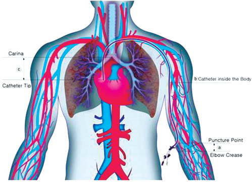
Cubital crease to carina length, CCL, d = a + b-c.
Equation of CCL through left upper arm vein in relation to patient height was obtained by these results.
Statistical Analysis
Student's t-tests were done if there were significant differences in age, height, weight, cubital crease to puncture point, catheter length inside the body, carina to catheter tip length, and CCL between male and female groups. Correlation analysis was performed using Pearson's test for evaluation of correlation between height and CCL. Linear regression analysis was done if height with or without weight is capable of predicting the CCL. Analysis of variance test was done if there was a significant difference of CCL among the groups of punctured veins (basilic, cephalic, and brachial veins), although the number of patients in whom cephalic and brachial veins were punctured was small. Finally, equation of CCL based on patient height in 165 cases was obtained.
Statistical analyses were performed using Sigma plot 2000 and SPSS version 10.0 (SPSS Inc., Chicago, Illinois, USA). P-value less than 0.05 was considered significant.
Results
Patient demographics were as follows; Male: female, 72: 79, mean age, male, 62.4 ± 19.3 years, range 20-89, female, 65.0 ± 15.2 years (range 32-87) (Tab. I).
Descriptive statistics of age, height, weight, cubital crease to puncture point, catheter length inside the body, carina to catheter tip length, and CCL are listed in Table I.
The height and weight of the patients were 168.1 ± 6.8 cm (range, 152 ~ 183) and 64.5 ± 13.7 kg (range, 42 ~ 140) in male, and 155.0 ± 5.4 cm (range, 140 ~ 168) and 54.5 ± 10.9 kg (range, 34 ~ 95) in female group, and they were significantly different in two groups (p<0.05) (Tab. I).
Cubital crease to puncture point and carina to catheter tip length were similar in male and female groups because we used the same method of PICC insertion.
The catheter length inside the body was 45.8 ± 2.4 cm (range, 40.0 ~ 52.0) in male and 43.0 ± 2.1 cm (range, 37.0 ~ 47.0) in female (p<0.01). The mean CCL through left upper arm vein was 47.1 ± 2.6 cm (range, 42 ~ 55) in male and 44.0 ± 2.9 cm (range, 34.0 ~ 50.0) in female patients and was longer in male (p<0.01) (Tab. I). The cubital crease to puncture point (mean 4.7 ± 1.4 cm, range, 2.0 ~ 8.0 in male, and mean 4.2 ± 1.6 cm, range, 1.0 ~ 9.0 in female) and carina to catheter tip length (mean 3.5 ± 2.5 cm, range, −2.0 to 9.0 in male and 3.3 ± 2.8 cm, range, −4.0 to 9.5 in female) were not significantly different in male and female patients (p>0.05).
Selected veins were basilic in 70.3%, cephalic in 13.3%, and brachial in 16.4% (Tab. II). There was no significant difference of CCL among the groups of punctured veins (basilic, cephalic, and brachial veins) (p>0.05).
CCL was significantly correlated with height (r = 0.54, p<0.0001). Using linear regression analysis, the equation was as follows:
CCL = height * 0.19 + 14 (r 2 = 0.22, p<0.0001);
this result is shown graphically in Figure 1.
Discussion
The distance from cubital crease to puncture point is variable inevitably due to patient characteristics and procedural difficulty, such as difficult arm abduction and supination and poor patient cooperation during the procedure, and initial venipuncture failure. The catheter length inside the body and CCL are different between male and female groups because the height and weight are different, that is, CCL was significantly correlated with patient height. In addition, no difference of CCL among the three selected veins (basilic, cephalic, and brachial) was present. So, we concluded than this equation of CCL determination from patient height could be used regardless of the sexuality and selected veins.
The mean carina to catheter tip length was 3.5 ± 2.5 cm in male and 3.3 ± 2.8 cm in female, although it is dispersed somewhat widely (about 11 cm in male and 13.5 cm in female). This dispersion is because we tried to locate the tip as deeply as possible near the cavoatrial junction, and trimmed the catheter somewhat longer than expected prior to catheter advancement worrying about the possible case of catheter malfunction due to its short length, and patient weight range was not considered that was dispersed widely about 98 kg in male and 61 kg in female group as well.
For multiple reasons, PICCs have become among the most frequently encountered central venous catheter in non-intensive care unit (ICU) patient. For instance, these devices are safer to insert than CVCs, eliminate the discomfort associated with phlebotomy and scheduled peripheral intravenous line changes, and provide extended and reliable venous access (7). Blind bedside insertion of PICC without the aid of fluoroscopy is necessary and sometimes inevitable, especially including ICU patients, so, many efforts should be done for correct central location of the catheter tip for long-term use of PICC because it resulted in as much as 60% of catheter tips located in a suboptimal position (6, 7, 9). Although not in all cases, incorrect estimation of the length of catheter is the main cause of low rates of successful central tip positioning (6, 8, 9). So, the venous length from the puncture point to carina should be estimated correctly in each patient before the procedure but is not easy because the patient's body size is quite different and venous puncture point is variable. In addition, direct measurement along the presumed course with tape ruler outside the body, as already incorporated in many institutions, is quite cumbersome and especially quite vulnerable to infectious complication. So, our study result is greatly useful in those situations of blind bedside procedure of PICC for the correct positioning of catheter tip because our equation can estimate the length of upper arm vein in relation to the patient's height and weight.
The recommended tip location of PICC is central vein, more specifically junction of SVC and right atrium, or distal SVC (16-19). Some argue that right atrium is a satisfactory position also if the tip does not abut the atrial wall or traverse the tricuspid valve or coronary sinus (20). Tip position of a central venous access is of paramount importance and should be verified before starting infusion. Because of difficulty to appreciate the junction of SVC and right atrium in the standard anterior-posterior chest radiograph, we decided to use the carina as the radiographic landmark of measurement. Considering the recommendation of Food and Drug Administration guidelines for central venous catheter placements that catheter tips never enter the atrium and a report about catheter migration with movement of the patient's arms or head 1-3 cm caudally, we tried to locate the tip of PICC in the lower SVC fast the carina near cavoatrial junction (19-21). In addition, when the catheter tip ends near the carina, the catheter tip tends to abut the right SVC wall with obtuse angle because of the natural anatomy of the junction of left innominate vein and SVC and stiffness of the catheter in left-side approach of PICC, so, we tried to insert the carina more deeply and fast as possible to lower SVC near the cavoatrial junction. Possibility of catheter tip movement according to phase of respiration, catheter type, insertion site, body habitus, and body position also contributed to this decision (22-24).
Although some debates about the preferential laterality of PICCs are present, we selected mostly left basilic vein as a first choice because of various causes such as patient preference, non-dominant arm selection, higher rates of venous thrombosis in cases of cephalic vein selection, and median nerve bisection in case of brachial vein selection (25-27).
When we determined the catheter length inside the body, we used the formula of our previous report of ECL through right basilic vein based on patient height, and considered the report that left innominate vein is 3.5-4 cm longer than the right one (7, 14).
Although there are documentations about the length of upper extremity veins including cephalic, basilic, axillary, subclavian, and innominate veins as well as SVC, those are only for the average-sized adult (5, 10). So, we think that our equation is more useful because it reflects the patient height statistically significantly. Although the estimation from the equation is not used initially during peripherally implantable venous access port insertion, we believe that this equation would be helpful for the calculation of the length of upper arm vein to carina in the case of exchange.
Among the intra-procedural methods for correct catheter tip positioning without the aid of fluoroscopy, the electrocardiography (EKG) method was proved to have many advantages, as it is as accurate as fluoroscopy, simpler, more readily available, less expensive, safer, and more cost-effective (28). So, the rate of successful central location of PICC tip could be greatly increased when EKG method is combined with the method of catheter length determination using our equation in cases of bedside insertion of PICC.
PICC tip movement of an average of 3.2 cm on changing position is more pronounced in overweight patients and with larger catheters (24). This is one of the reasons of a wide variation of CCL in same height in our patients.
There are several limitations in our study. Proper estimate of catheter length does not mean proper position of the tip, and this study has several bias, as all patients were in supine position with abducted arm and many of the observations were performed using the cephalic vein (which is an atypical vein to use for PICCs). And, all radiological measurements were performed by one radiologist, by subjective criteria. Finally, the equation is valid exclusively for left arm insertion.
Conclusion
There was a significant correlation between the height of the patient and the length of left upper extremity vein from cubital crease to carina. We could get equations of CCL from patient height and weight, and estimation of CCL from height might be useful in the following way: final catheter length = CCL - length between emergency site and cubital crease + 3 cm (which is the mean distance between carina and cavo-atrial junction in adults).
The results of our study offer a simple and reliable method for estimating catheter length when inserting PICCs on the left side, though proper central position of the tip obviously will require additional methods for tip location (radiology, intracavitary electrocardiography, or echocardiography).
Footnotes
Financial support: The authors have no financial disclosures to make.
Conflict of interest: The authors have no conflict of interest.