
Abstract
Purpose
To increase awareness of peripherally inserted central catheter (PICC) fracture and necessary nursing assessment to identify development of nerve injury after removal of the PICC fracture.
Methods
This is a case review of a cancer patient with fractured PICC and the postoperative symptoms leading to nerve injury.
Results
The reason for PICC fracture is the fragility of silicon. Secondary surgical intervention of a PICC fragment resulted in nerve damage from a hematoma placing pressure on the median nerve in the arm.
Conclusions
It is necessary to use power injectable polyurethane PICCs. It is vital to have a clear understanding of signs and symptoms of nerve impingement in the arm when monitoring a post-operative patient. Assessment of neurological status, circulation, swelling and patient complaints of pain are all necessary functions of the nurse in caring for this type of patient.
Introduction
Peripherally inserted central catheter (PICC) has been widely used in clinical practice, especially for patients needing periodic chemotherapy for tumors, long-term parenteral nutrition, and intravenous injection (1). Despite all its merits, PICC is associated with several catheter-related complications. Among them, catheter fracture is a rare one, with an estimated incidence rate of 0.69% in our hospital. It may happen during catheter insertion, high-pressure flushing, and removal (2, 3). The incidence of catheter fracture in the UK ranges from 0.2% to 9.7%, which is consistent with our data (4).
Generally, PICC fractures are removed using the radiographic method (5) or vascular incision. A radiographic method is used mainly when the catheter fracture retracts into the body and migrates to the superior vena cava or atrium without vein thrombus, while vascular incisions are applicable under the conditions of a breakage located in the upper limb vein or appeared vein thrombosis or adherence to the blood wall. The interventional method has been widely reported with no post-operative complication noted in all of these cases. There has been no report of the vascular incision and the possible complications.
On March 20th, 2014, catheter fractures occurred during extraction of a PICC from a cancer patient in our department. The ultrasound results of the patient in this report showed the PICC remnant located in her left brachial vein and there was no venous thrombus. The fracture could be removed by either of the methods (radiographic method or vascular incision) according to the invasive technology department. The patient requested removal by vascular incision and signed the informed consent. We successfully performed vascular incision to remove the fracture, after which a nerve injury unfortunately occurred. The treatment process is presented below and the related literature is reviewed.
Case Presentation
A 67-year-old woman was hospitalized for the eighth cycle of chemotherapy after surgical resection of moderately differentiated adenocarcinoma of sigmoid. A single-lumen 18-guage (4-Fr) silicon PICC (Bard Groshong, USA) was successfully placed in the left brachial vein under ultrasound guidance on October 21, 2013. The inserted portion of PICC was 36 cm in length, while 6 cm was left outside. The procedure was uneventful without catheter occlusion. Five months later, the patient finished the eighth cycle XELOX regime. A nurse performed the procedure according to institutional protocol, explained the indication and helped the patient to relax. Unfortunately, the catheter fracture occurred during extraction and a residue of 33 cm was deposited in the body (Figs. 1, 2). Vascular incision was performed to remove the fracture from the left brachial vein by the vascular surgery department. After the operation, the patient was transferred to the floor and received a thrombosis prophylaxis. On the following day, the patient complained of a mild pain at the surgical site with a numerical rating scale (NRS) score of 2. Physical examination showed no oozing bleeding from the incision, swollen but moveable limb, and touchable arteriopalmus. Slight elevation of the affected limb was initiated along with prescribed bed rest. On day 2 after surgery, the patient left hospital without discharge order that afternoon and did not return until the day 4. The pain worsened, with a NRS score of 5. The upper limb was obviously swollen with subcutaneous dotty ecchymosis and lump at the incision site. On the fifth morning after surgery, the swelling aggravated and numbness, paresthesia, and reduced activities of the left upper limb were noted. After vascular surgical consultation, anticoagulation treatment was discontinued and 1 g/day oral diosmin was started. Vascular ultrasound color Doppler examination showed a hematoma in the low echo area of subcutaneous layer of the left arm (Fig. 3).
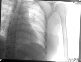
X-ray chest with fracture catheter.
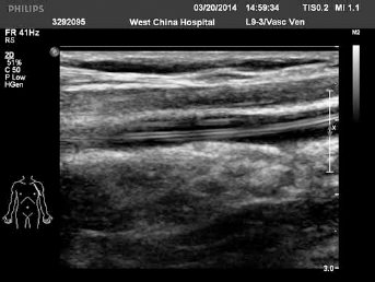
Intravenous ultrasound with fracture catheter.
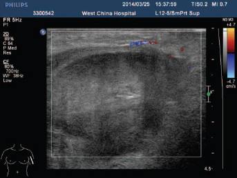
Vascular ultrasound color Doppler with a hematoma.
On day 6 after surgery, the second emergent operation was performed to clear the hematoma and suture the brachial vein. One day after that, the patient complained of dull pain and minor swelling. Diosmin treatment was continued while the uptake of low molecular weight heparin was suspended. The electromyography showed that the conduction volatility and velocity of median nerve of her left limb was reduced and the action of sensory nerve was slow. The results also indicted a possible neurogenic injury of short abductor muscle of the left thumb. After discussion with physicians from the vascular surgery, neurology and rehabilitation departments, pregabalin was added at a dose of 75 mg to relieve the pain with symptom improvement. Corresponding support therapies were carried out. In order to retrieve the function of the median nerve, a combination therapy of mecobalamin tablets, vitamin B12, and electro-acupuncture was needed. The patient was discharged and followed up regularly. Currently, the patient has improved perceptibly after 2 months of combination therapy.
Discussion
Reasons for PICC Fracture
The primary reason for the fracture in this case may be related to the catheter material. The catheter product we chose was a single-lumen 18-guage (4-Fr) PICC (Bard Groshong, USA) the material is silicone, which is fragile. A study showed that breakage occurred in 9 of 117 silicone catheters (8%) and none of 94 polyurethane catheters (p = 0.005). Cohen et al (6) also reported fewer catheter fractures with the polyurethane catheter compared with the silicone catheter. In addition, a probably inappropriate traction during some difficulty in removal and the poor compliance by this patient were also risk factors of catheter fracture.
Analysis of Nerve Injury
In this case, the nerve injury was primarily related to the hematoma compression, which may be caused by disrupted post-operative follow-up due to the poor compliance of the patient. Probably, the nerve injury was caused by oxaliplatin-induced peripheral nerve toxicity, a typical adverse reaction of XELOX regime. It has been reported that 68% of the patients suffered touch pain and 92% complained of acroparesthesia after six cycle of XELOX regime (7). Although there wasn't any evidence during/after chemotherapy showing that this was experienced by our patient, eighth cycle chemotherapy of XELOX regime may induce some degree of neurotoxicity. Finally, secondary operations also added to the risk of nerve injury.
Nursing Implications
First, nurses prefer polyurethane PICCs. Current practice with PICCs has moved to polyurethane as a safer material for the small diameter PICCs. Second, nurses should scan the arm using ultrasound before removal, possibly in any case, but absolutely in all cases of difficult removal. Then, if the PICC fragment is really stuck inside the vein let an experienced vascular surgeon do the maneuver. Finally, do not use antithrombotic prophylaxis after such an intervention. Nurses should do well in the health education for patients inserted with PICCs and catheter maintenance during the intermittent period of chemotherapy. In our hospital, we provide a PICC Health Education Handbook to patients and their healthcare providers to let them know how to manage signs of complications with PICCs and improve the patient compliance.
Conclusion
Nurses must be highly cautious about the risks of catheter fracture, know the possible causes and take effective measures to prevent their occurrence and development. The conclusions are: (i) use power injectable polyurethane PICCs; (ii) before PICC removal, rule out PICC-related thrombosis; (iii) if there is some difficulty on removal, stop and use ultrasound to understand where the obstacle is thrombosed or the rolling of a fibroblast sleeve; (iv) if the PICC needs to be freed surgically, please let an experienced vascular surgeon do this.
Footnotes
Financial support: No grants or funding have been received for this study.
Conflict of interest: None of the authors has financial interest related to this study to disclose.