






Abstract
The use of the tunneled central venous catheter (CVC) is steadily increasing worldwide as a means of vascular access for hemodialysis. The increased use of these devices, which often outlive the patients, and the extended time they are used are associated with more frequent complications. Among these, one of the emerging complications is that of the “embedded” or stuck catheter. This term refers to when the catheter cannot be removed after detaching the retention cuff. In medical literature, experiences with the removal of stuck catheters are described with the use of several different methods. Currently the most commonly used technique also considered the safest is “endoluminal dilation” also known as Hong's Technique, recently modified by Quaretti and Galli. Below, a new technique using a Vollmar ring is described for removing a stuck catheter as an alternative to Hong's technique, or after a failed attempt at using Hong's technique.
Introduction
The use of the tunneled central venous catheter (CVC) is steadily increasing worldwide as a means of vascular access for hemodialysis (1). This increase is partly justified by the increased age of the incident patients and the progressive aging of the prevalent patients associated with the exhaustion of vascular endowment and the more serious comorbidities that the patients in question suffer from (2). The increased use of these devices, which often outlive the patients, and the extended time they are used are associated with more frequent complications.
Among these, one of the emerging complications is that of the “embedded” or stuck catheter. This term refers to when the catheter cannot be removed after detaching the retention cuff. Fluoroscopic observations show an abnormal movement toward the upper portion of the cardiac silhouette and large vessels when an attempt is made to remove the CVC, which, in turn, can cause intense retrosternal pain if it is pulled forcefully. There have even been registered cases of vasomotor collapse, non-ST-elevation myocardial infarction (NSTEMI), avulsion of the vena cava, damage to the tricuspid valve having fatal consequences and breakage of the CVC (3).
In medical literature, experiences with the removal of stuck catheters are described with the use of several different methods: by means of a gooseneck snare catheter, the peel-away sheath introducer and the use of laser technology. Currently, the most commonly used technique and also considered to be the safest is “endoluminal dilation” also known as Hong's Technique (46), recently modified by Quaretti and Galli (7).
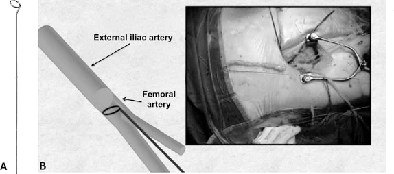
Below, a new technique using a Vollmar ring is described for removing a stuck catheter as an alternative to Hong's technique, or after a failed attempt at using Hong's technique (Fig. 1A).
(
Patients and methods
During the two-year period 2013-2014, three separate cases of stuck catheters were dealt with using the Vollmar ring. This device is well known in the field of vascular surgery for its application in endarterectomy to remove atherosclerotic plaque (Fig. 1B).
Case report 1
A 72-year-old woman in dialysis since 2001 suffering from diabetes mellitus treated with insulin and with progressive exhaustion of both vascular and arterial endowment, the latter worsened by her disease. Over the course of several years, she had numerous tunneled CVCs which had to be replaced frequently due to malfunction and infection of the tunnel. The last attempt to replace the CVC in right internal jugular vein (IJV) failed due to a split tip catheter (Medcomp Split Stream) stuck in situ for one year.
Case report 2
A 74-year-old woman in dialysis since 2009. User of a CVC placed in right IJV necessitating replacement twice; the first time due to a malfunction and the second time due to an infection of the tunnel after two years of the CVC remaining in place. On performing the second replacement, the split tip catheter (Hemostar Bard) was found to be stuck.
Case report 3
A 69-year-old woman in dialysis since 2001. Due to the exhaustion of vascular endowment, a tunneled CVC (Split Stream Medcomp) was inserted into left IJV due to total occlusion of right IJV. After a period of 17 months the CVC was replaced due to a malfunction. On performing the replacement, the CVC was found to be stuck.
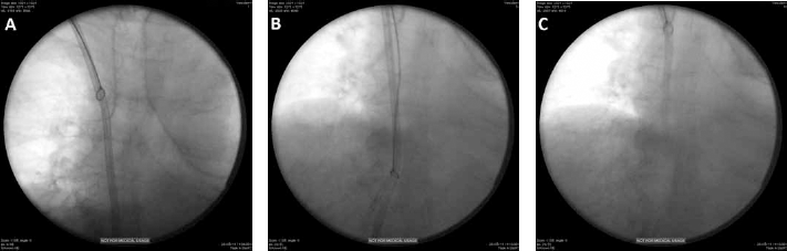
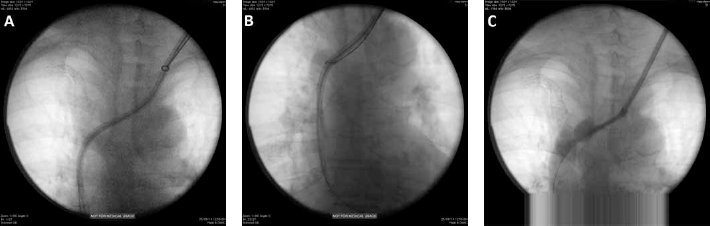
The removal of the stuck CVCs was carried out using an endarterectomy Vollmar ring. In the first case report, it was used after a failed attempt at Hong's technique of endo-luminal dilation while in the other two case reports it was used as a first choice because it was found to be more effective, faster and easier to use and was less time-consuming and costly. All procedures were carried out in the operating room while the patients were sedated but conscious. Their vital signs were being monitored and every maneuver was under control by means of radiological equipment. After cutting through the skin at the base of Sedillot's triangle where the CVC is inserted and sectioned off, a standard guide of 0.038” is inserted into one of the two lumens of the catheter. The Vollmar ring is then inserted onto the metallic guide and the sectioned-off end of the CVC (Fig. 2). The ring used must be slightly smaller than the catheter (one diameter size smaller) so that the opening of the catheter is forced. In order to facilitate the insertion, it is advisable to cut the catheter tip at an angle (like the mouthpiece of a recorder). Once the sectioned-off piece has passed through the ring, it is tightly kept into place by means of Crile forceps. By pulling slightly on the CVC, the ring is made to advance along the length of the catheter in rotary motion so as to progressively lyse the synechiae that keep the catheter anchored to the walls of the vessel. This operation is performed under continuous radiological observation in the entire length of the CVC, which can then be removed easily (Figs. 3A, B, C; Figs. 4A, B, C; supplementary video 1, 2, 3, 4, available online as supplementary material at www.vascular-access.info). The standard time it takes to remove the CVC is about 10 minutes. In two of the aforementioned cases, a new CVC was re-inserted. In one of the cases, the surgeon resorted to angioplasty due to stenosis where the superior vena cava and the left innominate vein meet in order to re-insert a CVC (Fig. 4C; supplementary video 5, available online as supplementary material at www.vascular-access.info).
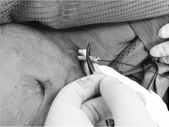
The Crile forceps prevent air from accidentally entering.
(
(
Discussion
The stuck catheter is an emerging complication in literature and probably an underestimated one. A recent review published by Forneris et al in the Journal of Nephrology focuses completely on factors that contribute to a stuck catheter and the possible pathogenesis that can derive from it (8). Among the contributing factors, it is likely that the amount of time the catheter remains in place plays an important role even though Forneris has stated (through personal experience) that complications may set in even at an early stage (8). The left internal jugular vein is particularly subject to complications given its tract is not straight and because of the three points of friction on the endothelium. Indeed, factors that cause damage to the walls of the endothelium, such as repeated replacements of the CVC and the infections that can arise, certainly tend to favor complications. The materials that the CVCs are made of do not seem to have any particular impact on the formation of adhesions, while it is still unclear as to whether the actual form of the CVC and above all its “side-holes” contribute to the formation of adhesions as Moore claims (9). There would appear to be more cases in women than in men: in fact, the three case reports analyzed in this study reflect the aforementioned contributing factors. The description of the technique used to remove the embedded CVC singles out Hong's technique, especially Hong's technique modified by Quaretti et al (7) to be the safest method with a success rate of 100%. In our experience, in the first case report, even when performing an intraluminal dilation by means of high-pressure balloons, the CVC did not dislodge. By way of a different approach, working outside the CVC but inside the vessel using a Vollmar ring enabled us to remove the CVC with no difficulty thanks to the breakage of the synechiae that kept it stuck to the walls. However, it is not possible to establish if this technique is superior to that of Hong's technique. It could be speculated that severing the adhesions with the Vollmar ring is perhaps preferable to forcefully removing the CVC with intraluminal dilation in terms of minimizing the damage and stress caused to the vessel walls when the device is pulled out. Pulling out the CVC might even cause the endothelium to detach leading to clotting. This is however a mere hypothesis that remains to be demonstrated. The relative simplicity and safety of the technique, the limited amount of time it requires, and (based on what the patients refer) the relatively painless procedure, has induced us to prefer it over the others in the other two case reports.
The Vollmar ring can be adapted to any type of CVC. They vary in diameter, and the conformation of the tip that is placed around the CVC can be either circular or elliptical. The device itself is flexible enough to guide through the winding course of the left IJV. During this insertion and subsequent progression, the Crile forceps prevent air from accidentally entering without having to resort to the use of valved introducers (Fig. 2). However, it is extremely important to remember that such an innovative technique must be carried out by expert hands in a suitable health facility having a radiological imaging system in place. The use of a Vollmar ring does not cause complications greater than those caused by use of valved peel-away introducers or other types of introducers. Furthermore, as vascular surgeons are well aware of through experience, the vena cava that has had a foreign body embedded in it for a long time, which has become stuck, will show signs of fibrotic modifications making its walls more resistant to possible perforations, therefore this reduces the risks in connection with the introduction and the progressive insertion of the Vollmar ring which must always be gently and expertly carried out.
Footnotes
Financial support: No grants or funding have been received for this study.
Conflict of interest: None of the authors has financial interest related to this study to disclose.