






Abstract
Purpose
To report the use of a balloon catheter to facilitate percutaneous ultrasound-guided thrombin injection of a subclavian artery pseudoaneurysm.
Case report
A 36-year-old man presented with hoarseness after the insertion of a right temporary internal jugular line. Arteriography showed a short neck right subclavian artery pseudoaneurysm. A 6 mm × 40 mm balloon catheter was positioned across the neck of the pseudoaneurysm to permit percutaneous ultrasound-guided thrombin injection. Immediate post-treatment angiogram demonstrated no filling of the pseudoaneurysm. Seven-month clinical and imaging follow-up confirmed resolution of the pseudoaneurysm and hoarseness.
Conclusions
This clinical vignette highlights the potential of balloon catheter-assisted percutaneous ultrasound-guided thrombin injection as an alternative to open or endovascular repair of pseudoaneurysms of the subclavian arteries.
Keywords
Introduction
Central venous cannulation is a common procedure for the diagnosis and treatment of multiple medical conditions (1). An uncommon, yet possibly fatal, complication associated with central line insertion is the development of a subclavian artery pseudoaneurysm (SAP) after accidental puncture of the artery (1). Progression of the false aneurysm can lead to rupture, and potentially life-threatening hemorrhage (1). Classical treatment of pseudoaneurysms is open surgery for resection and end-to-end anastomosis, venous graft, primary suture repair, or bypass (2). However, this often requires access to the thoracic cavity and therefore is unsuitable for high-risk patients with multiple medical co-morbidities (2).
The advent of endovascular and percutaneous treatment modalities has transformed the assessment and management of iatrogenic and traumatic SAPs. Recent literature has described less invasive techniques of treating SAPs, such as thrombin injection (3) or endovascular repair (4). We present the case of an iatrogenic right SAP that occurred as a consequence of incorrect placement of a right internal jugular central venous catheter, successfully repaired with percutaneous ultrasound (US)-guided thrombin injection (PUGTI).
Case description
A 36-year-old man with class V lupus nephritis received a living unrelated paired exchange renal transplant and allo-stem-cell transplant in India. The patient had delayed graft function and presented to our institution, directly from the airport, with acute kidney injury and bicytopenia, consistent with a diagnosis of antibody mediated rejection. Immunosuppression had to be reduced in hospital for severe Clostridium difficile-associated diarrhea, leading to accelerated thrombotic microangiopathy. In the context of two competing life-threatening events, the patient underwent graft nephrectomy. A right internal jugular central venous catheter was placed as a requirement of his perioperative care. Vessel access was obtained with standard Seldinger technique for triple-lumen catheter placement. After the central venous catheter was removed, the patient developed persistent hoarseness in the post-operative period without focal neurologic deficits or hemodynamic instability. He was seen promptly by the otolaryngology service who documented a right recurrent laryngeal nerve palsy. A non-contrast computed tomography (CT) scan of the neck and thorax revealed a heterogeneous high density mass measuring 4.1 × 4.2 × 5.5 cm (anteroposterior [AP] x medial-lateral [ML] × craniocaudal [CC]) in the right supraclavicular/paratracheal region neighboring the subclavian vessels and exerting a mass effect on the adjacent trachea. A subsequent contrast-enhanced CT scan (Fig. 1) confirmed this to be a 1.9 × 2.6 × 3.0 cm (AP × ML × CC) false aneurysm of the proximal right subclavian artery with surrounding hematoma. The neck of the pseudoaneurysm was within 5 mm of the origin of the right vertebral artery. Notably, both vertebral arteries were co-dominant on the CT angiogram.
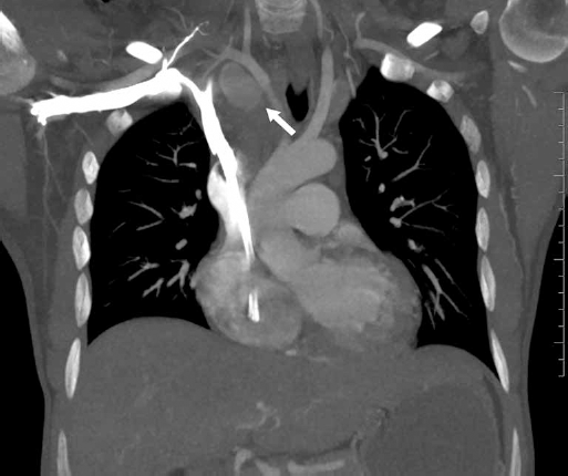
Enhanced arterial phase coronal computed tomography (CT) maximum intensity projection (MIP) of the chest shows a pseudoaneurysm of the proximal right subclavian artery (arrow).
Arteriography performed through a trans-radial 65 cm 5-F Kumpe catheter (Cook Medical, Bloomington, Indiana) showed a short neck right subclavian pseudoaneurysm (Fig. 2A). A 6 mm × 40 mm Mustang balloon (Boston Scientific, Marlborough, Massachusetts) was positioned across the neck of the pseudoaneurysm over a Rosen wire (Cook Medical). A test occlusion produced no neurological symptoms. The right subclavian pseudoaneurysm was directly punctured under ultrasound (US) guidance from the neck. Contrast was injected to demonstrate adequate positioning in the pseudoaneurysm (Fig. 2B). The balloon was inflated across the pseudoaneurysm neck and 500 IU of thrombin (Tisseel, Baxter, Mississauga, Ontario) was injected (Fig. 2C). After a 2-minute period, the balloon was deflated and exchanged for a Kumpe catheter. Another angiogram was performed demonstrating no filling of the pseudoaneurysm (Fig. 2D). US examination revealed echogenic material within the pseudoaneurysm sac consistent with fresh thrombus formation.
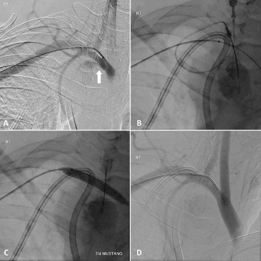
(
A chest CT performed one day after the procedure showed a mixture of clotted contrast-enhanced blood within the pseudoaneurysm (Fig. 3A). A follow-up duplex US performed two days’ post-procedure demonstrated no residual pseudoaneurysm. A follow up CT chest performed 7 months later showed complete resolution of the pseudoaneurysm (Fig. 3B). Clinically the patient's voice has returned to normal.
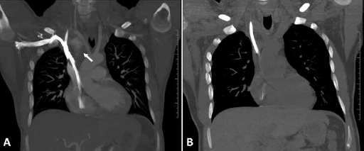
(
Discussion
SAPs are relatively uncommon and are often caused by atherosclerotic disease (5). Other etiologies include, but are not limited to, trauma, infection, thoracic outlet syndrome and congenital conditions such as Marfan and Turner's syndrome (6). Inadvertent puncture of the subclavian artery during central venous cannulation is another rare cause of SAP (7). While there is very little literature regarding the prevalence and pathogenesis of SAP, a review of almost 400 case reports of subclavian artery aneurysms showed the risk of life- and limb-threatening complications, such as rupture, embolization and thrombosis, is unrelated to the diameter of the aneurysms with thrombo-embolic complications occurring in aneurysms as small as 12 mm (8).
False aneurysms of the subclavian artery can be adequately imaged with contrast-enhanced CT, which is useful in diagnosis and in assessing mass effect on surrounding neurovascular structures. Angiography can also be used to accurately establish a diagnosis, and is often utilized in endovascular treatment options, such as intraluminal stenting or embolization (7). In this case, CT was a source of useful information regarding the size and relationship of the pseudoaneurysm to surrounding structures. Adequate imaging, however, requires intravascular contrast agents, which can be nephrotoxic in patients with acute renal injury (9), especially in our patient with pre-existing renal dysfunction. Renal complications however, can be limited with adequate hydration, use of low doses of contrast agent, and choosing less toxic alternatives (9).
SAPs are amenable to a variety of treatment options including US-guided compression, open surgical repair, endovascular repair and PUGTI. US-guided compression is usually unsuitable for SAPs because of the close proximity of the clavicle, which makes it difficult to compress the subclavian artery (10). We considered an open sternotomy and suture repair of the proximal subclavian artery with control of the innominate, subclavian, vertebral, and common carotid arteries. The patient was believed to be at high risk for open surgical exploration, in light of his renal dysfunction and protracted medical history. Endovascular options were also considered, but placement of a covered endoluminal subclavian artery stent would have necessitated covering and compromising the right vertebral artery in this relatively young patient. Additionally, there was a risk of subsequent systemic sepsis secondary to stent infection in this immunocompromised host. Thrombin injection is most appropriate in patients who are hemodynamically stable with a small pseudoaneurysm (11). Our patient, in fact, was hemodynamically stable, and the location of the pseudoaneurysm enabled safe US-guided percutaneous access for thrombin injection. Although relatively safe and effective, there are numerous risks associated with PUGTI. These include, but are not limited to, the risk of arterial thrombosis and embolization, which is of particular concern due to the potential for cerebral ischemia. The use of balloon assistance during the procedure was aimed at reducing the risk of systemic thrombin leading to clot formation while maintaining a high concentration of thrombin in the pseudoaneurysm sac. The advantages of thrombin injection, in relation to other modalities such as US-guided compression, include its higher success rate, greater patient comfort, and the ability to treat non-compressible pseudoaneurysms. It also does not require discontinuation of anticoagulants prior to treatment (12).
This case also confirms the importance of US guidance in reducing complications in central catheter placement. However, even with US guidance, complications may arise: care must be taken to evaluate the depth of the jugular vein to avoid inadvertently advancing the needle beyond its expected location and puncturing the subclavian artery.
Conclusion
In conclusion, PUGTI is an acceptable alternative to open or endovascular repair of iatrogenic pseudoaneurysms of the subclavian artery.
Footnotes
Financial support: No grants or funding have been received related to this study.
Conflict of interest: None of the authors has financial interest related to this study to disclose.