






Abstract
Introduction:
Valvular disease and pulmonary hypertension are common conditions in haemodialysis patients. In presence of tricuspid regurgitation, an increased retrograde blood flow into the right atrium during ventricle systole results in a typical modification of the normal venous waveform, creating a giant c-v wave. This condition clinically appears as a venous palpable pulsation within the internal jugular vein, also known as Lancisi’s sign.
Case report:
An 83-year-old woman underwent haemodialysis for 9 years. After arteriovenous fistula thrombosis, a right internal jugular vein non-tunnelled central venous catheter (CVC) was placed. About one month later, the patient was referred to our facility for the placement of a tunnelled CVC. Neck examination revealed an elevated jugular venous pulse, the Lancisi’s sign. Surprisingly, chest x-ray posteroanterior view showed the non-tunnelled catheter tip in correspondence with the right ventricle. She underwent surgery for temporary to tunnelled CVC conversion using the same venous insertion site (Bellcath®10Fr-length 25 cm to Mahurkar®13.5Fr-length 19 cm). In the postoperative period, we observed a significant reduction of the jugular venous pulse.
Discussion:
The inappropriate placement of a 25-cm temporary CVC in the right internal jugular vein worsened the tricuspid valve regurgitation, which became evident by the Lancisi’s sign. Removal of the temporary CVC from the right ventricle resulted in improved right cardiac function. Safe approaches recommended by guidelines for the CVC insertion technique and for checking the tip position should be applied in order to avoid complications.
Introduction
Valvular disease and pulmonary hypertension are common conditions in haemodialysis patients. Generally, tricuspid valve disease occurs secondary to left-side heart valve disease, in particular mitral valve disease. End-stage renal disease (ESRD) patients have clinical features such as anaemia, chronic volume overload and arteriovenous fistula that lead to a preload increase on the right heart chambers with adverse effects on their performance. Right heart structures lose their distensibility with tricuspid regurgitation and further right heart volume overload (1). Lancisi’s sign occurs in presence of severe tricuspid regurgitation and it clinically appears as a venous palpable pulsation within the internal jugular vein. In this condition, an increased retrograde blood flow into the right atrium during ventricle systole results in a typical modification of the normal venous waveform, creating the giant c-v wave (2) (Fig. 1).
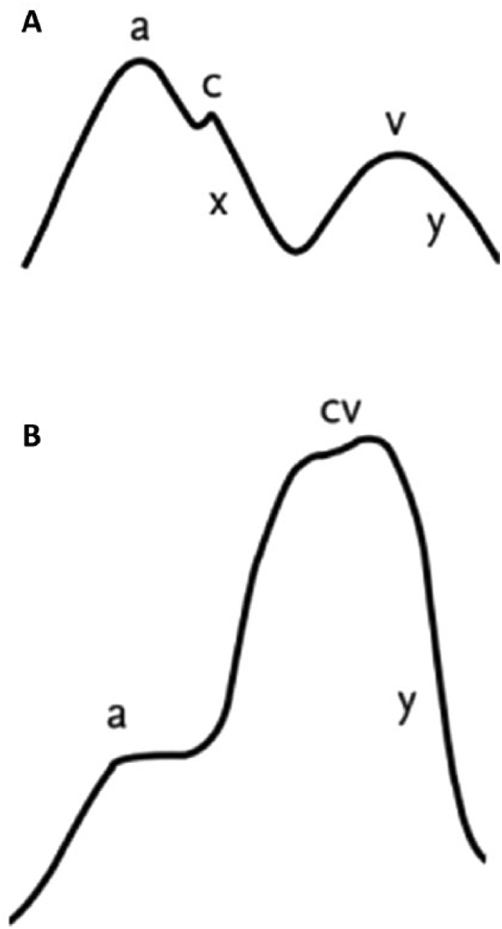
Lancisi’s sign venous waveform. Normal venous waveform (
Case presentation
An 83-year-old woman underwent haemodialysis for 9 years. After arteriovenous fistula thrombosis, a right internal jugular vein non-tunnelled central venous catheter (CVC) was placed. About one month later, the patient was referred to our facility for the placement of a tunnelled CVC. She presented with NYHA (New York Heart Association) class III breathlessness and mild lower limb oedema. Neck examination revealed an elevated jugular venous pulse, the Lancisi’s sign (see video, available online as Supplementary material at www.vascular-access.info). On cardiac auscultation, she presented an apical mid-diastolic rumble and a holosystolic murmur at the left lower sternal border. Surprising, the chest x-ray posteroanterior view showed the non-tunnelled catheter tip in correspondence with the right ventricle (Fig. 2). Doppler transthoracic echocardiography demonstrated a moderate/severe tricuspid regurgitation (Peak Tricuspid Regurgitation Velocity [TRV] ≥2.9 m/s). She underwent surgery for temporary to tunnelled CVC conversion using the same venous insertion site (Bellcath®10Fr-length 25 cm to Mahurkar®13.5Fr-length 19 cm) (3). In the postoperative period, we observed a significant reduction of the jugular venous pulse. Chest x-ray showed the catheter tip in correspondence with the mid-right atrium and echocardiography demonstrated improving function of the right chambers.
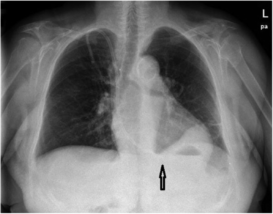
Preoperative chest x-ray. Right internal jugular vein non-tunnelled CVC tip in correspondence with the right ventricle.
Discussion
Maintenance haemodialysis patients (MHD) have a high incidence of cardiovascular diseases (CVDs), which is the first cause of morbidity and mortality in this population. Tricuspid regurgitation is a marker of adverse outcome, and patients with moderate/severe tricuspid regurgitation have a worse prognosis. Clinical conditions such as anaemia, inflammation and fluid overload act on global and right cardiac function leading to heart structural changes. Therefore, in MHD patients a regular cardiac ultrasound with attention to right chambers is helpful for the early diagnosis of cardiac dysfunction. The Lancisi’s sign represents a useful clinical feature of tricuspid valve and right heart chamber impairment. In normal conditions, the venous waveform is characterized by three peaks (a, c and v) and two slopes (x and y). In patients with severe tricuspid regurgitation the loss of the x slope creates the fusion of the c and v waves, which results in a prominent c-v wave (2). It is evident on neck examination as a large pulsation of the right internal jugular vein. In literature, there are few reports of Lancisi’s sign, none involving MHD patients (4, 5). In this case, the Doppler transthoracic echocardiography performed before the AVF thrombosis did not show right chamber function impairment. Therefore, the inappropriate placement of a 25-cm temporary CVC in the right internal jugular vein worsened the tricuspid valve regurgitation, which became evident by the Lancisi’s sign. Removal of the temporary CVC from the right ventricle resulted in improved right cardiac function. After tunnelled CVC insertion chest x-ray was performed. Guidelines suggest a postoperative control of the tip position by chest or abdomen x-ray for non-tunnelled CVC and by x-ray or fluoroscopy for tunnelled CVC (6). In conclusion, safe approaches recommended by guidelines for the CVC insertion technique and for checking the tip position should be applied in order to avoid severe complications.
Footnotes
Disclosures
Financial support: No grants or funding have been received for this study.
Conflict of interest: None of the authors has financial interest related to this study to disclose.
Meeting presentation: The manuscript was presented as Poster at the 10th Congress of the Vascular Access Society, April 5-8, 2017, Ljubljana, Slovenia.