






Abstract
Thymoma is the most common primary neoplasm of the anterior mediastinum. At diagnosis, up to 40% of patients present with advanced disease. Because advanced thymomas receive neoadjuvant chemotherapy, diagnostic imaging is crucial to plan the correct treatment. For characterizing thymomas, CT is the first choice modality, whereas 18F-FDG/PET is reserved for questionable cases and MRI is not routinely employed. Hereby, we describe a case of thymoma with a single contralateral pleural implant in a 30-year-old woman. The small pleural thickening detected at CT was correctly interpreted as pleural seeding related to thymoma at diffusion-weighted (DW)-MRI after a negative 18F-FDG/PET scan, and was subsequently confirmed at surgery. Precise diagnosis and accurate preoperative staging are crucial in managing thymic epithelial tumours in order to design the appropriate treatment and improve prognosis. Indeed, when stage IVa for pleural seeding is diagnosed preoperatively, a multimodality approach including primary chemotherapy followed by surgery and postoperative radiotherapy/chemotherapy is recommended. This is the first report that used DW-MRI for the characterization of pleural seeding in thymoma and demonstrates that DW-MRI could be useful for the correct pre-operatory staging in thymoma patients, especially in cases with indeterminate pleural thickenings at CT, in order to define the correct management.
Introduction
Thymoma is the most common primary neoplasm of the anterior mediastinum. It arises from the thymic epithelium and represents 20% of all mediastinal tumors in adults (1–4). Although usually asymptomatic, 15% of patients present with myasthenia gravis (5, 6). At diagnosis, approximately 15%-40% of patients have advanced disease (Masaoka-Koga stage III-IV) for invasion of adjacent structures, pleural/pericardial seeding, and metastatic disease (5–7). The preoperative distinction between non-advanced and advanced thymomas is essential to plan the proper treatment, because patients with advanced disease receive neoadjuvant chemotherapy before surgery (8). Hence, diagnostic imaging has a crucial role in the assessment of patients with thymoma. Computed tomography (CT) is the first-choice modality in the identification, staging, and follow-up of patients with thymoma, although it has limited value in differentiating among the various histological subtypes and in detecting residual or recurrent disease after treatment (9–13). In addition, CT has limitations in the characterization of indeterminate tiny pleural thickenings, especially when ispilateral to the primary mediastinal tumour, since it may be difficult to report these findings to pleural implants or non-calcified benign plaques (6, 14). 2-[18F]-fluoro-2-deoxy-D-glucose ( 18 F-FDG)/positron emission tomography (PET) is very helpful for the characterization of thymomas, particularly in suggesting the aggressiveness of thymoma, in detecting metastatic lesions, and in evaluating residual or recurrent disease (15). Currently, magnetic resonance imaging (MRI) is not routinely employed in the assessment of patients with thymoma and is generally reserved for further evaluation of equivocal CT findings of local invasion because of its better contrast resolution, or in case with suspected vascular invasion in which iodine contrast cannot be administered (6, 16–19). Nevertheless, newer MRI techniques like diffusion-weighted (DW) imaging could help in differentiating non-advanced from advanced thymomas in order to plan the correct management in a multimodality approach (4, 6, 8). Hereby, we present a case of advanced thymoma in which the single small pleural implant, contralateral to the primary tumour, was detected at DW-MRI after a negative 18 F-FDG/PET.
Case report
A 30-year-old woman with recent onset of myasthenia gravis underwent CT. Chest CT (64-row MDCT; section-thickness, 1.5 mm) revealed a left anterior mediastinal mass with soft tissue attenuation, that measured 65 mm in maximal diameter, resembling a thymoma (Fig. 1A–B). In addition, a small nodular pleural thickening of the right diaphragm, that measured 7 mm in maximal diameter, was appreciable in the anterior recess of the lung (Fig. 1C), contralateral to the primary tumour, with no other apparent relevant findings in the thorax and abdomen. For further characterization both of the mediastinal lesion and of the single pleural thickening, the patient underwent 18 F-FDG/PET. 18 F-FDG/PET demonstrated an area of intense and homogeneous 18 F-FDG uptake in the left mediastinum that did not cross the midline (Fig. 2, upper images), corresponding to the soft tissue mass detected at CT. No other areas of increased 18 F-FDG uptake were seen, especially in the location of the nodular pleural thickening of the right diaphragm (Fig. 2, lower images). In consideration of the 18 F-FDG/PET findings and of the contralateral location compared to the primary tumour, the focal thickening of the right diaphragm was initially interpreted as a benign non-calcified pleural plaque. At last, chest MRI (1.5-Tesla unit; section-thickness, 2.5 mm) was performed and confirmed the anterior mediastinal lesion that was characterized by restricted diffusion at DW imaging with a low apparent diffusion coefficient (ADC) of 0.56 × 10⁻³mm2s−1 (Fig. 3A–C). Moreover, DW-MRI showed the tiny focal thickening of the diaphragm in the anterior recess of the right lung as a nodule of high signal intensity, even at high b values, suggesting the diagnosis of small pleural implant, with a low ADC value similar to that of the primary tumour (Fig. 4A–B). Hence, the patient was preoperatively diagnosed at stage IVa for evidence of pleural seeding and consequently treated with the choice of a multimodality approach. After primary chemotherapy, the patient underwent extensive surgical resection through median sternotomy and right intercostal thoracotomy for access to the diaphragm. At surgery, thymomectomy with extended complete thymectomy, through the en bloc resection of the thymus and all the adipose mediastinal tissue (including the removal of the upper cervical poles and surrounding mediastinal fat), and the discrete resection of the individual pleural nodule, with macroscopically complete tumour removal, were performed and followed by postoperative consolidation therapy. The patient was confirmed to have a thymoma with a single pleural metastasis (WHO type, B3; Masaoka-Koga stage, IVa). Contrast-enhanced CT of the chest and upper abdomen performed one year later did not demonstrate any site of disease.
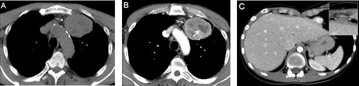
A-B) Unenhanced and contrast-enhanced chest CT scan obtained at the level of the aortic arch (soft tissue window, axial plane) demonstrates an unilateral oval-shaped soft-tissue mass (thin arrows) that arises in the left lobe of the thymus, anterior to the aorta and superior vena cava. The mass exhibits smooth and well-circumscribed borders, slightly heterogeneous attenuation on unenhanced scan, and noticeable diffuse heterogeneous enhancement with hypodense areas consistent with necrotic areas within the lesion (thick arrow). C) Contrast-enhanced CT scan (mediastinal window, axial plane) shows a small nodular pleural thickening of the diaphragm in the anterior recess of the right lung, contralateral to the primary tumour.
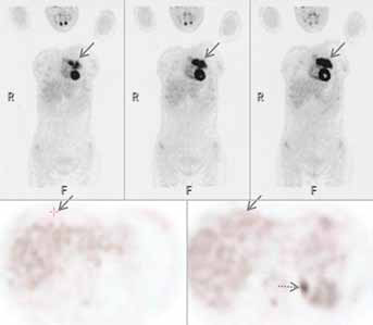
Maximum intensity projection 18 F-FDG PET images (coronal views; upper images) demonstrate a large area of increased 18 F-FDG in the mediastinum on the left side, superior to the heart (arrows), corresponding to the anterior mediastinal lesion detected at CT. No other area of abnormal uptake was seen, especially in the location of the small nodular pleural thickening of the right diaphragm appreciable on CT (arrows; axial views; lower images). The small area of increased 18 F-FDG uptake (dotted arrow; left axial image) represents an upper calyx of the left kidney.
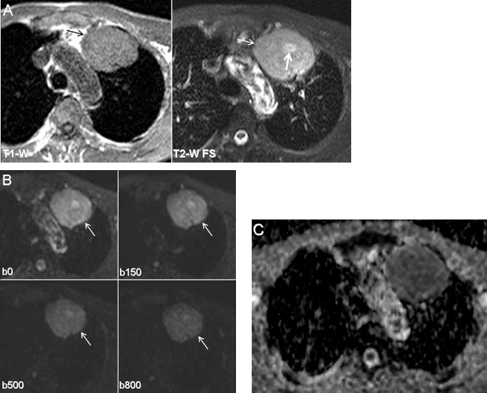
A) MR images (axial plane) confirm the large unilateral soft-tissue mass of the anterior mediastinum (thin arrows), adjacent to the aortic arch, that shows intermediate signal intensity on T1-weighted images (left image), similar to that of muscle, and a high signal intensity on T2-weighted images that remains unchanged on fat suppressed T2-weighted sequence (right image). T2-weighted image clearly detects a high signal intensity region which corresponds to cystic area (thick arrow), that is not clearly detectable on T1-weighted images (T1-W = T1-weighted; T2-W FS = fat suppressed T2-weighted). B) Diffusion-weighted MRI (axial plane) at the level of the mediastinal lesion was obtained at different b values of 0, 150, 500, and 800 s/mm2, respectively. The transverse image obtained at a b value of 0 s/mm2 demonstrates a high signal intensity within the lesion (arrow). At higher b values, the lesion presents a progressive slight attenuation of the signal intensity (arrows) that is not completely suppressed, even at the b800 gradient image. This appearance suggests a restricted diffusion within the tissue. C) At ADC-MAP, the lesion exhibits heterogeneous low signal intensity with a low mean ADC value of 0.56 ± 0.15 × 10⁻³mm2s−1, consistent with the hypothesis of a soft tissue lesion with high cellularity, suspicious for a thymic epithelial tumour.
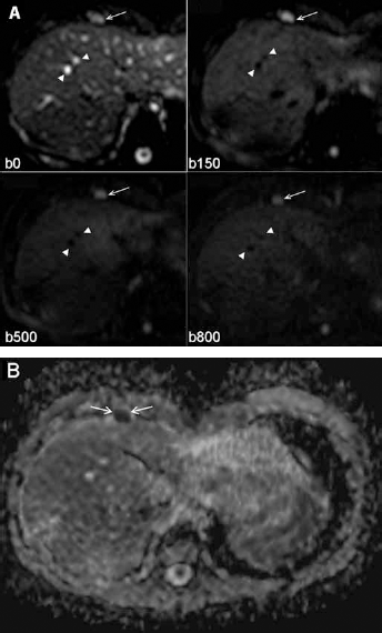
A) Diffusion-weighted chest MRI (axial plane) at the level of the anterior recess of the right lung in the location of the nodular thickening of the right diaphragm was obtained at different b values of 0, 150, 500, and 800 s/mm2. The transverse image obtained at a b value of 0 s/mm2, that results in conventional anatomic T2-weighted images with no vessels signal attenuation (arrowheads), demonstrates a high signal intensity lesion in the anterior recess of the right lung, close to the anterior margin of the liver (arrow), corresponding to the site of the nodular pleural thickening on CT. At higher b values, where vessels signal attenuation is seen (arrowheads), the lesion presents similar signal intensity (arrows); especially, the b800 gradient image shows a high signal intensity that means restricted diffusion of the nodule and suggests the diagnosis of a pleural implant. B) At ADC-MAP, the low signal of the nodule and the low mean apparent diffusion coefficient value of 0.52 ± 0.11 × 10⁻³mm2s−1, similar to that of the primary tumor, demonstrates restricted diffusion within the tissue and strengthens the hypothesis of metastatic pleural lesion related to thymoma.
Discussion
CT is currently considered the cross-sectional imaging modality of choice in the identification, staging, as well as in the follow-up of patients with thymic epithelial tumors (1–6, 20, 21). However, CT has limitations in differentiating among the various histological subtypes of thymomas and in defining locally advanced disease (stage III and IVa), especially for stage IVa when pleural or pericardial implants are represented by a very small amount of tissue, as in our case (9–13). In addition, CT does not allow to distinguish between pleural implants related to thymoma (that may appear as discrete soft tissue nodules or as circumferential diffuse pleural thickening) and non-calcified benign pleural plaques (1–6). 18 F-FDG/PET has been reported as an accurate tool in preoperative staging of thymic epithelial tumours, although it has limits in detecting very small lesions and imparts a substantial dose of radiation (15). In our case, 18 F-FDG/PET did not demonstrate an increased uptake of the contralateral small pleural implant, maybe due to the limited size of the pleural nodule. In addition, considering the negative finding obtained at 18 F-FDG/PET, and since pleural seeding typically appears ipsilateral to the primary mediastinal tumour, the contralateral pleural thickening was wrongly interpreted as a benign pleural thickening (4–6).
In the last years, DW-MRI has come as a novel radiologic modality for assessment of disease extent in various malignancies and is already being incorporated into general oncologic imaging practice for various tumours (22–26). DW-MRI, that is sensitive to thermally driven molecular water motion (which in vivo is impeded by cellular packing, intracellular elements, membranes and macromolecules), is able to characterize malignant tumours and metastases that show restricted diffusion by appearing as high signal intensity lesions even at high b values, with a low ADC (Fig. 3B–C, 4A–B) (22, 24, 27). This appearance reflects a combination of high cellularity, tissue disorganization, and increased extracellular space tortuosity inside the tissue as can be usually seen in malignant lesions and in our case of pleural implant from thymoma (22–27). Therefore, although MRI is not routinely employed in the assessment of thymic epithelial tumours, DW-MRI could be useful in patients with thymoma that exhibit indeterminate ipsilateral or contralateral pleural thickening at CT for differentiating benign pleural plaques from pleural implants related to thymoma. Gill et al (28), in an attempt to differentiate the various histological subtypes of malignant pleural mesothelioma by using the ADC value obtained from DW-MRI, included in their cohort two cases of benign pleural plaques confirmed at histology. They found an average ADC of 0.85 ± 0.17 × 10⁻³mm2s−1 for benign plaques that was lower than that of all the histological subtypes of mesothelioma and similar to that of the adjacent muscles. Hence, the authors supposed that the cellular structure in the benign plaque was similar to the structure of the normal tissue determined with the ADC value. These data were not confirmed in a subsequent study by Coolen et al (29) that found, in 17 benign pleural lesions, a lower ADC value of 1.521 × 10⁻³mm2s−1 (talc pleurodesis) and a higher ADC value of 1.969 × 10⁻³mm2s−1 (sarcomatoid mesothelioma) in the remaining 14 malignant pleural lesions. Thus, since malignant lesions can exhibit low ADC values, within a wide range, we believe that overlapped values between malignant lesions of the chest (such as pleural implants in thymoma or malignant pleural mesothelioma) and benign pleural plaques cannot be excluded (22–29). Nevertheless, the mean ADC of the pleural implant in our patient was lower than the average ADC of benign plaques reported by Gill et al (28). Furthermore, in our case the mean ADC of the metastatic pleural nodule was similar to that of the primary tumour. This finding may reflect a similar tumour cell density and cellular architecture of the small pleural implant compared to the associated thymoma, although we cannot exclude that the similar ADC value between the pleural implant and the thymoma is a casual observation. Hence, it would be beneficial to add the information obtained by DW-MRI in order to rule out benign pleural disease in patients with thymoma, before that invasive procedures are considered. In addition, any non-calcified nodular or band-like thickening of the pleura detected at CT in thymoma patients, that shows a similar ADC to the primary tumour at DW-MRI, should be considered as a potential pleural implant and further investigated, although further studies with a greater number of subjects with pleural implants in thymoma and benign pleural plaques are needed to demonstrate the usefulness of DW-MRI in distinguishing benign plaques from pleural implants. In the present case, DW-MRI was superior to 18 F-FDG/PET in characterizing the tiny pleural thickening detected at CT, providing the correct preoperative staging through the detection of a high signal intensity small lesion that was proved to be a pleural implant at surgery. In conclusion, this report suggests that DW-MRI can be an useful and novel tool for the accurate staging of thymic epithelial tumors, although it needs further validations, representing an effective radiation-free alternative to CT and 18 F-FDG/PET.
Footnotes
Financial support: None.
Conflict of interests: None.