






Abstract
Aims and background
Hypoxia inducible factor 1α (HIF-1α) and vascular endothelial growth factor (VEGF) have been deemed as key in angiogenesis of lung cancer. The aim of this study was to investigate diagnostic and prognostic values of HIF-1α and VEGF in patients with lung cancer.
Methods
From May 1, 2011, to April 20, 2014, blood samples and/or pleural effusions were collected from 100 patients with lung cancer, 18 patients with tuberculosis, 47 patients with community-acquired pneumonia, and 29 healthy controls. The pretreatment levels of HIF-1α and VEGF were measured by enzyme-linked immunoassays. Patients with lung cancer were followed up during the period of this study and survival times were recorded for analysis.
Results
We detected that the levels of serum and pleural HIF-1α in lung cancer were significantly higher than those in the tuberculosis population, and that the VEGF expressions were not significantly different between malignancy and benign diseases. An area under the curve of pleural HIF-1α (0.877 ± 0.053) showed a high ability to differentiate lung cancer from benign diseases. The significant negative predictors of survival in the univariate analysis were performance status (>1), no anticancer therapy, low serum albumin, advanced stage, and serum high level of VEGF (>324.17 pg/mL), while in the multivariate Cox regression analysis, only the pretreatment serum level of VEGF, stage, and anticancer therapy were identified as independent prognostic factors.
Conclusions
The overexpression of HIF-1α especially in pleural effusion may be an angiogenic factor for distinguishing malignancy from tuberculosis, and the pretreatment level of serum VEGF may be an independent predictor of survival.
Keywords
Introduction
Lung cancer is the most common malignancy and the leading cause of cancer death worldwide. The epidemiologic situation is especially severe in China. The total new cases of lung cancer were 536,407 in 2005 and the incidence is increasing, with mortality increasing by 464.84% during the past 30 years (1). The median survival of patients presenting advanced disease with standard therapy is 8 to 10 months, which demonstrates the urgent need to explore the mechanisms of lung cancer and new approaches of treatment for this disease (2).
Intratumoral hypoxia has been thought to be an essential characteristic of solid tumors, and results in activation of hypoxia inducible factor 1 (HIF-1), the key element in the process of tumor neovascularization. Hypoxia inducible factor 1 is a heterodimer consisting of 2 subunits, HIF-1α and HIF-1β. Hypoxia inducible factor 1α is the functional subunit regulated by oxygen, and plays critically important roles in maintaining the energy metabolism of tumor cells, tumor angiogenesis, accelerating tumor proliferation, and metastasis (3). Furthermore, HIF-1α can regulate more than 60 downstream genes, including vascular endothelial growth factor (VEGF) (4). As one of the most potent angiogenic factors, VEGF mediates the proliferation of endothelial cells, promotes vascular permeability, and has become an important therapeutic target in lung cancer (5). Therefore, more attention is being placed on the intricate relationship among hypoxia, angiogenesis, and the development of cancer.
Increased levels of HIF-1α and VEGF are implicated in small-cell lung cancer (SCLC) and non-small-cell lung cancer (NSCLC). They may be correlated with pathologic stage, treatment response, and decreased survival (6-9). However, most of these studies focused on expression of HIF-1α or VEGF separately; few researchers combined them. Moreover, there has been controversy concerning the prognostic values of HIF-1α and VEGF overexpression in patients with lung cancer; conflicting results have been reported from different laboratories (6, 10-12). Additionally, almost all studies have been performed in cell lines and tumor tissues. According to previous reports, circulating serum angiogenic molecules using enzyme-linked immunosorbent assays (ELISA) were identified in clear correlation with microvessel density of the resected tumor specimens (11), and compared with immunohistochemistry, sampling from peripheral blood is more easily assessable with less bias and time required. This method can be used in almost all circumstances, especially for patients with advanced disease who might not be available for biopsies (13).
In the present study, we evaluated serum and/or pleural concentrations of HIF-1α and VEGF in pretreatment patients with lung cancer, patients with nonmalignant disease, and healthy individuals, and analyzed the clinical implications of HIF-1α and VEGF for differential diagnosis of lung cancer and association with overall survival (OS). We hoped to find some biological markers in differential diagnosis and forecasting prognosis of lung cancer.
Patients and Methods
Patient selection
We mainly studied patients with lung cancer, and also selected patients with pulmonary active tuberculosis (TB), patients with community-acquired pneumonia, and healthy subjects as nonmalignant and normal controls in our hospital (a moderate Grade A Class 3 and teaching hospital in China) from May 1, 2011, to April 20, 2014. All patients with lung cancer were followed up until April 20, 2014, or to the date of death. The study was approved by our ethics committee and all patients gave informed consent.
The pathologic diagnosis of lung cancer was made if there was direct evidence of malignant cells, i.e., cancer cells, by bronchoscopy or percutaneous biopsy. Tumor staging assessment was based on the staging system of the American Joint Committee on Cancer. Performance status (PS) was evaluated according to the Eastern Cooperative Oncology Group. The treatment of patients was conducted according to the lung cancer guidelines in China. Overall survival times were calculated from the day of first sampling to death by any cause.
In the control group, the diagnosis of TB was identified according to guidelines for pulmonary TB diagnosis and therapy published by the Tuberculosis Branch Association of the Chinese Medical Association. The following diseases were excluded: acute exacerbation of chronic obstructive pulmonary disease, severe asthma, cor pulmonale, respiratory failure, other hypoxic diseases, and severe comorbidities. Another 29 healthy individuals who underwent physical examination during the same period were chosen as the normal group.
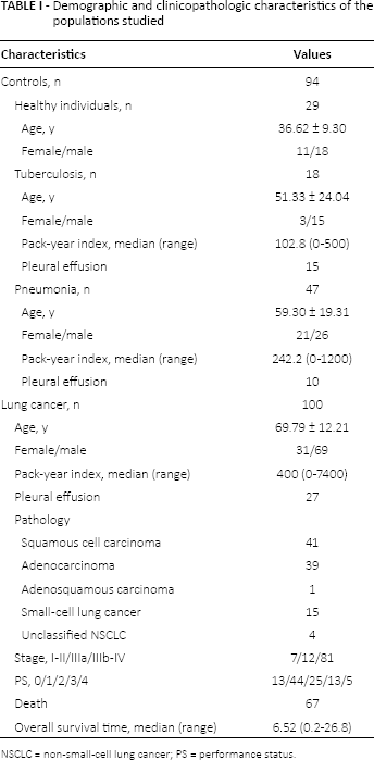
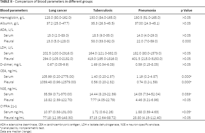
The demographic data and biochemical parameters of all patients and healthy individuals in the blood and/or pleural effusion were extracted from the clinical files (Tabs. I and II).
Demographic and clinicopathologic characteristics of the populations studied
NSCLC = non-small-cell lung cancer; PS = performance status.
Comparison of blood parameters in different groups
ADA = adenosine deaminase; CEA = carcinoembryonic antigen; LDH = lactate dehydrogenase; NSE = neuron-specific enolase.
p analyzed by nonparametric test.
Data are median (range).
Samples and measurement of HIF-1α and VEGF
Within 24 hours after hospitalization, 5 mL of venous blood were taken from fasting patients under static status in the morning for measurement of HIF-1α and VEGF. If pleural effusion was present, the same volumes of specimens were extracted by thoracentesis. Next, serum and/or pleural effusion without anticoagulant were centrifuged at 3000 rpm/min for 10 minutes at 4°C. Cell-free supernatants of serum and fluid were transferred to Eppendorf tubes and preserved at −80°C. The concentrations of HIF-1α (Cloud-Clone Corp., Houston, TX, USA) and VEGF (Wuhan Boster Biological Technology, Wuhan, China) were determined using ELISA kits according to the manufacturer's instructions. Absorbance values of standard products and samples were measured at 450 nm with an enzyme labeling device. Standard curves were drawn and the levels of HIF-1α and VEGF were calculated from them. Each sample was tested at least 2 times and the average value was adopted.
Statistical analysis
All statistical tests were performed by IBM SPSS software version 20 (IBM, Armonk, NY, USA). Continuous data were presented as mean ± SD unless otherwise noted. Differences among multiple groups were performed by one-way analysis of variance or by the nonparametric Kruskal-Wallis test depending on equal or unequal variance. Receiver operating characteristic (ROC) curves, areas under the ROC curves, and their 95% confidence intervals were calculated using standard techniques. To examine the cutoff values for serum levels of HIF-1α and VEGF, we calculated the total sensitivity and specificity for each cutoff value and then chose the cutoff values that maximized each factor as cutpoints. Correlation analysis was used with the Spearman method. Probability of survival was estimated by the Kaplan-Meier method and compared by log rank. Multivariate variable survival analyses were done with the Cox model. Statistical significance was set at p<0.05.
Results
Clinical characteristics of subjects
We recruited a total of 100 subjects with lung cancer, 18 with pulmonary TB, 47 with community-acquired pneumonia, and 29 healthy individuals (Tab. I). The mean age of patients with lung cancer was 69.79 years, there were 69 men and 31 women, and 27 cases were accompanied by malignant pleural effusion.
Of the 100 cases with malignancy proven by pathology, the predominant histologic type was squamous cell carcinoma. After evaluation, only 9 patients could be operated and 8 of them received surgical resection. The majority of the patients (90%) could not be resected by surgery because of locally advanced or distant metastases; among them, 42 received standard combination therapy or radiotherapy. The other 50 cases did not receive anticancer therapy or surgery due to practical factors. By the end of the study, 30 patients were alive, 67 were dead, and 3 were lost to follow-up. The median survival time was 6.52 months (range 0.2~26.8). Clinical characteristics of all subjects are shown in Table I.
There were several differences among multiple groups. The pack-year index (the average root numbers per day multiplied by smoking years) from the lung cancer group was significantly higher than that from benign groups by non-parametric test (p = 0.000). The pleural adenosine deaminase (ADA) was significantly elevated in patients with TB when compared to those of pneumonia (p = 0.001) and lung cancer groups (p = 0.000). As for tumor markers, the levels of carcinoembryonic antigen (CEA) in serum and pleural effusion were highest in patients with lung cancer compared to the other groups (p = 0.000). There was a significant increase of serum concentrations of neuron-specific enolase (NSE) and CYFRA 21-1 in malignancy. No differences were found in hemoglobin, serum albumin, serum ADA, serum and pleural lactate dehydrogenase, d-dimer, pleural NSE and CYFRA 21-1, or serum and pleural CYFRA 21-1 among the 4 groups (Tab. II).
Concentrations of HIF-1α and VEGF in serum and pleural fluid
To assess the effects of HIF-1α and VEGF involved in lung cancer, we measured their expression in peripheral blood supernatant from different groups, summarized in Figure 1. Our data show that the level of HIF-1α in the serum of malignancy was significantly higher (1.93 ± 1.16 ng/mL) than that in TB (1.27 ± 0.67 ng/mL) and healthy groups (1.18 ± 0.78 ng/mL) (p<0.05, p<0.01, respectively). Similar to serum results, the level in malignant pleural effusion was 1.63 ± 1.03 ng/mL, which was significantly elevated in comparison to that in the TB group (0.46 ± 0.28 ng/mL, p<0.001). Increased secretion of serum HIF-1α in patients with pneumonia (1.71 ± 1.01 ng/mL) was also observed as compared with healthy controls (p<0.05) (Fig. 1, A and B).
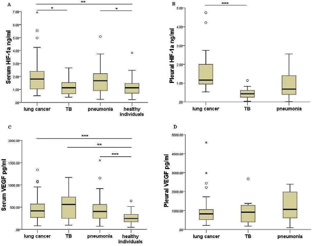
Hypoxia inducible factor 1α (HIF-1α) and vascular endothelial growth factor (VEGF) concentrations in serum and pleural effusion of lung cancer, tuberculosis, pneumonia, and healthy controls. (
The findings of VEGF were different from the results of HIF-1α. Patients with TB (562.74 [98.48-1171.31] pg/mL), pneumonia (403.91 [72.73-1555.29] pg/mL), and lung cancer (418.52 [84.04-1341.55] pg/mL) produced higher levels of VEGF in serum compared to the healthy controls (247.77 [47.80-641.39] pg/mL) (p<0.01), while there were no differences between benign and malignant diseases. Similarly, the difference of VEGF in pleural effusion among various groups was not statistically significant (Fig. 1, C and D).
To analyze the sensitivity and specificity of serum and pleural effusion of HIF-1α and VEGF for diagnosing malignancy, ROC curves were constructed. It was shown that an area under the curve (AUC) for serum HIF-1α was 0.634 in comparison to benign and normal controls (p = 0.002). At the optimal cutoff value of 1.55 ng/mL, the diagnostic sensitivity and specificity were 60.2% and 62.6%, respectively (Fig. 2A). When the curve of pleural HIF-1α was established, we observed a high AUC value of 0.877 (Fig. 2B, p = 0.000). At a cutoff value of 0.8541 ng/mL, the diagnostic sensitivity and specificity were 88.9% and 83.3%, respectively. On the other hand, AUC for VEGF in serum was 0.586 (p = 0.04) in comparison to benign and normal controls, with the cut-points set at 324.17 pg/mL, sensitivity was 69.0%, and specificity was 52.2%. However, VEGF in pleural effusion did not reveal that it can discriminate lung cancer from benign diseases (p = 0.654).
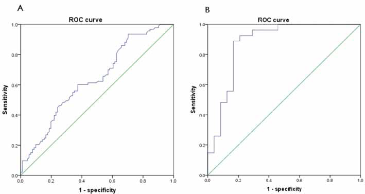
Sensitivity and specificity of serum and pleural hypoxia inducible factor 1α (HIF-1α) for distinguishing lung cancer from nonmalignant pulmonary disease. (
Relationship of HIF-1α and VEGF with clinicopathologic factors
To investigate interactions between angiogenic factors (HIF-1α or VEGF) and the other endpoints such as age, sex, pack-year index, stage, PS, and CEA, the possible relationship was further analyzed. Only 2 significant linear correlations were found using a Spearman nonparametric test. Serum HIF-1α values were positively correlated with its concentrations in pleural effusion (r = 0.548, p = 0.000) (Fig. 3). Serum VEGF was clearly negatively correlated with serum albumin (r = −0.281, p = 0.000) (Fig. 4). However, no significant correlation existed between serum HIF-1α and VEGF (r = 0.093, p = 0.376). Also, no clear correlations were found between serum HIF-1α or VEGF and other clinicopathologic factors.
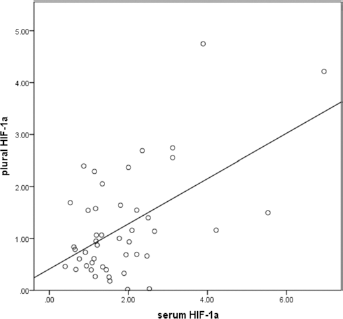
Correlation between hypoxia inducible factor 1α (HIF-1α) in serum and pleural effusion (r = 0.548, p = 0.000).
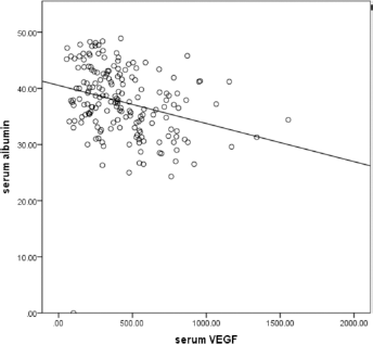
Correlation between the level of serum vascular endothelial growth factor (VEGF) and albumin (r = −0.281, p = 0.000).
Survival analysis
To observe the potential factors affecting OS, we selected some possible variables in clinics and analyzed the differences for the whole patient population using the Kaplan-Meier method. As shown in Table III, PS, stage, serum albumin, and anticancer treatment were statistically significant survival factors. Moreover, we observed that patients with pretreatment serum VEGF level ≤324.17 pg/mL had significantly longer survival time than those with a high serum level (>324.17 pg/mL) (p = 0.045) (Fig. 5). On the other hand, the serum HIF-1α did not exhibit significant prognostic value in our data (p = 0.637). Other variables that could not be prognostic determinants of a poor outcome by univariate analysis were as follows: sex, age, pack-year indexes, serum CEA, histologic types, and pleural effusion or not (data not shown).
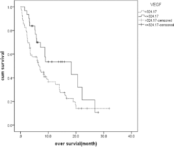
Kaplan-Meier survival curves for serum vascular endothelial growth factor (VEGF) in patients with non-small-cell lung cancer (p = 0.045).
Univariate survival analysis in lung cancer by Kaplan-Meier
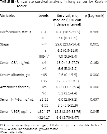
CEA = carcinoembryonic antigen; HIF-1α = hypoxia inducible factor 1α; VEGF = vascular endothelial growth factor.
One patient died.
The independent prognostic factors were analyzed by multivariate analysis in confounder models after adjusting the effects of different factors, such as age, sex, smoking, stage, treatment, PS, serum albumin, and VEGF (Tab. IV). According to Cox proportional model, disease stage, anticancer therapy, and serum VEGF level exhibited statistically significant differences (p = 0.039, p = 0.000, p = 0.019, respectively). When patients with low levels of serum VEGF (≤324.17 pg/mL) were compared with those with high levels (>324.17 pg/mL), the survival curves were drawn as in Figure 6.
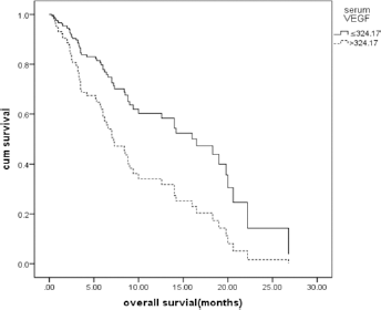
Cox proportional survival curves of 100 patients with lung cancer stratified according to pretreatment vascular endothelial growth factor (VEGF) serum levels (p = 0.019).
Cox proportional hazard estimation of overall survival between low and high levels of serum vascular endothelial growth factor (cutoff 324.17 pg/mL)
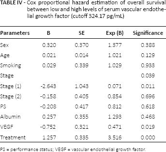
PS = performance status; VEGF = vascular endothelial growth factor.
Discussion
The morbidity and mortality rates of lung cancer in Chinese populations are much higher than the worldwide average, presenting a major public health issue. Our clinical data partly reflected the real aspects of lung cancer in China, such as high tobacco consumption, late diagnosis, low rate of treatment, limited effective treatment, and short survival times. As a result, efficient and practical serum biomarkers are required to aid the diagnosis and to assess the progression of lung cancer.
Angiogenesis induced by HIF-1α and VEGF in lung cancer
High expression of HIF-1α was reported to be involved in the growth and progression of both NSCLC and SCLC (8, 9, 14, 15), which was also demonstrated in our study. Additionally, no difference in HIF-1α between lung cancer and pneumonia can be explained by the fact that the most common causes of tissue hypoxia are inflammation and/or malignant solid tumor, both of which can result in the elevation of HIF-1α (3, 4), whereas such elevation in pneumonia was not as high as the level in lung cancer in the present study. Furthermore, ROC curves revealed that the expression of HIF-1α particularly in pleural effusion might have clinical value in the differential diagnosis of malignancy. Under hypoxic conditions, HIF-1α can move into the nucleus, where it freely forms an active complex with HIF-1β. This complex binds to the hypoxia response elements and drives the transcription of downstream genes, including VEGF (16).
Vascular endothelial growth factor-induced angiogenesis has a pivotal role in the growth and progression of malignancies. All lung cancers (NSCLC and SCLC) aberrantly express higher levels of VEGF (7, 13). In agreement with previous studies, the levels of serum VEGF from lung cancer were higher than in healthy controls, while VEGF was also highly expressed in benign diseases (TB and pneumonia). Several reports showed the high levels of VEGF in TB and infectious diseases (17, 18), and association with hyperpermeability to fluid and protein (19). The ROC curves in the present study also revealed the low ability of serum VEGF in the differential diagnosis of malignant and benign diseases. Thus, we can draw the conclusion that as angiogenesis is not only exists in cancers, but also in inflammation, VEGF cannot be used as a marker to differentiate benign disease from malignancy.
Several previous studies had demonstrated that HIF-1α positively correlated with expression of VEGF (8, 14, 15). In contrast to such results, we did not find such significant correlations. The conflicting results might be due to the complex mechanism of HIF-1α and VEGF regulation. Some researchers found that biopsy that was positive for VEGF staining was negative for HIF-1α (14). Besides the traditional HIF-1-mediated pathway, VEGF expression can be regulated by other factors, such as cytokines and activation of certain oncogenes (5, 16). On the other hand, in addition to VEGF, HIF-1α may enhance the angiogenic potential of malignancy by regulating some other angiogenic genes (9, 20).
Negative predictors for survival
Some possible prognostic factors have been suggested in various studies, such as smoking, serum albumin, PS, disease stage, and chemotherapy (21, 22). In partial agreement with others, we found that high level of VEGF, advanced stage, and without therapy were independent poor prognostic factors, but not PS or serum albumin, in univariate analysis. These results can be explained by the following possibility. For albumin, a significant negative correlation was noted between serum VEGF and albumin; we speculated that the absence of albumin for predicting survival time maybe be affected by the high independent prognostic factor of VEGF. As for PS, because patients with low scores of PS (≤2) would receive chemotherapy or other therapy, no significant prognosis of PS may be affected by the strong predictive factor of anticancer therapy.
Reports of the relation between the expression of HIF-1α and prognosis are not consistent. Two recent Chinese meta-analyses demonstrated that high expression of HIF-1α in NSCLC predicted a poor prognosis for survival, particularly in Asia (6, 23); however, some other studies did not prove this (10, 15). Our data clearly indicated that serum HIF-1α was not a predictive biomarker; low levels of HIF-1α seemed to have decreasing OS as opposed to high levels, which was supported by a previous study that showed that HIF-1α-negative carcinomas resulted in shorter median survival times than HIF-1α-positive carcinomas (10). The above inconsistent results might be due to the different types of HIF (HIF-1α, HIF-2α, and HIF-3α) (16, 24); only elevated HIF-2α expression might be an independent prognostic indicator (25).
With regard to the prognosis of VEGF, a few scholars claimed that a higher circulating VEGF did not independently determine prognosis of NSCLC (12, 22, 26). However, we observed that patients with lung cancer with pretreatment serum VEGF concentrations ≤324.17 pg/mL had a significantly longer survival time, which was supported by previous studies (7, 11, 13, 27). For example, a review of 5386 patients from 51 studies showed that VEGF overexpression had a significant correlation with poor survival in patients with NSCLC and SCLC from different countries (7). A recent analysis focused on the prognostic value of serum circulating VEGF had similar results (13). Based on these data, it can be suggested that circulating VEGF might provide a simple and practicable method to predict OS in lung cancer.
In conclusion, we clarified that serum HIF-1α and VEGF had different characters involved in lung cancer. The levels of HIF-1α particularly in pleural effusion had obvious value in differential diagnosis of malignancy and TB. Furthermore, our investigation indicated that the pretreatment VEGF level in serum could be identified as an independent prognostic factor for patients with lung cancer. In the future, further investigations on the mechanism of HIF-1α and VEGF involving diagnosis and survival are warranted.
Footnotes
Acknowledgment
The authors thank Professor Giuseppe Marciano (Azienda Ospedaliera Universitaria Senese, Italy) for critical reading and correction of the manuscript.
Financial support: Supported by grants from the major projects for science and technology development of Nanjing Medical University (no. 2010njmuz53).
Conflict of interest: None.