






Abstract
Purpose
The FAT1 gene is involved in some cancers; however, its role in medulloblastoma is less clear. This study investigated the effects of FAT1 expression on the prognosis of medulloblastoma patients.
Methods
Whole exome sequencing was undertaken in 40 medulloblastoma patient samples. FAT1 mRNA and protein expression levels in normal and brain tumor tissues were determined by fluorescence quantitative PCR and immunohistochemistry, respectively. The association of FAT1 expression with overall survival (OS) was examined by Kaplan-Meier curve analysis with a log-rank test. Following lentiviral-mediated FAT1 knockdown using shRNA in Daoy cells, proliferation, Wnt signaling, and β-catenin protein expression were determined.
Results
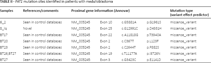
Eight FAT1 missense mutations were detected in 7 patients. FAT1 mRNA expression in tumors was significantly lower than in adjacent normal tissue (p = 0.043). The OS of patients with high FAT1 protein expression was significantly longer than that of patients with low FAT1 protein expression (median survival time: 24.3 vs 4.8 months, respectively; p = 0.002). shFAT1 cells had significantly higher proliferation rates than shControl cells (p≤0.028). Furthermore, the mRNA expression of LEF1, β-catenin, and cyclin D1 was significantly upregulated in shFAT1-Daoy cells (p≤0.018).
Conclusions
Low FAT1 expression was associated with poor prognosis in children with medulloblastoma. Furthermore, FAT1 may act on Wnt signaling pathway to exert its antitumor effect.
Introduction
Medulloblastoma is an invasive malignant embryonal tumor originating in the cerebellum or fourth ventricle and often found in children aged 4-7 years. It is the most common malignancy in childhood and accounts for 12%-25% of central nervous system tumors in children. In children aged <15 years, the annual incidence of medulloblastoma is 5/100,000 (1). Surgery remains the major treatment strategy for medulloblastoma, although its impact on the prognosis of patients is controversial, especially given that the tumor is adherent to the brainstem in some cases, rendering complete surgical resection difficult. Although cancer cells may metastasize through the cerebrospinal fluid, resulting in a poor prognosis and high mortality (2, 3), the 5-year overall survival for medulloblastoma in developed countries can reach as high as 65%-71% (4). However, an analysis of 67 Chinese patients with medulloblastoma showed a 3-year survival of 55.1%, which may be largely due to early termination of treatment resulting from misunderstanding of the disease by parents as well as financial limitations and transportation difficulties (5).
Recent studies reveal that gene mutation and abnormal intracellular signal transduction are important causes of medulloblastoma occurrence and development (6, 7). For example, the oncogene DJ-1 plays a key role in medulloblastoma pathogenesis, regulating cell proliferation and differentiation (8). Thus, investigating the mechanism underlying the pathogenesis of medulloblastoma may prove helpful for the development of nonsurgical therapeutic strategies and may also have great clinical importance in risk stratification and monitoring of prognosis (9).
Previous studies have shown that the FAT1 gene is repressed in some cancers, including oral cancer, and upregulated in others, such as leukemia (10). Its regulatory roles in the pathogenesis of cancers, including suppression of ductal carcinoma in situ progression to invasive breast cancer (11), have also been reported (12). In a pilot study, we previously employed exome sequencing of cancer-related genes in 26 patients with medulloblastoma, and identified candidate gene mutations. A FAT1 missense mutation was found, and SIFT and Polyphen2 were used subsequently employed for prediction (unpublished data). Seven mutation sites in 8 cases were found to be the major pathogenic sites and subjected to further validation. Thus, we speculate that FAT1 gene alterations may be involved in the pathogenesis of medulloblastoma.
The FAT1 gene, which is mutated in many types of cancer, including glioblastoma, colorectal cancer, and head and neck cancer (12, 13), is mapped to chromosome 4q35 (between 187,508,937 and 187,644,987 bp) and has 27 exons, including 26 encoding exons. FAT1 protein is a member of the cadherin superfamily, a group of transmembrane proteins expressed in epithelium that functions as adhesion molecules and/or signal transduction receptors (14). FAT1 is also involved in the formation of intercellular adhesion, and its silencing significantly reduces the stability of intercellular connections and disrupts cell polarity (15), which is known to enhance cancer cell migration and invasion. The expression of FAT1 mRNA in embryonic stem cells and neural tissues is suggestive that it also has a role in normal cerebellum embryogenesis (16). However, the role of FAT1 in the pathogenesis of medulloblastoma remains unknown.
FAT1 mutations in some somatic cells activate the Wnt signaling pathway in cancers (12, 13), which is a characteristic biological feature of WNT type medulloblastoma (6). Therefore, the present study was undertaken to further explore the relationship between FAT1 and the Wnt signaling pathway in medulloblastoma. FAT1 expression was analyzed in medulloblastoma samples from patients, and the association between its expression and patient prognosis was evaluated. These results may be useful for predicting the prognosis of patients with medulloblastoma as well as the molecular mechanisms underlying the actions of FAT1.
Methods
Study Participants
A total of 40 children with pathologically proven medulloblastoma were recruited from the Children's Hospital of Fudan University between January 2004 and June 2014. Medulloblastoma tissues were collected during surgery, fixed in 10% neutral formalin, and embedded in paraffin. In addition, 26 fresh samples were harvested for DNA extraction, along with 10 normal peritumoral brain tissues as controls. This study was approved by the Institutional Review Board of the Children's Hospital of Fudan University, and informed consent was obtained from the patients’ parents or guardians.
Patients with medulloblastoma were enrolled if they met the following inclusion criteria: receipt of macroscopic total or subtotal surgical resection for medulloblastoma between 2004 and 2014, the absence of other severe diseases before the surgery, the presence of complete follow-up data and a complete medical record, and death due to disease progression or recurrence if the patients died during the study.
Follow-Up
All patients received follow-up care every 3 months. The presence of cystic changes in the medulloblastoma was determined by magnetic resonance imaging.
Exome Sequencing
Total DNA was extracted from the 26 freshly isolated medulloblastoma samples, and sequencing was performed in the Beijing Genomics Institute (Shenzhen, China). Each sample was analyzed for variations using the Genome analysis toolkit (GATK) (Broad Institute, Cambridge, MA, USA), and a total of 70,000-80,000 single nucleotide variants (SNV). After quality control screening (SNV quality ≥20; SNV genotype quality ≥20; INDEL quality ≥50; INDEL genotype quality ≥20; number of covered segments ≥4), each sample contained about 1,200-1,500 variants, which were then annotated with different software and databases. For mutations, SIFT (http://sift.bii.a-star.edu.sg/) and PolyPhen-2 (http://genetics.bwh.harvard.edu/pph2/) were used to predict the type of mutation and the impact of the mutation on the protein structure and function.
Further screening was conducted according to the mutation frequency and type. The candidate mutation sites were acquired, and each sample had about 300-500 variants. Whole exome sequencing was employed for sequencing (mean length of sequence: 88; mean depth of sequencing: 70×; original number of reading: 9.5 × 10⁷). After removal of data with low quality, the number of effective data read was approximately 9.0 × 10⁷.
The FAT1 mutations were validated by Sanger sequencing using Primer3 to design the primers for polymerase chain reaction (PCR). The PCR products were subjected to Sanger sequencing at the Beijing Genomics Institute (BGI), China.
Quantitative PCR (qPCR) Analysis
The tissues and cells were processed for the extraction of RNA with Direct-zol RNA MiniPrep (Zymo Research, Irvine, CA, USA) according to the manufacturer's instructions. Reverse transcription was performed in a 20-μL reaction mixture with ABM 5× All-In-One RT MasterMix (Applied Biological Materials, Richmond, Canada). qPCR was performed in a 20-µL reaction containing 10 µL of Kapa SYBR Fast qPCR Master Mix (2×), 2 µL of cDNA (10 ng/µL), and 7.2 µL of RNase-free H2O with the following primers at a final concentration of 10 μM (0.4 µL each): GAPDH sense, 5’-CTCTCTGCTCCTCCTGTTCGAC-3’ and antisense, 5’-TGAGCGATGTGGCTCGGCT-3’ (69 bp); FAT1 sense, 5’-TCAGCAGATCCAAACGCCAT-3’ and antisense, 5’-CTGATTGCTGAATTCGGGCG-3’ (135 bp); β-cadherin sense, 5’-CTCTTACACCCACCATCCCAC-3’ and antisense, 5’-GATGTGCACGAACAAGCAACT-3’ (145 bp); LEF1-F sense, 5’-TCCCGTGAAGAGCAGGCTAA-3’ and antisense, 5’-AGGCAGCTGTCATTCTTGGAC-3’ (172 bp); cyclin D1 sense, 5’-GCTGCGAAGTGGAAACCATC-3’ and antisense, 5’-CCTCCTTCTGCACACATTTGAA-3’ (135 bp). The PCR reaction was incubated at 95°C for 3 minutes, followed by 40 cycles of 95°C for 5 seconds and 60°C for 30 seconds, and one final cycle at 60°C for 3 minutes and 94°C for 10 seconds.
Western Blot Analysis
Total protein was extracted with RIPA lysis and extraction buffer (Beyotime Institute of Biotechnology, Shanghai, China). The protein concentration was determined using the BCA method (Thermo Fisher Scientific, MA, USA) according to the manufacturer's instructions, and 30 µg was separated in 10% sodium dodecyl sulfate polyacrylamide gel electrophoresis. After transfer to polyvinylidene difluoride membranes, the membranes were blocked with 5% nonfat milk in PBS-Tween 20 for 1.5 hours at room temperature and incubated in anti-FAT1 antibodies (1:100; Abcam, Cambridge, UK) or anti-GAPDH antibodies (1:10,000; Abcam) at 4°C for 12 hours and then incubated with a horseradish peroxidase–conjugated secondary antibody (1/2,000; Abcam) at room temperature for 1 hour. The bands were visualized using the ECL method, and GAPDH was used as an internal control.
Immunohistochemistry Analysis
Paraffin-embedded tissue sections of 4 μm were routinely processed using the EnVision 2-step immunohistochemistry system, and visualization was performed with DAB following antigen retrieval with boiling 0.05 M citrate buffer, pH 6.0 for 5-10 minutes. The primary antibody was anti-FAT1 antibody (1:100; GeneTex, San Antonio, TX, USA).
For semiquantitative analysis, 5 fields were randomly selected at a magnification of 200, and positive cells were counted to obtain an average. Sections undergoing hematoxylin & eosin staining also served as controls.
The pathological evaluation was performed by 2 pathologists in a blind manner under a light microscope, and FAT1 expression was semiquantitatively detected (positive or negative) in medulloblastoma and normal brain tissues. Positive cells had brown granules in the cytoplasm or nucleus; negative cells had no granules. At a high magnification (400×), the field with high staining intensity was selected for observation, and positive cells were counted. High FAT1 expression was recorded if >50% of cells were positive for FAT1; low expression was indicated if ≤50% of cells were positive for FAT1. Any discrepancies between the pathologists were resolved by consultation.
Cell Culture
Daoy cells were obtained from American Type Culture Collection (Manassas, VA, USA) and maintained in Dulbecco's Modified Eagle Medium (Gibco, Carlsbad, CA, USA; 11995065) containing 10% fetal bovine serum (Gibco; 10099141), 4 mM L-Glucose, 1 mM sodium pyruvate, and 1% antibiotics (Gibco) at 37°C in an environment with 5% CO2.
FAT1 Silencing
Lentivirus vectors were purchased from Jeayea Biotechnology (Shanghai, China). Daoy cells were independently transduced with Lenti-shControl, Lenti-shFAT1-1 (5’-GCCTGTGGGTTCCAGTGTAA-3’), Lenti-shFAT1-2 (5’-GCAACCGGCTCTCTCTATAC-3’), and Lenti-shFAT1-3 (5’-GGAGCAAGCTGTTTATCATC-3’), and all subsequent experiments were conducted after 3 days.
Cell Proliferation Assay
Transduced Daoy cells were seeded into 96-well plates at a density of 3,000 cells/well and divided into 4 groups (15 wells/group). The viable cells in 3 wells of each group were measured once daily, and the medium was refreshed once every 2 days.
At 1, 2, 3, 4, and 5 days after incubation, cell viability was determined using the MTS method. In brief, 20 µL of MTS (CellTiter 96 Aqueous One Solution Cell Proliferation Assay, Promega, Madison, WI, USA) was added to each well, followed by incubation at 37°C for 2 hours. The optical density was measured at 490 nm (OD490).
Statistical Analyses
The continuous data were expressed as mean and standard deviation (SD); the differences between 2 groups and more than 2 groups were tested with independent 2-sample t tests and one-way analysis of variance (ANOVA), respectively. The Bonferroni correction was performed for the post hoc tests using one-way ANOVA. Categorical data were expressed as number with percentage in each group, and their association with FAT1 expression was tested with the chi-square test or Fisher exact test (when at least one of the cells had an expected count that was <5). A Kaplan-Meier curve with a log-rank test was performed to analyze association of overall survival with FAT1 expression. A repeated measurement ANOVA test with post hoc tests and Bonferroni correction was also performed to compare the protein expression over time among the groups. Survival status was summarized as n (%), except for mean ± SD for age. The association with survival time was analyzed using univariate and multivariate Cox regression model and results were shown as hazard ratio and corresponding 95% confidence intervals and p values. A 2-tailed p value <0.05 indicated statistical significance. Statistical analyses were performed with IBM SPSS statistical software version 22 for Windows (IBM Corporation, Armonk, NY, USA).
Results
Identification of FAT1 Missense Mutations
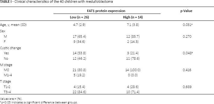
The characteristics of the 40 study participants, including 29 boys and 11 girls with a mean age of 5.5 years (SD 3.4), are shown in Table I. Whole exome sequencing analysis identified 8 FAT1 missense mutations found in 7 patients (Tab. II).
Clinical characteristics of the 40 children with medulloblastoma
Values are n (%).
p<0.05 indicates a significant difference between groups.
FAT1 mutation sites identified in patients with medulloblastoma
FAT1 Expression in Normal Brain Tissues was Higher than in Medulloblastoma
To elucidate whether FAT1 mRNA and protein expression levels are altered in medulloblastoma, qPCR and immunohistochemistry analyses were carried out in normal brain and medulloblastoma tissues. FAT1 mRNA expression in tumors was significantly lower than in adjacent normal brain tissue (p = 0.043, Fig. 1A). Representative immunohistochemical images of FAT1 staining are shown in Figure 1, B-C-D-E, which showed that adjacent normal tissues expressed a dramatically higher level of FAT1 protein than cancer tissues.
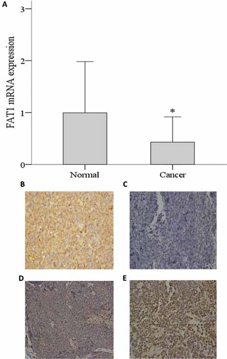
FAT1 mRNA and protein expression in adjacent normal tissues and cancer tissues. (
Associations between FAT1 Expression and Patient Clinicopathological Characteristics and Overall Survival
We next sought to examine whether FAT1 expression was associated with patient characteristics and prognoses. The children with high FAT1 protein expression were significantly older than those with low FAT1 protein expression (mean age 7.1 vs 4.7 years, p = 0.031). Of the 17 patients with cystic changes in their tumors by imaging examination, low FAT1 expression was found in 14 (82.4%), and high FAT1 expression in 3. Of cases without cystic changes, low FAT1 expression was found in 12 patients, and high FAT1 expression in 11. FAT1 expression was significantly different between patients with and without cystic changes (p = 0.048). As shown in Figure 2, log-rank analysis showed that the overall survival of patients with high FAT1 protein expression was significantly longer than that of patients with low FAT1 protein expression (median survival time 24.3 vs 4.8 months, respectively; p = 0.002).
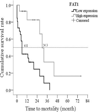
The association of overall survival with FAT1 expression. The overall survival of patients with high FAT1 expression was significantly longer than that of patients with low FAT1 expression (median survival time 24.3 vs 4.8 months, p = 0.002 by log-rank test).
High FAT1 expression and treatment with radiation, chemotherapy, or both were associated with better survival by univariate analysis (all p≤0.005; Tab. III). Disease recurrence was associated with poor survival (p<0.034). Treatment with both radiation and chemotherapy or chemotherapy alone remained associated with better survival outcomes in multivariate analysis (Tab. III).
Univariate and multivariate Cox regression analysis of factors associated with overall survival in patients with medulloblastoma
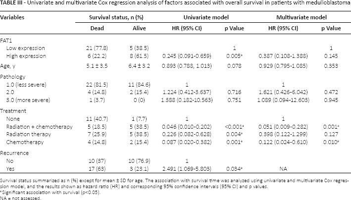
Survival status summarized as n (%) except for mean ± SD for age. The association with survival time was analyzed using univariate and multivariate Cox regression model, and the results shown as hazard ratio (HR) and corresponding 95% confidence intervals (95% CI) and p values.
Significant association with survival (p<0.05).
NA = not assessed.
Low FAT1 Expression Promoted Daoy Medulloblastoma Cell Proliferation
To investigate the influence of FAT1 expression on medulloblastoma cell proliferation, FAT expression was silenced in Daoy medulloblastoma cells by shRNA. The effectiveness of 3 distinct shRNAs against FAT1 was first compared. As shown in Figure 3, A and B, all 3 shRNAs inhibited FAT1 mRNA (p<0.001) and protein expression in Daoy cells.
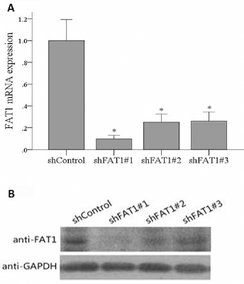
The effects of FAT1 knockdown on FAT1 mRNA and protein expression. (
The effect of FAT1 knockdown on Daoy cell proliferation was subsequently investigated. Although the proliferation in each group continuously increased from day 2 to day 5, the shFAT1#1, shFAT1#2, and shFAT1#3 groups had significantly higher proliferation rates than the shControl group over this period (p≤0.028; Fig. 4), indicating that loss of FAT1 promotes cell proliferation.
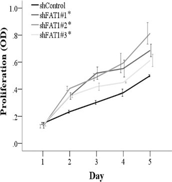
FAT1 knockdown enhanced proliferation of Daoy cells. *The shFAT1#1, shFAT1#2, and shFAT1#3 groups had significantly higher proliferation rates than the shControl group from days 2 to 5 (p≤0.028). Data presented as mean with standard deviation (n = 3 per group).
FAT1 suppresses the WNT/ β-catenin Signaling Pathway to Exert its Effects
To further explore the signaling pathway affected by FAT1, the expression of proteins in the Wnt signaling pathway was investigated after FAT1 knockdown. The mRNA expression of 3 genes, including LEF1, β-catenin, and cyclin D1, was significantly upregulated in the shFAT1-Daoy cells (p≤0.018; Fig. 5, A-B-C).
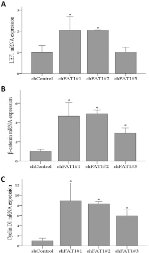
The mRNA and protein expression of molecules in the WNT signaling pathway. (
Representative Western blots of β-catenin protein expression in Daoy cells are shown in Figure 6A. LGK974 is an inhibitor of the Wnt/β-catenin signaling pathway, which suppresses β-catenin protein expression (Fig. 6A). β-catenin protein expression in Daoy cells treated with combination of LGK974+shFAT1 from day 1 to day 5 was significantly lower than those treated with normal control and shFAT1, but was significantly higher than those treated with LGK974 (all p<0.05; Fig. 6B).
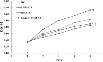
Effect of shFAT1 and/or LG-974 on β-catenin protein expression in Daoy cells. (
Discussion
In the present study, FAT1 expression was analyzed in medulloblastoma samples from patients to identify mutations and determine if its expression was associated with patient characteristics and overall survival. FAT1 mRNA expression in tumors was reduced as compared to that observed in adjacent normal brain tissue. In addition, children with high FAT1 protein expression were significantly older and had longer overall survival than those with low FAT1 protein expression. In vitro analyses using Daoy medulloblastoma cells revealed higher proliferation rates following FAT1 knockdown. The mRNA expression of LEF1, β-catenin, and cyclin D1 was significantly upregulated in the shFAT1-Daoy cells. These results may be useful for predicting the prognosis of patients with medulloblastoma as well as the molecular mechanisms underlying the actions of FAT1.
FAT1 mutations have previously been identified in bladder cancer (17). We identified 8 FAT1 missense mutations at chromosome 4q35.2. The FAT1 gene is mapped to chromosome 4 and is a fat homologous gene in Drosophila. Chromosome 4q35 deletion is frequently found in human cancers, and chromosome 4q35 is also known as an antioncogene (18, 19). Previous studies have reported that FAT1 is an antioncogene, playing important roles in the pathogenesis of some cancers, including glioma and colon cancer (12). Specifically, FAT1 plays a crucial role in the proliferation, movement, and invasion of cancer cells (10, 14). Similarly, in the present study, repression of FAT1 expression in Daoy cells resulted in increased proliferation, and an inverse correlation between FAT1 level with Ki67 expression was observed (64.7 ± 22.3% in patients with low FAT1 expression vs 32.1 ± 23.1% in patients with high FAT1 expression, p<0.001; data not shown). These results are consistent with Settakorn et al (20), in which FAT protein expression was inversely associated with Ki67 proliferative index in intrahepatic cholangiocarcinoma. It is also consistent with Morris et al (13), in which inactivation of FAT1 increased the proliferation of SF295, U87, HS683, and IHA cancer cells. Further studies are necessary to determine the full impact of each FAT1 missense mutation detected on the protein function and to determine if its loss of expression is related to medulloblastoma tumor growth.
Previous studies have shown that oncogenes, including DJ-1, may have prognostic value for patients with medulloblastoma (8). In addition, epidermal growth factor receptor overexpression was associated with poor outcomes in a retrospective study of 44 patients with medulloblastoma (21). In the present study, high FAT1 expression was associated with greater patient age and longer overall survival. This is in contrast to a study analyzing FAT1 expression in hepatocellular carcinoma, in which FAT1 expression was associated with tumor stage and proliferation (22), and another in which high FAT1 expression was associated with shorter relapse and overall survival in patients with acute lymphoblastic leukemia (23, 24). Thus, the role of FAT1 as an oncogene versus tumor suppressor may be tumor type–dependent.
Morris et al (12, 13) showed that FAT1 inactivation may activate Wnt signaling pathway in the pathogenesis of cancers. The Wnt signaling pathway activation is closely related to the occurrence of medulloblastoma, and is a characteristic biological feature of WNT type medulloblastoma (25). The Wnt signaling pathway is an evolutionally conservative pathway that regulates cell differentiation, proliferation, polarity, and migration. In the classic Wnt signaling pathway, β-catenin translocation from the cytoplasm to the nucleus is a marker of signaling activation (6, 26, 27). Because FAT1 can specifically bind to β-catenin (14) to regulate a series of biological behaviors of cells, we speculated that it may regulate the Wnt/β-catenin signaling pathway in medulloblastoma cells. We showed that the mRNA expression of key molecules in Wnt signaling pathway, LEF1, β-catenin, and cyclin D1, was increased significantly following FAT1 knockdown. Although this further confirms that FAT1 regulates Wnt signaling, the specific mechanism is unknown. Thus, further studies are necessary to examine if this effect is directly mediated through interaction with β-catenin or through inhibition of the expression of Wnt signaling mediators.
The present study is limited by its small sample size. Although an association between FAT1 and overall survival was observed, long-term studies with larger numbers of patients are required to confirm our findings and determine if there is a correlation between FAT1 expression and medulloblastoma histotypes or subgroups. Additional studies are also required to examine the clinical impact of monoclonal antibody therapy targeted toward FAT1 as described for colon cancer (28). Furthermore, the impact of the identified missense mutations on FAT1 protein structure and activity as well as medulloblastoma growth was not analyzed. In addition, it is not known if the influence of FAT1 on medulloblastoma proliferation is mediated through Wnt signaling. Thus, further studies are required. However, we speculate that FAT1 plays important roles in the pathogenesis of medulloblastoma.
Conclusion
FAT1 may act as an antioncogene in the pathogenesis of medulloblastoma, repressing the activation of the Wnt signaling pathway and cell proliferation. FAT1 expression may also reflect the prognosis of patients with medulloblastoma as those with low FAT1 expression had poorer prognoses. Further long-term studies with larger numbers of patients are required to confirm our findings.
Footnotes
Financial support: No financial support was received for this submission.
Conflict of interest: None of the authors has conflict of interest with this submission.