






Abstract
Introduction
Leiomyomas are rare benign tumors that can occur in the male urinary tract. We present a unique case of leiomyoma of the vas deferens.
Case Description
We present the clinical case of a 69-year-old patient with a suspected bulk close to the right epididymis, which turned out to be a leiomyoma of the vas deferens. To our knowledge, it is the fourth case in literature.
Conclusions
A proper identification and the knowledge of this pathology, even if it is a very unusual event, is necessary to avoid a surgical over treatment and preserve the testicle, by removing only the tumor.
Introduction
Leiomyomas are rare benign tumors that can occur in the male urinary tract (1). The majority of these have their roots in the kidney. There are also reports that reveal leiomyomas in other organs of the genitourinary tract, such as the urinary bladder, glands, and testicle (2, 3).
Our aim is to present this rare disease and its management according to the literature.
Case Description
A 69-year-old man turned to our department for the presence, close to the right epididymis, of a neoformation, which, by palpation, resulted rough, nontender neither painful, and which, by an objective consideration, seemed to belong to the tail of the right epididymis. The man denied recent trauma, fever, or other symptoms. He also had a case of right epididymitis about a year before. The patient underwent a scrotal sonography that showed the presence of a right testicle with regular echogenicity and normal dimensions. In the right epididymis, evidence was found of a bulk about 3 cm in size. It was found to be vascularized at the color Doppler sonography.
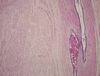
The patient did not report chronic pathology in his medical history and he was in good health. In the past, he was subject only to appendectomy, cholecystectomy, and lumbar discectomy for a dischernia. The preparatory blood exams were within normal range. The patient was also subjected to the dosage of the tumor markers, which resulted to be lactate dehydrogenase (LAD) 182 U/l; beta-human chorionic gonadotropin (B-Hcg) 1.1 IU/l; Alpha fetoprotein (AFP) 3.46 μg/l. Subsequently, he was subjected to an inguinal testicular exploration. Intraoperatively, a multi-lobed bulk had its roots in the proximal portion of the vas deferens and was entirely removed, preserving the testicle (Fig. 1). Histologically, the lesion was composed of variously twisted bundles of bland spindle cells with eosinophilic cytoplasm and elongated ‘cigar-shaped’ nuclei. It arose from the muscular wall of the vas deferens, with eccentric growth over the same (Fig. 2). No cyto-karyological atypia, karyokinetic figures (with low proliferation index assessed by Ki67), or areas of necrosis were observed. At immunohistochemistry, these cells were positive for antibodies to the determinations with desmin and smooth muscle actin confirmed the differentiation of the tumor leiomuscolar line. The final diagnosis, therefore, was of a leiomyoma originating from the muscular wall of the vas deferens.
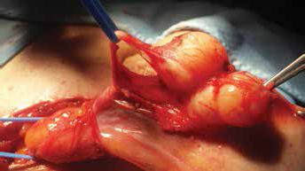
Intraoperative appearance of leiomyoma of the deferens.
The figure shows a detail of the vas deferens in the right side and of the leiomyoma in the left side.
Conclusions
Leiomyomas are benign tumors originating from the smooth muscle tissue. They can arise in virtually any organ and are quite common at the level of the uterine myometrium, where they are the most frequent tumors in females. Leiomyomas of the urogenital tract are rare formations (1), and their location in the vas deferens is exceptional. To our knowledge, our case is the fourth case of leiomyoma of the vas deferens ever reported in literature (1, 4, 5). The rarity in this occasion could be attributed to a smaller proportion of smooth muscular cells at deferential level compared with the other organs, such as the uterus or the gastrointestinal system, where it is surely more represented.
They are benign tumors that do not need an aggressive therapeutic approach, and a correct treatment should allow, where possible, the preservation of the testicle. The greatest difficulty is the clinical distinction between these benign tumors and the malignant neoplasms with their epididymis and with their testicle. Because of the rarity of this pathology, there are not clear and known sonographic features that could allow to certainly distinguish these benign formations from malignant tumor formations. The recommended surgical approach, which we have realized, takes place through an inguinal incision. In the case of a clinical doubt, in order to preserve the testicle, an extemporaneous examination could be operationally required. In view of a benign pathology, treatment or any accurate postoperational follow-up is not necessary.
The leiomyomas of the vas deferens are extremely rare benign tumors, which can be easily confused with malignant tumors. A proper identification and the knowledge of them is necessary to avoid a surgical overtreatment and preserve the testicle, by removing only the tumor.
Footnotes
Financial support: The authors did not receive financial support.
Conflict of interest: There are no conflicts of interest.