Select search scope: search across all journals or within the current journal


 Available accessReview articleFirst published November, 2015pp. 609-619
Available accessResearch articleFirst published November, 2015pp. 621-627
Available accessResearch articleFirst published November, 2015pp. 629-633
Available accessResearch articleFirst published November, 2015pp. 635-644
Available accessResearch articleFirst published November, 2015pp. 645-649In the present study, we aimed to examine the changes in the thickness of the transversus abdominis (TrA) and internal oblique (Io) muscles using ultrasonography in adults who performed bridge exercises with the abdominal drawing-in maneuver and a sling and received micro vibrations. In total, 32 subjects were divided into a therapy (n= 16) and control (n= 16) groups. The therapy group completed nine sets, with four repetitions, of bridge exercises with the sling and received vibrations. The control group completed nine sets, with four repetitions, of bridge exercises with the sling and did not receive vibrations. The thicknesses of the TrA and Io muscles were measured in both groups using ultrasonography before and after therapy. According to the pressure applied to the biofeedback unit, both groups showed significant changes in the thicknesses of the TrA and Io muscles after therapy (P< 0.05). The change in the thickness of the TrA muscle after therapy was significantly different between the 2 groups when the pressures applied at 38, 42, and 46 mmHg (P< 0.05). Moreover, the change in the thickness of the Io muscle did not significantly different between the 2 groups at any of the pressures applied (P> 0.05). These findings indicate that approximately 15 minutes of vibrations during bridge exercises on unstable surfaces with a sling facilitates the activation of the deep trunk muscles and further enhances the activation of the TrA.
Available accessResearch articleFirst published November, 2015pp. 651-660
Available accessResearch articleFirst published November, 2015pp. 661-673
Available accessResearch articleFirst published November, 2015pp. 675-680
Available accessResearch articleFirst published November, 2015pp. 681-687
Available accessResearch articleFirst published November, 2015pp. 689-697
Available accessResearch articleFirst published November, 2015pp. 699-707
Available accessResearch articleFirst published November, 2015pp. 709-719
Available accessResearch articleFirst published November, 2015pp. 721-730
Available accessResearch articleFirst published November, 2015pp. 731-737
Available accessResearch articleFirst published November, 2015pp. 739-747
Available accessResearch articleFirst published November, 2015pp. 749-753
Available accessResearch articleFirst published November, 2015pp. 755-759
Available accessResearch articleFirst published November, 2015pp. 761-767
Available accessResearch articleFirst published November, 2015pp. 769-782
Available accessResearch articleFirst published November, 2015pp. 783-787
Available accessResearch articleFirst published November, 2015pp. 789-795
Available accessResearch articleFirst published November, 2015pp. 797-802
Available accessResearch articleFirst published November, 2015pp. 803-810
Available accessResearch articleFirst published November, 2015pp. 811-825
Available accessResearch articleFirst published November, 2015pp. 827-832
Available accessResearch articleFirst published November, 2015pp. 833-840
Available accessResearch articleFirst published November, 2015pp. 841-847
Available accessResearch articleFirst published November, 2015pp. 849-858
Available accessResearch articleFirst published November, 2015pp. 859-864
Available accessResearch articleFirst published November, 2015pp. 865-871
Available accessCase reportFirst published November, 2015pp. 873-875
Available accessCase reportFirst published November, 2015pp. 877-882A 22-year-old patient undergoing unilateral surgical reconstruction of the anterior cruciate ligament (ACL) of the right knee volunteered for the research project and followed an established contemporary hospital-based rehabilitation programme. The patient was supervised post-surgically by an experienced and clinically specialized physiotherapist. The clinical outcomes of rehabilitation were assessed by selected validated patient-reported and objectively-measured outcomes of functional performance capability on four different occasions (pre-surgery, 6th, 12th and 24th week post-surgery). The patient scored 30, 56, 60 and 85 on IKDC(maximum score, 100); 46, 53, 90 and 91 on Lysholm (maximum score, 100); 141, 73, 128 and 175 on K-SES (maximum score, 220); 17, 12, 6 and 6 on the symptom subsection of KOOS (maximum score, 28); 7, 7, 5 and 5 on the pain subsection (maximum score, 36); 1, 0, 3 and 1 on the daily function subsection (maximum score, 68); 0, 0, 5 and 5 on the sport and recreation function subsection (maximum score, 20); 13, 11, 15 and 13 on the quality of life subsection (maximum score, 16) of KOOS at pre-surgery and at the 6th, 12th and 24th week following ACL reconstruction, respectively. Moreover, the patient scored 1.96 m, 1.92 m and 1.99 m on single-leg hop (injured leg) when assessed at pre-surgery and at the 12th and 24th week post-surgery, respectively, following ACL reconstruction. The total time spent in supervised rehabilitation by the patient (675 minutes) was computed as the aggregate patient-reported time spent in exercise during each hospital-based rehabilitation session(verified by physiotherapist evaluation) across the total number of sessions. The patient managed to return to the sport in which he had participated prior to the injury, immediately after the completion of the contemporary rehabilitation programme, at 24 weeks post-surgery. A total of fifteen physiotherapy sessions supervised by the physiotherapist, were attended by the patient during the 24 week rehabilitation period. The latter number of physiotherapy sessions was substantially less than the average supervised physiotherapy sessions reported in the literature.
Available accessCase reportFirst published November, 2015pp. 883-885Most cases of scapular winging (SW) are attributed to either long thoracic or spinal accessory nerve lesions. Dorsal scapular nerve lesions are quite rare and the literature contains very few case reports of SW secondary to rhomboid paralysis. We are reporting the unusual case of a young patient who developed right-side scapular winging due to dorsal scapular neuropathy and rhomboids palsy, and we highlight the role of conservative treatment and rehabilitation for cases of mild/medium injury to the dorsal scapular nerve or to the rhomboid muscles. For those cases, physiotherapy is recommended, and this is mainly aimed at strengthening the trapezius in order to compensate for rhomboids weakness.
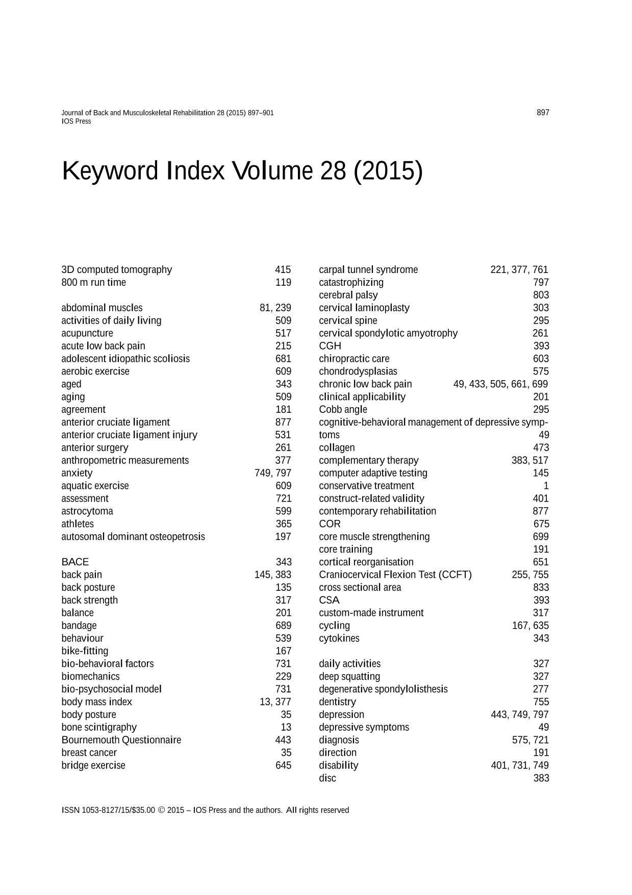
Free accessOtherFirst published November, 2015pp. 887-896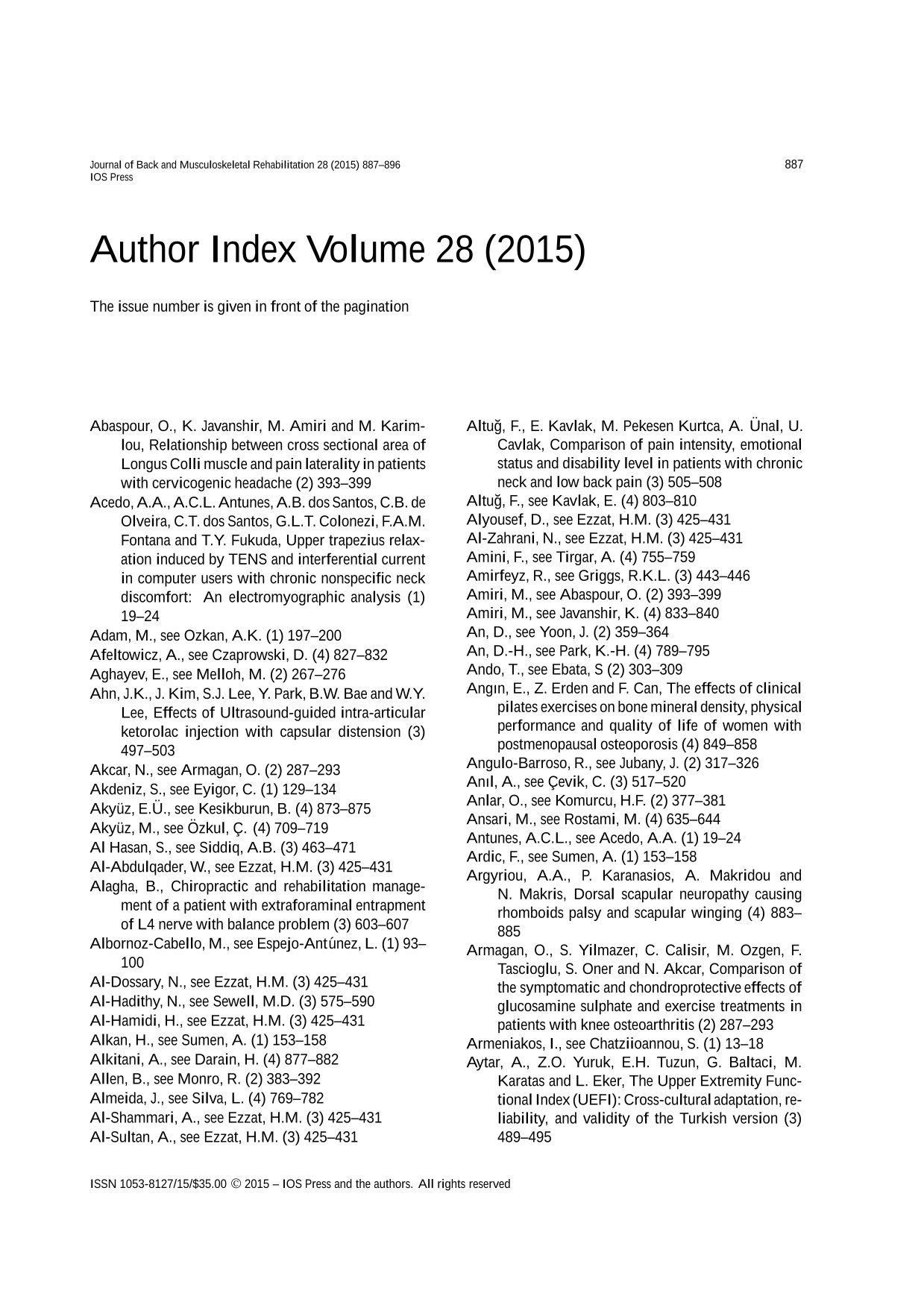 Free accessOtherFirst published November, 2015pp. 897-901