
Abstract
The tenosynovium in the human carpal tunnel is connected to the flexor tendons and the median nerve by the subsynovial connective tissue (SSCT). The most common histological finding in carpal tunnel syndrome (CTS), a compression neuropathy of the median nerve, is noninflammatory fibrosis of the SSCT. The relationship, if any, between the fibrosis and nerve pathology is unknown, although some have speculated that a change in the SSCT volume or stiffness might be the source of the compression. Yet, while animal models have been used to study the physiology of nerve compression, so far none have been used to study the relationship of the SSCT pathology to the neurophysiological abnormalities. The purpose of this study was to identify animal models that might be appropriate to study the interaction of SSCT and nerve function in the development of CTS. The front paws of a rat, rabbit, dog, and baboon were dissected. The carpal tunnel anatomy and SSCT of these animals were also examined by light and scanning microscopy and compared to the relevant human anatomy and ultrastructure. The carpal tunnel anatomy and contents of the baboon and rabbit are similar to humans. The canine carpal tunnel lacks the superficial flexor tendons and the rat carpal tunnel is very small. The human, baboon, and rabbit specimens had very similar organization of the SSCT, and content of the carpal canal. We conclude that, while both the baboon and rabbit would be good animal models to study the relationship of the SSCT to CTS, the rabbit is likely to be more practical, in terms of cost and animal care concerns.
Introduction
Carpal tunnel syndrome (CTS), a compression neuropathy of the median nerve, occurs frequently, and has been studied by many investigators [2, 8, 9, 11, 25]. Despite the prevalence and economic impact of CTS [4, 8], it is remarkable how little is known concerning its etiology. The majority of CTS cases are still described as being idiopathic [9, 11, 39, 40], and the most common histological finding in CTS is noninflammatory synovial fibrosis [1, 11, 25].
Many animal models have been used for CTS research [43]. In these models, CTS is induced by tightening the flexor retinaculum [25], nerve banding with a silastic tube [16, 26, 28], inserting an inflatable device [9] or fluid into the tunnel [23, 32, 37], or placing a tourniquet around the limb [14, 31]. Most carpal tunnel studies focus on histomorphologic changes of the median nerve [7, 17, 18, 26–28, 35]. These animal studies may be more appropriately characterized as compression neuropathy models, rather than models designed to test hypotheses related to the specific etiology of CTS.
The tenosynovium in the human carpal tunnel is connected to the flexor tendons and the medial nerve by the subsynovial connective tissue (SSCT). The SSCT serves as a sliding unit to reduce the friction and to protect the blood supply to the tendon and synovium [15]. In previous studies, histological and biological changes have been noted within the SSCT of patients with CTS [11, 20]. Several investigators have suggested that the nerve compression may actually be secondary to an initial change in SSCT stiffness, volume, or permeability [25, 40]. Recently, a scanning electron microscopy (SEM) study has shown that the most severe changes in the SSCT in patients with CTS [12] were found close to the tendon, suggesting that these changes may be attributable to a shearing injury.
Based on this evidence, we also believe that the etiology of CTS might be related to an injury of the SSCT. To study this possibility, an animal model with a similar anatomy and structure to the human carpal tunnel, including a similar SSCT organization, is essential, yet no studies to date have systematically compared these features between human and putative animal models. The objective of this study was, therefore, to identify a potential in vivo animal model with similar anatomic features to the human carpal tunnel, including, for the first time, consideration of the structure of the SSCT. To accomplish this objective, we investigated the anatomy of the carpal tunnel contents in five different species (human, rat, rabbit, dog, and baboon) and compared the morphology of the SSCT in these species by light and SEM.
Materials and Methods
The front limbs from fresh cadaver rat, rabbit, dog, and baboon specimens were obtained from our institutional Section of Veterinary Medicine. The animals had all been sacrificed in the course of other experiments. In each case, the animals were euthanized by anesthetic overdose and the front paws were harvested from the elbow.
An upper extremity from a human female cadaver (age, 38 years) and one upper extremity from a human male cadaver (age, 75 years) were also used for this study. A medical record review was performed on the human cadavers before the study, to be sure that there had been no antemortem diagnosis of CTS. Exclusion criteria also included a history of diabetes, glucose intolerance, thyroid disease, rheumatoid arthritis, osteoarthrosis, flexor tendinitis, gout, hemodialysis, BMI of >30, sarcoidosis, amyloidosis, peripheral nerve disease or traumatic injuries to the ipsilateral arm.
In addition to the anatomic specimens, we used veterinary anatomy texts [5, 10, 13, 30, 33] to assist in the dissection and to help identify the anatomy of the carpal tunnel. In each of the five species studied, in addition to the anatomic dissections we harvested tendon and tenosynovial tissue for histology and SEM. In addition, one upper limb of each species was deep-frozen (−20°C) and transverse sections were made through the carpal tunnel.
This study was approved by our Institutional Review Board and Institutional Animal Care and Use Committee (IACUC).
Light Microscopy
In the rat, rabbit, baboon, and human specimens, the middle digit flexor digitorum superficialis tendon and its tenosynovium was dissected. This tendon was chosen because it is adjacent to the median nerve and connected to the SSCT and visceral synovium within the carpal tunnel. In the dog, the flexor digitorum profundus tendon was used, because the superficial flexor tendons do not pass within the carpal tunnel. The SSCT biopsies were formalin-fixed and paraffin-embedded. Sections (5 μm) were made and standard hematoxylin and eosin (HE) staining procedures were performed in our department (Department of Laboratory Medicine and Pathology).
Scanning Electron Microscopy
Scanning electron microscope imaging was used to determine the ultrastructrural morphology of the tenosynovium in all five species. The contents of the carpal tunnel were excised en bloc in the rat and rabbit, and the middle digit flexor digitorum superficialis tendon was marked with a marker pen, to mount the dried tissue with the superficial layer up. In the dog, we collected the tenosynovium and approximately 2 cm of the middle digit flexor digitorum profundus tendon within the carpal tunnel. In the baboon and human cadaver, we collected the middle digit flexor digitorum superficialis tendon and its tenosynovium.
The SSCT tissue was fixed in Trump's fixative (1% gluteraldehyde and 4% formaldehyde in 0.1 M phosphate buffer, pH=7.2 [29]). The biopsies were dehydrated through a graded series of ethanol solutions in a critical point dryer. Tissue was then rinsed for 30 min in two changes of 0.1 phosphate buffer (pH=7.2). The tissue was dehydrated in progressive concentrations of ethanol to 100% and either critical point dried. The specimens were then mounted on aluminum stubs and sputter coated with gold-palladium. Images were captured on a cold-field emission scanning electron microscope operating at 2KV (Hitachi S-4700, Hitachi High Technologies America, Inc., Pleasanton, CA, USA).
Results
Human
The cross section of the carpal canal in the human is shown in Fig. 1a. The human carpal canal forms a rigid passageway, bounded dorsally, medially, and laterally by the carpal bones and palmarly by the flexor retinaculum. The contents of the carpal tunnel include the flexor digitorum profundus tendons, the flexor digitorum superficialis tendons, the flexor pollicis longus tendon, the SSCT, the radial and ulnar bursa, and the median nerve. The SSCT loosely connects the finger flexor tendons and the synovial membrane, which in turn encloses the tendons within the ulnar tenosynovial bursa. The SSCT consists of fibrous bundles parallel to the tendon, interconnected by smaller microfibrillar fibers (Figs. 2 and 3).
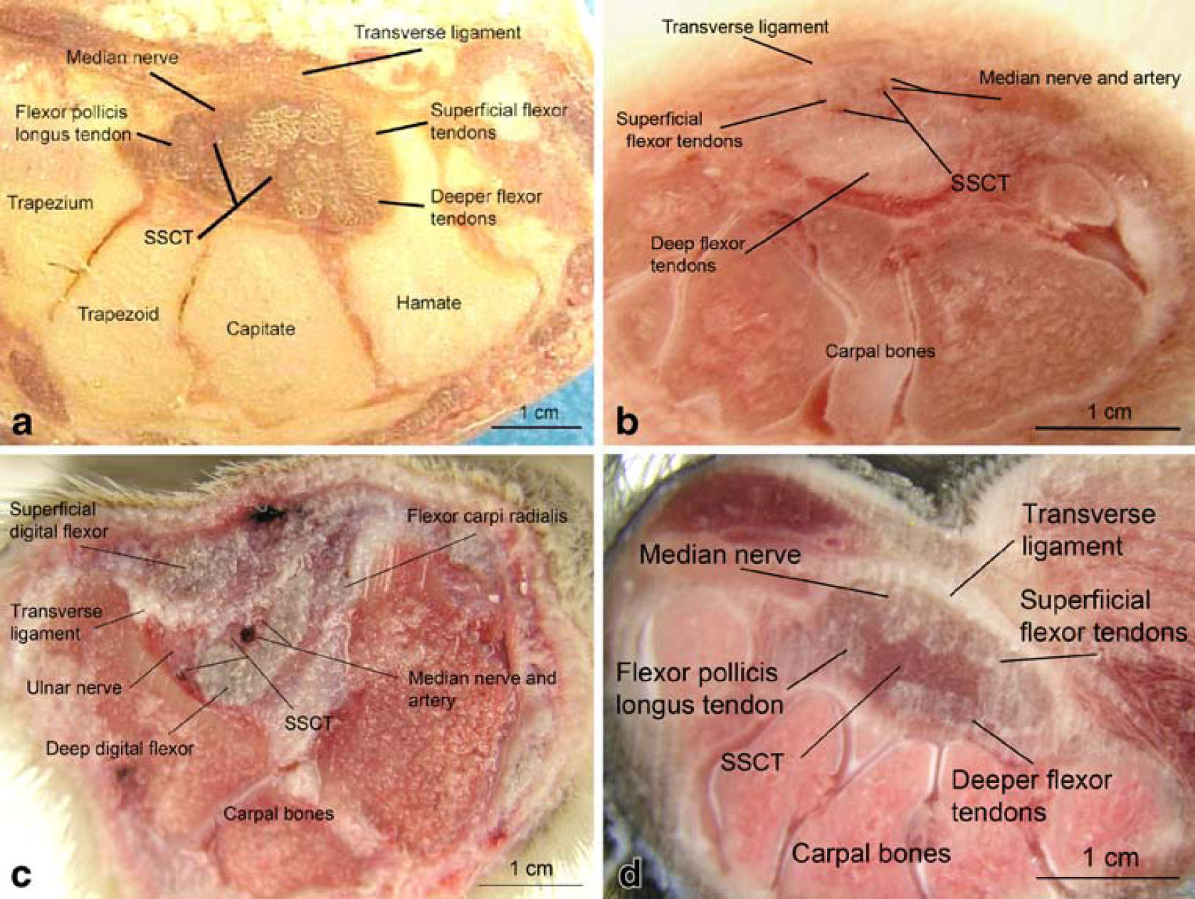
Cross section of the carpal canal in the (a) human, (b) rabbit, (c) dog, and (d) baboon.
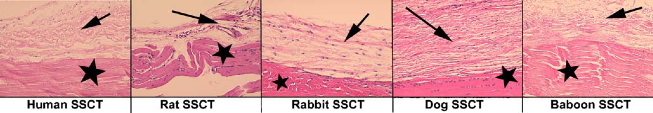
Microscope images of the subsynovial connective tissue (SSCT) of the carpal tunnel in a human, rat, rabbit, dog, and baboon (H&E; original magnification, 200x). Note SSCT (arrow) and tendon (star).
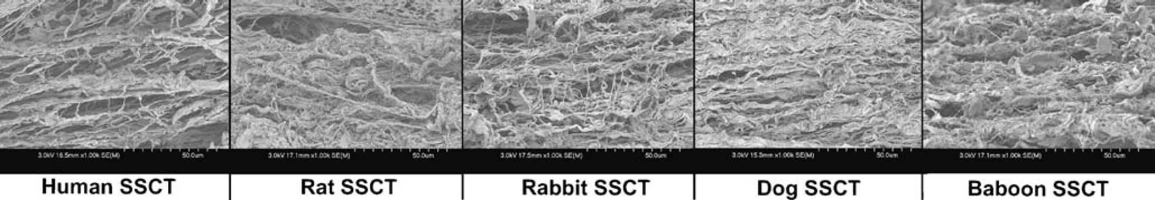
Scanning electron microscope images of the subsynovial connective tissue (SSCT) of the carpal tunnel in a human, rat, rabbit, dog (original magnification, 1000x).
Rat
The front paws of five Sprague-Dawley male rats (weight, 0.35–0.5 kg) were used. Six front paws were used for dissection and four front paws were used to make cross sections.
After making a longitudinal incision in the palmar skin, the median nerve and flexor tendons were exposed. A flexor retinaculum was present. The superficial and deeper flexor tendons were separated structures within the carpal tunnel. An SSCT was present. Light and scanning electron microscopic images of the rat SSCT are shown in Figs. 2 and 3. The SSCT of the rat does not show the typical construction of parallel fibrous cables with interconnections as seen in the human, but rather shows a meshwork of fine fibers that tend to form braids but lack the interconnecting fibrous structures. Cross-sectional illustrations of the rat carpal tunnel did not provide sufficient information for presentation.
Rabbit
Both front paws from five New Zealand White rabbits (weight, 4.0–4.5 kg) were used for dissection of the anatomy. Two additional front paws were used for frozen and transverse sections. The cross section of the carpal canal in the rabbit is shown in Fig. 1b.
In the New Zealand rabbit, the carpal bones and the transverse carpal ligament form a rigid passageway at the wrist through which the flexor tendons and the median nerve travel. The carpal bones consist of three proximal bones, and a small accessory carpal bone and a distal carpal series (I–IV), which is attached to each metacarpal except the fifth. The transverse ligament contains pencil-shaped or triangular-shaped cartilage disks, which can be palpated and used as a marker for surgery or injections.
The rabbit's median nerve, median artery, flexor digitorum profundus tendons, and flexor digitorum superficialis tendons all lay inside the carpal tunnel (Fig. 1b). The superficial flexor tendons are separate tendons. The profundus flexor tendons of the four digits are fused in the carpal tunnel. The flexor pollicis longus of the rabbit is also within the carpal tunnel. The median nerve originates from the sixth and seventh cervical nerves in the brachial plexus and on the caudal side of the humerus and continues across the elbow to the lateral surface of the forearm. In four rabbits (both front paws), we found that the median nerve passed through the carpal tunnel with the median artery on the radial volar side, while in both paws of one rabbit, the median nerve split about 2 cm proximal to the flexor retinaculum into a ramus medialis and ramus ulnaris, both of which went through the carpal tunnel. The small medial ramus of the median nerve ran superficial to the flexor tendons on the radial side adjacent to the medial artery. The ulnar ramus of the median nerve ran at the ulnar side in the carpal tunnel, also superficial to the tendons.
Standard hematoxylin and eosin staining of the SSCT shows that rabbit SSCT is similar to human SSCT (Fig. 2). By SEM, we found that the SSCT of the rabbit consisted of fibrous bundles that run parallel to the tendon interconnected by smaller microfibrillar collagenous fibers, similar in structure to that of humans (Fig. 3).
Canine
The front paws from five mongrel dogs (approximately 30 kg in weight) were used for dissecting the anatomy and biopsies, and one for transverse sections.
In the forepaw, the carpus includes seven small, irregular bones arranged into two rows. On the palmar side of the wrist there is a fat pad, which is covered with the skin typically seen on the volar side of the digits. The carpal canal is formed by the accessory carpal bone laterally, the palmar carpal ligament and the carpal bones dorsally, and the flexor retinaculum on the palmar surface (Fig. 1c). All the metacarpal bones are fused.
The superficial flexor tendon lies beneath the skin and antebrachial fascia on the caudomedial side of the forearm. The tendon is at first single, then crosses the palmar surface of the carpus medial to the accessory carpal bone in the carpal canal, and finally divides into four tendons of nearly equal size. The deep digital flexor has three heads of origin that fuse at the carpus to form a single tendon. This tendon is held in place in the carpal canal by the thick, deep part of the fibrous flexor retinaculum. The flexor retinaculum lies between the superficial and the deeper flexor tendon. The median nerve and median artery pass under the ligament (Fig. 1c). The light and scanning electron microscopic investigation of the SSCT showed that canine SSCT is similar in structure to the human SSCT (Figs. 2 and 3), and that the SSCT of the canine consists of fibrous bundles that run parallel to the tendon interconnected by smaller microfibrillar collagenous fibers.
Baboon
Both upper extremities from a female baboon (age, approximately 4 years; weight, 11.3 kg) were used. One upper extremity was used for dissection of the anatomy and biopsy and one for the transverse section. The cross section of the carpal canal in the baboon is shown in Fig. 1d. The baboon's hand is similar to that of the human. On the proximal palmar side, the baboon has a large fat pad covered with the skin typically seen on the palm of the hand. When dissecting the skin, we found the transverse ligament going diagonally across the palm. It was approximately 2.5 cm long. Under the ligament, the median nerve was present and was approximately 2 mm thick. The four superficial flexor tendons and the four profundus flexor tendons are separate structures within the carpal tunnel. We also found a flexor pollicis longus tendon within the carpal canal.
The SSCT lies between the carpal tunnel structures and consists of fibrous bundles parallel to the tendon, interconnected by smaller microfibrillar fibers that appear similar to human tissue (Figs. 2 and 3).
Discussion
The choice of an animal model to study CTS depends on the purpose of the study design. For example, to study the sequence of structural alterations in the median nerve due to mechanical compression, any animal with myelinated mixed nerves could be used. The study of the etiology of CTS is a more complex endeavor, and requires the ability to observe the interaction of the median nerve in situ with the surrounding tendons and tenosynovium, in an arrangement similar to that in the target human population.
To assess the utility of various animals to study the etiology of the CTS, we therefore focused on the comparability of the anatomy of the animal carpal tunnel and the morphology of the SSCT The criterion considered most essential for proposing an appropriate animal model of CTS etiology was the morphology of the SSCT and, in particular, its relationship with the superficial flexor tendons and the course of the median nerve, since disruption of this structure and relationship is the principal nonneuropathologic finding in CTS. We also considered the relative independence of the superficial flexor tendons in these animals as compared to the human because this may be of special interest when it comes to simulating shearing injury to the SSCT, a possible repetitive use mechanism for the etiology of CTS.
In all of these animals, except partially in the baboon, the front paw is a weight-bearing extremity. This means that the position of the wrist is in hyperextension. Neither rat, rabbit, nor dog had an opposable thumb. Food-holding motions are accomplished by pressing material between the forepaws in the rat and rabbit, whereas the dog does not typically use this behavior. All three species can perform a unified flexing of the digits, e.g., the dog can flex the digits when digging a hole in the ground, but the baboon is the only animal (in this investigation) that can perform grasping and differential finger movement. Yet, ultimately, none of the animals use their forelimbs as humans do. Thus, even an analogous anatomy does not imply a perfect model, and any conclusions in the model must still be validated clinically.
Rat
The rat has been used for a model of CTS [7] and has been shown to be a good model to investigate the neuropathological changes induced by nerve compression [18, 27], especially the sciatic nerve. However, the small size of the front paw and the difference of the SSCT structure make this animal less appropriate for the study of the etiology of CTS.
Rabbit
The New Zealand rabbit has been commonly used as an animal model for CTS, so that there is extensive data available on the histopathology of the rabbit median nerve [9, 32, 34, 35]. We used New Zealand White rabbits weighing 4.0–4.5 kg because the median nerve is better visualized when the rabbit is bigger, and also because there is more tenosynovium present. The rabbit has a nearly identical situation as the human regarding the osseous and connective tissue formations in the carpal canal [21, 30, 33], and there is extensive data available describing median nerve compression in the rabbit carpal tunnel [9, 32, 34, 35]. We found an anatomical variation in the course of the median nerve, but this high bifurcation is similar to variations also found in humans [22, 24]. The tenosynovium in the rabbit carpal tunnel is similar to human SSCT tissue, and is of sufficient volume to be examined for the purpose of etiologic studies. The lower costs and availability as compared to the primate makes this animal model attractive for study.
Canine
The canine model has been widely used for flexor tendon research [3, 6, 38, 44, 45], because it has a flexor tendon anatomy and functional structure similar to human and it is adaptable for surgery and therapy. However, the canine model for CTS has only been described in a few recent studies [41, 42]. Although the dog has an SSCT structure similar to that of humans, and the median nerve passes under the flexor retinaculum [13], there is no flexor digitorum superficialis tendon in the carpal tunnel in the canine model. This is an important drawback in etiological studies.
Baboon
The primate hand is closely related to the human hand, in terms of anatomy and function [19, 36], and the carpal tunnel is identical to the human. The primate has been used in a few studies on CTS, by directly compressing the median nerve with the use of a silastic tube [26, 28] or by supplying fluid into the carpal canal [37]. We found that the baboon's SSCT consisted of fibrous bundles parallel to the tendon, interconnected by smaller microfibrillar fibers, and looks similar to the human SSCT.
The primate would be the obvious first choice for studies regarding repetitive movement and CTS. The limitations are mainly those of costs and regulatory requirements associated with primate studies.
Of all four animals investigated, the baboon and rabbit have osseous and connective tissue formations in the carpal canal that are closest to those of the human situation. Among these, the rabbit carpal canal is recommended as the most practical model for most studies, with the baboon or similar primate could be reserved for more complex studies involving the effect of specific tasks on the SSCT and median nerve function.
Footnotes
Acknowledgments
This study was funded by grants from NIH (NIAMS AR 49823) and Mayo Foundation. The authors gratefully acknowledge Jon Charlesworth for his help with the scanning electron microscopic investigation and imaging during this study.