
Abstract
Objectives:
The objective of the present study was to investigate whether moderately experienced meditators activate hippocampus and the prefrontal cortex during silent mantra meditation, as has been observed in earlier studies on subjects with several years of practice.
Methods:
Subjects with less than 2 years of meditation practice according to the Kundalini yoga or Acem tradition were examined by functional magnetic resonance imaging during silent mantra meditation, using an on–off block design. Whole-brain as well as region-of-interest analyses were performed.
Results:
The most significant activation was found in the bilateral hippocampus/parahippocampal formations. Other areas with significant activation were the bilateral middle cingulate cortex and the bilateral precentral cortex. No activation in the anterior cingulate cortex was found, and only small activation clusters were observed in the prefrontal cortex.
Conclusions:
In conclusion, the main finding in this study was the significant activation in the hippocampi, which also has been correlated with meditation in several previous studies on very experienced meditators. We propose that the hippocampus is activated already after moderate meditation practice and also during different modes of meditation, including relaxation. The role of hippocampal activity during meditation should be further clarified in future studies, especially by investigating whether the meditation-correlated hippocampal activity is related to memory consolidation.
Introduction
Different brain-imaging technologies such as SPECT (single photon emission computed tomography), PET (positron emission tomography), and fMRI (functional magnetic resonance imaging) have been deployed to study the neural correlates to meditation. In an early study by Newberg et al., 9 Tibetan Buddhist meditators with more than 15 years of experience were examined with SPECT during meditation. The authors found increased regional cerebral blood flow in the cingulate gyrus and the dorsolateral prefrontal cortex (DLPFC) as well as in the thalamus and the inferior and orbital frontal cortices during meditation as compared to baseline scans. Lou et al. 10 investigated the neural response to meditation in Yoga Nidra meditators with more than 5 years of practice. The subjects performed an auditory-guided relaxation meditation during PET scanning. In that study, meditation focusing on bodily sensations was correlated with activation in the parietal lobe and in the supplementary motor area. Meditation generating a sensation of joy induced activation in the left parietal and the superior temporal lobes. In addition, activation in bilateral hippocampi was observed at all conditions. However, no activation in prefrontal areas and in the anterior cingulate cortex (ACC) was observed in that study. This absent activation was interpreted to be caused by the limited volitional influence during auditory-guided meditation.
In a pioneering fMRI study by Lazar et al., 11 Kundalini meditators with more than 4 years of practice were examined. When comparing the meditative state (passively observing the breath and repeating a mantra) with the control state (generating a random list of animals), activation in the parahippocampal/hippocampal formation, putamen, the midbrain, and ACC were observed. In another fMRI study by Brefczynski-Lewis and colleagues, 12 brain activation in expert meditators (Tibetan Buddhists) during meditation with focused attention was compared to novice meditators. In that study, both groups elicited activation in frontoparietal regions and cerebellum as well as in the parahippocampal, temporal, and the posterior occipital cortices. In addition, the authors observed more activation in the medial frontal cortex and ACC in novice compared to expert meditators. This result was, however, contradicted in a recent fMRI study by Hölzel et al. 13 in which Vipassana meditators were investigated during mindfulness on breathing. In this study, activation in the bilateral medial prefrontal cortex and bilateral ACC was observed when comparing meditators to novice controls in a meditation versus arithmetic calculation task. Furthermore, a recent fMRI case study at our laboratory on one expert Tibetan Buddhist meditator performing compassion meditation demonstrated left-sided activation in the medial prefrontal cortex and ACC. 14
Previous imaging studies have shown somewhat mismatching results, which is probably explained by differences in study design as well as differences in the meditation experience among the participants.
14
Despite this, some brain regions have been identified as significantly correlated with meditation in more than one study. Most studies report activation in hippocampal and parahippocampal areas. In addition, studies involving volitional-controlled meditation have shown activation in prefrontal areas (i.e., DLPFC and the medial prefrontal cortex). Activation in the ACC has been attributed to meditation, albeit it is disputed whether this activation predominantly occurs in expert or in novice meditators. A large majority of earlier imaging studies on meditation have been performed on very experienced subjects with more than 5 years of daily meditation practice.
2,15
For the clinical aim to use meditation for alleviation of different medical conditions, it would be helpful to obtain imaging evidence of altered brain function even after a short time of practice. In the present study, the neural correlates to meditation in subjects with no longer than 2 years of meditation practice were investigated by fMRI. Participants practicing either Acem meditation (
The aim of the present study was to investigate whether moderately experienced subjects display activation in the hippocampal/parahippocampal formation, the ACC, and in the prefrontal cortex during silent mantra meditation, as has been observed in earlier studies on meditators with several years of practice.
Materials and Methods
Subjects
Subjects were recruited from a cohort of Acem and Kundalini yoga meditators. Inclusion criteria were right-handed subjects who had been practicing meditation at least 2 times a week for a minimum of 6 months and a maximum of 24 months. Minimum age was set to 18 years and subjects above 60 were excluded. According to these criteria, 12 subjects were recruited to the study. Three (3) subjects were excluded due to movement during scanning. Another subject fell asleep during the fMRI session. In the present study, results are reported for 8 subjects: 5 women and 3 men with a mean age of 35 years (range: 21–50 years). The mean meditation experience was 14 months (range 6–24 months) and the subjects meditated a mean of 8 times (range 2–20 times) per week. The time for a normal meditation session was a mean of 32 minutes (range 15–45 minutes). Of the 8 subjects, 5 practiced Kundalini yoga meditation and 3 practiced Acem meditation. Demographic data of the subjects are found in Table 1.
F, female; M, male.
Written information about the study including details about the fMRI scanning session was sent to each subject in advance. Each subject completed a questionnaire with general health questions, including questions about current medication, prior or current neurological and psychiatric disease, and head/neck trauma. None of the subjects declared that they had any prior or current health issues that could affect the study. The local Ethical Committee approved the study, and written informed consent was obtained from each subject.
Study design
The study was designed as an on–off block-design with two alternating blocks: meditate (on) where the subjects were instructed to meditate (using a mantra/method-word as a key) and word (off) where the subjects were instructed to silently (in the same general way as with the mantra/method-word) repeat the short phrase “table and chairs” (bord och stolar in Swedish). This phrase was selected to be as neutral as possible in order not to evoke an emotional response from the subject during the word block. The choice of a language-related control task was motivated by the intention to subtract language components originating from the mantra during meditation. Each block was set to 2 minutes and a total of 8 blocks, alternating meditate and word, were used (4 meditate and 4 word). The total scan time for each subject was 16 minutes.
The subjects were instructed to use a mantra/method-word that they were familiar with, but they were not asked to reveal the phrase they were using. The subjects were instructed not to vocalize either the mantra or the control phrase in order to avoid any movement of the head and neck area during fMRI scanning. In addition, the subjects were instructed to breathe normally throughout the scanning session (i.e., they were instructed not to use any kind of breathing techniques during meditation).
fMRI acquisition
In order to achieve a very quiet and calm environment, we opted to perform the fMRI scanning during weekends where no clinical activity was in progress at the MRI facility. When the subjects arrived at the facility, verbal instructions regarding the alternating two blocks meditate and word were repeated. The subjects were placed in the MR scanner (Achieva 1.5 T, Philips Medical Systems) with heads fixated using the headrest and fixation pillows. The subjects were issued with headphones where the verbal commands were presented preceded by a short auditory signal (440 Hz for 0.2 seconds) by a Windows XP computer running SuperLab Pro 2.0. The scanning was initiated and two anatomical scans were acquired. The fMRI part of the examination consisted of a blood oxygen level dependent (BOLD) sensitive echo planar imaging (EPI) sequence. The following parameters were used: repetition time = 2.7 seconds, echo time = 40 ms, matrix = 80 × 80, field of view = 24 cm, slice thickness = 3 mm, number of slices = 31. Each scanning session lasted for approximately 30 minutes.
Image analysis
The fMRI images were preprocessed and analyzed using SPM5 (Wellcome Department of Imaging Neuroscience, University College, London, UK) according to the following steps: (1) movement correction (re-alignment), (2) normalization to the Montreal Neurological Institute EPI template included in the SPM5 package, and (3) image smoothing applying a 8-mm Gaussian kernel. Each subject was first analyzed individually to check for inconsistency, failed scans, or errors during preprocessing. Realignment parameters from the movement correction were included in the analysis, and the hemodynamic response function implemented in SPM5 was used as a model for the BOLD response. The contrast vector was set to [1 − 1], applying the null hypothesis that brain activation during meditation was not different from the control condition.
Conjunction analysis applying a multisubject design matrix (e.g., ref. 16) was performed in order to make inferences on brain activation at the group level. 16 Areas in the whole brain that were activated during both the word and the meditate conditions were investigated using a threshold of p = 0.001 (uncorrected). The WFU PickAtlas tool 17 was used to create regions of interest (ROIs) from the Automated Anatomical Labeling atlas. 18 The following ROIs were used in the analysis: the hippocampus/parahippocampal formation, ACC, the medial prefrontal cortex, the opercular and triangular part of the inferior frontal cortex, and the middle frontal cortex. The hippocampus/parahippocampus ROI was masked for overlap with the lateral ventricles. This is because spurious activation in adjacent parts of the lateral ventricles, which was interpreted to be caused by the spatial smoothing procedure, was found. A threshold of p = 0.05, corrected for familywise error, was applied in the ROI analysis.
Results
The most significant activation in this group of moderately experienced meditators during the meditate condition (silent mantra meditation) was found in the right hippocampus (Table 2). Other areas with common activation were the middle cingulate gyrus (bilateral), the precentral gyrus (bilateral), and the right precuneus. During the word condition, this group elicited activation in the left and right superior temporal gyrus and in the left superior frontal gyrus.
The table shows coordinates in Montreal Neurological Institute space (x, y, z) for the most significant voxels, the number of significant voxels (#), and statistical value (Z).
R., right; L., left.
A ROI analysis was performed in order to further investigate whether moderately experienced meditators activate similar areas during meditation, as has been found in previous studies on more experienced meditators. The ROI analysis revealed significant activation in both the left and right hippocampus/parahippocampal formations (Table 3 and Fig. 1). Small clusters of activation were also found in the right inferior and medial prefrontal cortices. The activation in the medial frontal cortex was divided into 4 smaller clusters containing 1–5 significant voxels. No significant activation was found in ACC or in the middle frontal cortex.
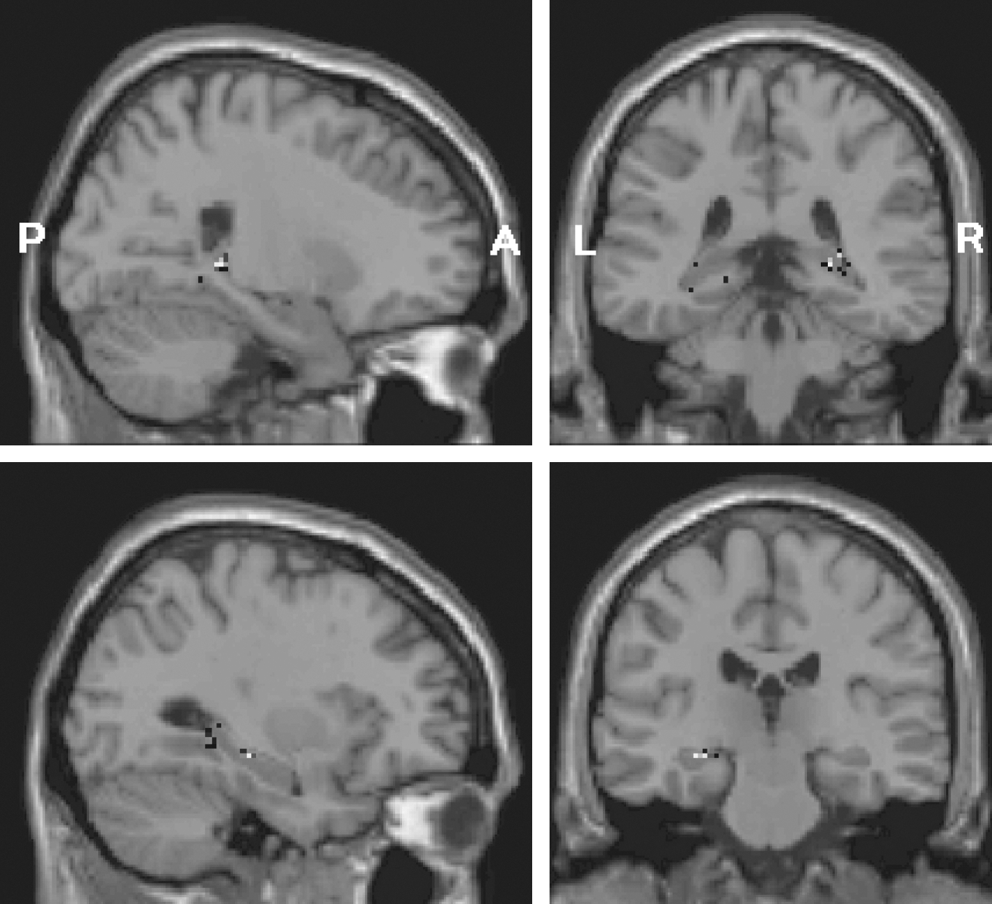
Results from the region-of-interest analysis showing activation in sagittal (left panels) and coronal (right panels) images in the right (upper panels) and left (lower panels) hippocampus/parahippocampal formations during silent mantra meditation. P, posterior; A, anterior; L, left; R, right.
The table shows coordinates in Montreal Neurological Institute space (x, y, z) for the most significant voxels, the number of significant voxels (#), and statistical value (Z).
R., right; L., left.
Discussion
The main result of the investigation was that moderately experienced meditators elicited significant activation in the bilateral hippocampi during silent mantra meditation. Previous studies have also reported hippocampal activation to be associated with meditation, including the relaxation response. 10 –12 However, the reason for hippocampal involvement during meditation has not been discussed. What could be the implications of hippocampal activation? The involvement of the hippocampus in memory is well documented, although its precise role remains elusive. 19 Some theories claim that the hippocampus is the storage and/or consolidation site of memories. Other theories propose that the hippocampus serves as the librarian for memories and may also be tagging memories with respect to context; that is, the hippocampus keeps track of where memories are stored and in what context the memories were originally acquired. 20 Thus, memory consolidation could be one possible explanation of hippocampal activation during meditation. However, despite the known hippocampal correlations with both meditation and memory, the literature on meditation and concomitant memory effects is sparse. One recent study reports the effects of meditation on visuospatial memory, a function that is clearly related to the hippocampus. 21 In the study by Kozhevnikov et al., 22 it was found that visuospatial memory was enhanced after a session with Buddhist deity meditation. It has also been shown that mindfulness cognitive therapy increases specific retrieval of autobiographical memory and reduces overgeneral memory in depressed patients. 23 In addition, meditation training significantly enhances working memory capacity. 24 The role of the hippocampus in working memory is, however, ambiguous because working memory primarily is attributed to activity in the prefrontal, parietal, and cingulate cortices. 25
In this context, the relations between, on the one hand, meditation and sleep and, on the other hand, between memory and sleep are noteworthy. Several electroencephalography studies have reported sleeplike stages during meditation as well as alterations in sleep cycles as an effect of meditation (see Cahn and Polich 14 and the references therein). In a recent study by Nagendra et al., 26 Vipassana meditators showed significantly increased rapid eye movement (REM) and slow wave sleep (SWS) compared to a control group.
The relation between sleep and memory consolidation is well documented. 27 –29 Hippocampus-dependent consolidation of declarative memories benefits particularly from SWS, whereas procedural memories are primarily consolidated during REM sleep. 27,29 Future studies on meditation and declarative memory might elucidate the role of hippocampal activity during meditation in more detail and serve to clarify whether there is a correlation between meditation and memory consolidation.
Other areas with significant activation in the present study were the middle cingulate cortex and the precentral cortex. These areas are regarded to be involved with motor control and execution. The middle cingulate cortex is thought to be involved in orienting the body position in response to sensory stimuli, 30 and the precentral gyrus is defined as the motor cortex. Thus, activation in these areas could be involved with awareness of bodily sensations during meditation. Noteworthy, when the subjects were instructed to silently repeat a short phrase without attempting to meditate (the word block), activation was found in the bilateral temporal lobes and in the right frontal lobe. These areas are clearly associated with language function.
Several studies have indicated prefrontal areas and ACC to be involved in inducing and also maintaining meditation. 31,32 These areas are also regarded to be important for sustained and selective attention. 25 In particular, the right hemisphere has been designated to be involved with sustained attention. 25,32 In the current study, only small clusters of activation were found in the right DLPFC and the medial prefrontal cortex. These scarce findings might be a result of the difference in meditation method and experience among subjects. Three (3) subjects practiced meditation according to the Acem method, and 5 subjects were practicing Kundalini Yoga (Table 1). We performed an additional statistical analysis to search for differences in brain activation between Acem and Kundalini Yoga meditators. However, the sample size was too small to obtain conclusive results. In addition, 5 subjects had practiced meditation for 6–12 months, and 3 subjects had been practicing for 18–24 months. Half of the subjects meditated 7 times or more each week, and the other subjects meditated more seldom. Yoga meditators had an overall longer time of meditation experience compared to Acem meditators, and more experienced meditators meditated in general fewer sessions per week compared to more novice meditators (Table 1). We believe that prefrontal activation is individually distributed and very much dependent on both experience and the actual meditation practice.
A limitation of the present study was the relative small group size. Due to the selected inclusion criteria, few subjects could be recruited locally. Of 12 recruited subjects, only 8 were retained in the study. Three (3) subjects (corresponding to 25%) had to be excluded after scanning due to movement artifacts. Problems with movement are general in fMRI; however, we consider this percentage of excluded subjects to be comparatively high. A specific reason for the abundant movement artifacts could be the different breathing patterns between the meditate and the word state. Inclusion of movement parameters from the re-alignment procedures ameliorated the images for the group as a whole; still, 3 subjects had to be excluded.
Conclusions
In conclusion, the main result was that subjects with less than 2 years of meditation experience during silent mantra meditation activated the bilateral hippocampi, which also has been correlated with meditation in several previous studies on very experienced meditators. We propose that the role of hippocampal activity during meditation should be further clarified in future studies, especially by investigating whether the meditation-correlated hippocampal activity is related to memory consolidation.
Footnotes
Acknowledgments
The County Council of Östergötland and the strategic research area of Medical Image Science and Visualization are acknowledged for financial support.
Disclosure Statement
No financial conflicts exist.