
Abstract
While the most prevalent skin conditions in the paediatric age group include acne, dermatitis/eczema, bacterial and fungal infections, recent articles published in InnovAiT cover these topics in detail. This article aims to provide an overview of some of the conditions frequently encountered in the paediatric dermatology clinic, considering in particular birthmarks, vascular malformations, neonatal skin conditions, paediatric skin tumours, mucocutaneous syndromes and infections, including viral exanthems.
The GP curriculum and childhood skin rashes
Demonstrate a reasoned approach to the diagnosis of skin symptoms using history, examination, incremental investigations and referral
Intervene urgently when patients present with an emergency skin problem
Work with patients to empower them to look after their own health and take responsibility for managing their skin problems
Promote skin well-being by applying health promotion and disease prevention strategies appropriately
Make timely appropriate referrals on behalf of patients to specialist services
Appreciate the importance of the social and psychological impact of skin problems on the patient's quality of life and on the patient's family
The knowledge base of
Birthmarks
Skin disorders in children
Viral exanthems in children
Birthmarks and vascular lesions
Birthmarks indicate an excessive local collection of one or more normal skin components, such as blood vessels, melanocytes, sebaceous glands or collagen. Vascular birthmarks in children may be categorized into infantile haemangiomas and vascular malformations.
Mongolian blue spot (congenital dermal melanocytosis)
A Mongolian blue spot is usually present from birth, although may appear within the first few weeks after delivery. It refers to a steel blue patch on the lumbosacral region, caused by entrapment of dermal melanocytes during migration into the dermis. Although Mongolian blue spots can occur in all races, they are much more common in Asian or black-skinned children. They usually regress spontaneously by 4 years of age.
Naevi of Ota and Ito
Naevi of Ota and Ito are typically blue—brown patches, with naevi of Ota affecting the face over the distribution of the first and second branches of the trigeminal nerve and naevi of Ito affecting the shoulder or upper arm. As with Mongolian blue spots, these pigmented lesions are more common in black or Asian skin. As neither naevi of Ota nor Ito regress spontaneously, management involves cosmetic camouflage, laser or intense pulsed light (IPL) treatment. Glaucoma is a rare complication of naevi of Ota; therefore, regular eye examinations should be undertaken if there is hyperpigmentation of parts of the eye.
Infantile haemangiomas
Infantile haemangiomas are benign vascular neoplasms and the most prevalent soft tissue tumours of infancy. The most common presentation (Fig. 1) is a superficial, bright red nodular lesion with a lobulated surface (strawberry haemangioma). Around 30% are present at birth.
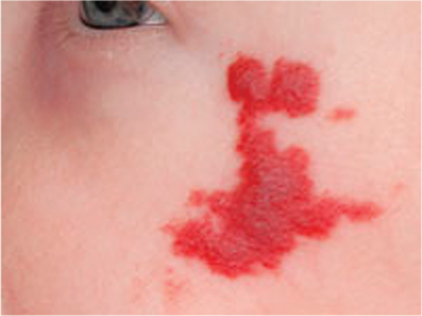
Infantile haemangioma.
Infantile haemangiomas follow a characteristic clinical course of rapid proliferation during the first few months, followed by regression over several years:
30% involute by 3 years
50% involute by 5 years
70% involute by 7 years
90% involute by 9 years
Most infantile haemangiomas are not medically significant, require no treatment and resolve spontaneously. However, lesions with concerning features (Box 1) should be referred for paediatric dermatology advice. Treatment of medically significant haemangiomas is usually with steroids (topical, intralesional or oral), oral propranolol or laser.
Features of haemangiomas for which referral should be considered
Ulceration
Recurrent bleeding
Large size, causing coagulopathy
Large facial haemangiomas with a deep component
Regionally significant haemangiomas because of possible interference with vision, hearing, breathing, feeding or other bodily functions, e.g.
peri-ocular
nasal tip
lips
pinna
anogenital region (possible interference with vision/hearing/breathing/feeding)
Vascular malformations
Unlike infantile haemangiomas, vascular malformations are always present at birth and progress slowly, in proportion with the general growth of the child. The two most common malformations are the salmon patch (naevus simplex) and port-wine stain (naevus flammeus).
Salmon patches are pink or red patches that occur in approximately 40% of neonates. They may be on the nape of the neck (stork bite), glabella (angel's kiss) or eyelids. These lesions fade spontaneously, although remnants, particularly of stork bites, may persist into adult life. They do not require treatment.
A port-wine stain occurs in about 0.3% of neonates; at birth, it is a well-demarcated dark red or purple patch, while later, it may become more bumpy or lobulated. Port-wine stains may present at any body site, although usually occur on the face. Most do not fade or regress, and some even deepen in colour with time. Sturge-Weber syndrome occurs in 10% of neonates with a port-wine stain involving the ophthalmic branch (V1) of the trigeminal nerve. Individuals with Sturge-Weber syndrome have a port-wine stain associated with intracranial haemangiomas and often arteriovenous malformations.
Sturge-Weber syndrome is associated with glaucoma, epilepsy and developmental problems. Therefore, all children with such port-wine stains involving V1 should be referred for assessment. Treatment is with laser, often pulsed dye laser (PDL).
Common skin conditions in neonates
Milia
Approximately 40% of neonates have multiple white 1–2 mm papules on the forehead, nose and cheeks (milia), and 60% of neonates have these lesions in the oral cavity, where they are referred to as Epstein's pearls. The papules rupture and extrude their contents onto the skin surface within a few weeks of birth.
Sebaceous gland hyperplasia
In 50% of infants, 1 mm yellow macules or papules may be apparent over each pilosebaceous follicle on the nose and cheeks. These are caused by maternal androgenic stimulation and spontaneously recede by 6 months of age.
Erythema toxicum neonatorum
Another common transient dermatosis in neonates is erythema toxicum neonatorum. This affects around 50% of full-term neonates, usually 24–48 hours after birth. Lesions may vary in morphology, including erythematous macules, papules, pustules and wheals (Fig. 2). They begin on the face and spread to the trunk and limbs, sparing the palms and soles. The eruption resolves spontaneously within 5 days.

Erythema toxicum neonatorum.
Infantile acne
Infantile acne usually manifests at 3 months of age as lesions typical of acne, including comedones, papules and pustules. It can persist to 3 years and is thought to have a genetic aetiology. Treatment is initially with topical benzoyl peroxide and erythromycin, with topical tretinoin or adapalene for comedones. Severe or refractory infantile acne may require oral erythromycin and even isotretinoin. Tetracycline antibiotics should be avoided as they may cause staining of teeth.
Eczema
Atopic eczema is a chronic pruritic inflammation of the epidermis and dermis that is frequently associated with a personal or family history of atopy. As one of the most prevalent childhood skin conditions, it affects approximately 12–15% of infants, usually within the first 6 months of life. Less than half of these children continue to be symptomatic at 18 months and thereafter. Remission occurs by the age of 15 years in 75% of children with the condition.
In infancy, eczema appears as a pruritic vesicular rash on the face and hands, often with exudate and/or secondary bacterial infection. After 18 months, the rash becomes more classical, affecting the flexures, wrists and ankles. Erythema, lichenification, excoriations and post-inflammatory hyperpigmentation are all frequently occurring features after infancy.
Management is holistic and includes parent education, avoidance of precipitants, the application of emollients, topical steroids, tacrolimus, wet-wrap bandages for exudative eczema and systemic antihistamines. For further detail on this significant condition, please refer to the InnovAiT article ‘Childhood atopic eczema’ (Simon, 2008).
Common skin tumours in children
Epidermal naevus
Epidermal naevi arises from a defect in ectoderm causing overgrowth of the epidermis. There are many types, classified according to the cell type involved. The lesions are often unilateral and linear (Fig. 3), following Blashko's lines, and while initially fat, they become thickened and more verrucous with age.
Epidermal naevus.
Epidermal naevi are difficult to treat medically, although topical calcipotriol may reduce the verrucous appearance to an extent. Larger lesions should be referred for biopsy, and removal of naevi can either be surgical or by laser.
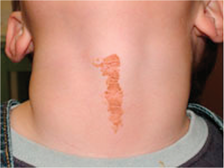
Naevus sebaceous
Naevus secaceous is a variant of the epidermal naevus, presenting at birth as a hairless well-demarcated yellow patch on the scalp, face or neck that in puberty can become more raised and verrucous. Rarely, neurological complications may ensue, notably epileptic seizures. A total of 10–15% develop a secondary tumour within the lesion, most often basal cell carcinoma (BCC).
Management of these lesions is generally by excision during the teens or early adulthood. However, if the lesion is difficult to excise because of size or position, long-term regular observation may be considered.
Becker's naevus
Becker's naevus or Becker's melanosis is a late-onset variant of the epidermal naevus that occurs predominantly in males. It presents as a unilateral area of hyperpigmentation and hypertrichosis, commonly on the upper back and shoulders. The lesion appears during puberty secondary to circulating androgens. Treatment is by laser or electrolysis to remove hair and laser to reduce pigmentation.
Pilomatricoma
Pilomatricoma is a rare benign tumour, caused by the overgrowth of hair matrix cells. Characteristic features are calcification, tented skin and blue—grey discoloration. Occasionally, pilomatrical cancer has been reported. Treatment is by excision as the lesions do not resolve spontaneously.
Juvenile xanthogranuloma
Juvenile xanthogranulomas (JXGs) are benign, red, yellow or brown papules and nodules made up of histiocytic cells (Fig. 4). They develop in the first year of life and most fatten within 3–5 years, with residual hypopigmentation. Lesions can occur in the eyes in 0.3% and more rarely in the viscera. Associated conditions include Niemann—Pick disease, type 1 neurofibromatosis and juvenile myelomonocytic leukaemia (JMML).

Juvenile xanthogranuloma.
Skin lesions are often managed conservatively, while extracutaneous lesions may respond to steroids or radiotherapy. Excision, however, is usually curative.
Bacterial skin infections in children
Impetigo
Impetigo is a highly contagious condition caused by infection of the superficial layers of the epidermis by Staphylococcus aureus. It is characterized by vesicular lesions with fragile roofs that are quickly lost, leaving erosions covered by honey-coloured crusts. It most commonly affects exposed areas such as the face, nares and extremities.
Advice and treatment include prevention of transmission (avoiding close contact and using separate towels and temporary exclusion from school), debridement of crusted lesions with an antibacterial wash and topical antibiotics, e.g. fusidic acid. In widespread disease, oral antibiotics may be preferable.
Folliculitis, furunculosis and abscesses
Infection of the hair follicles or abscesses is usually caused by S. aureus, although Gram-negative organisms, such as Pseudomonas do occur. The presentation depends on level of infection of follicle—superficial folliculitis appears as tiny pustules; furunculosis (deep folliculitis) appears as tender erythematous nodules. Abscesses are commonly found on the trunk and buttocks.
Treatment for superficial folliculitis includes a 10–14 day course of topical antibiotics. Furuncles and abscesses should be treated by incision and drainage. Long-term systemic antibiotics may be required in severe cases.
Erysipelas
Erysipelas is an intradermal infection caused by toxin-producing Streptococcus pyogenes. Vesicles and bullae develop over the face and legs. Treatment is with intravenous antibiotics.
Ecthyma
Ecthyma is an infection characterized by a firm, dry dark crust with surrounding erythema and induration. Pressing the crust leads to extrusion of purulent material from underneath. These painful lesions most commonly occur on the buttocks and legs. Treatment is the same as for impetigo.
Serious skin rashes with a bacterial cause
These conditions (Table 1) include staphylococcal scalded skin syndrome, scarlet fever, meningococcal septicaemia (Fig. 5) and necrotizing fasciitis. Recognition of the urgency of hospital-based management is essential.
Serious skin rashes with a bacterial cause

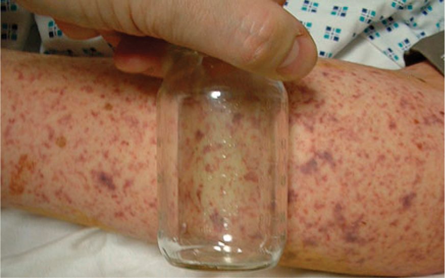
Meningococcal rash.
Fungal and yeast infections in children
Dermatophytoses
The dermatophytoses are filamentous fungi that infect the outer layer of the skin, hair and nails. Tinea capitis (scalp ringworm) is largely due to infection with Trichophyton tonsurans or Microsporum canis. Circular areas of alopecia with diffuse fine scaling are noted, often in association with a boggy inflammatory mass—a kerion.
In Tinea corporis, there are annular lesions with a central clearing and a palpable erythematous border. The characteristic ring shape gives this condition its common name of body ringworm.
Tinea pedis or athlete's foot is an itchy scaling and cracking of the skin of the feet. It primarily affects the web spaces between the toes.
Tinea unguium (onychomycosis) typically affects the toenails giving a white thickened appearance. Nails can also become brittle.
Candidiasis
Maceration of the skin or mucous membranes in warm moist parts of the body can lead to pathogenic Candida albicans. Oral candidiasis (white plaques on an erythematous base) and ‘nappy rash’ (large erythematous regions and satellite red plaques) are common manifestations in infants (Fig. 6). Intertriginous candidiasis involving the inframammary, axillary, neck and inguinal regions may be seen in obese infants, children and adolescents. Other presentations include paronychia, commonly seen as a result of thumb sucking and vulvovaginal candidiasis.
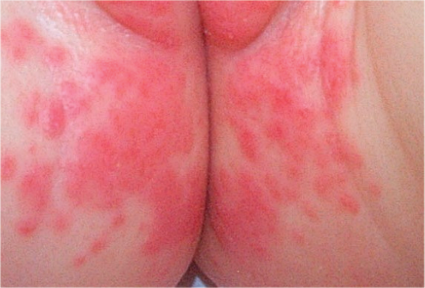
Fungal nappy rash (candidiasis).
Treatment
Topical anti-fungal therapy is usually sufficient with the exception of childhood scalp infection, which should always be treated with oral therapy. Topical treatments include nystatin, clotrimazole and miconazole. Patients should be educated on the importance of hygiene in preventing recurrence and transmission. Persistent or recurrent mucocutaneous infection should raise suspicion of immunodeficiency.
Viral skin infections in children
Molluscum contagiosum
Molluscum contagiosum is caused by mollusci poxvirus. It presents with smooth pearly papules with a central punctum, most commonly on the trunk. The lesions usually resolve spontaneously within 6 months to 2 years, although cryotherapy may be considered.
Warts
Warts are commonly seen skin growths caused by direct contact or autoinoculation with human papillomavirus. They are subclassified by region and morphology into common, plantar (verruca), mosaic, plane, filiform, oral and genital types. On exposed skin warts generally present with a hard surface and central black dots due to capillary thromboses.
Without treatment, 90% of warts in children resolve spontaneously within 2 years and often treatment is not pursued as it is inconvenient and sometimes more uncomfortable than the warts themselves. Treatment strategies include skin soaks followed by exfoliation, continuous occlusion (e.g. duct tape, verruca plasters), topical salicylic acid or podophyllin, imiquimod, cryotherapy, curettage and cautery. Unfortunately, warts often recur despite successful treatment.
Viral exanthems
A viral exanthem is any cutaneous eruption associated with acute viral infection, such as in rubella (Fig. 7). The presentation of the commonly encountered viral exanthems is summarized in Table 2.
Viral exanthems


Rubella PR. PH.
Infestations
Scabies
Scabies is caused by the mite Sarcoptes scabiei and transmitted by prolonged skin-to-skin contact. The first symptom is pruritus, worse at night, caused by a hypersensitivity reaction to the mite or its faeces. Burrows on the anterior aspects of the wrists or interdigital webs are pathognomic. Itchy papules or blisters can develop and become secondarily infected. Permethrin cream 5% spread all over the whole body (including the scalp, neck, face and ears) should be prescribed as treatment for those affected and close contacts. Pruritus can be managed with antihistamines and topical steroids.
The mucocutaneous syndromes
Erythema multiforme
Erythema multiforme may be classified as either
minor with classic target lesions or raised acral oedematous papules or
major with typical targets or papules and involvement of one or more mucous membranes; epidermal detachment involves less than 10% of total body surface area (TBSA).
Stevens—Johnson syndrome
In Stevens—Johnson syndrome (SJS), widespread blisters occur on the trunk and face, presenting with erythematous or pruritic macules and one or more mucous membrane erosions.
SJS has now been classified as a variant of toxic epidermal necrolysis (TEN); epidermal detachment is less than 10% TBSA for SJS and 30% or more for TEN. Approximately 50% of cases are idiopathic while other factors commonly implicated are drug reactions, bacterial infection and viruses, principally herpes simplex virus. Patients should be referred urgently for paediatric review and supportive treatment.
Kawasaki disease
Kawasaki disease is a systemic vasculitis, which is the most common cause of acquired heart disease in children. Patients are miserable with a fever unresponsive to antipyretics. Dermatological manifestations include a polymorphous rash, erythema of the palms and soles of the feet, red, dry or cracked lips and ‘strawberry’ tongue. Immediate paediatric referral should be organized for intravenous immunoglobulin therapy.
Key points
Birthmarks and vascular malformations are common and most resolve spontaneously without requiring any treatment; haemangiomas with worrying features and port-wine stains involving the first branch of the trigeminal nerve should be referred for further assessment.
Rarely, benign skin tumours in children carry potential complications; secondary lesions within sebaceous naevi should be referred in view of the risk of malignancy.
Bacterial and fungal infections are very common and treatable.
Viral exanthems may appear difficult to distinguish; therefore, diagnosis should be based on the history and accompanying clinical features.
Mucocutaneous syndromes require urgent paediatric referral and supportive management.