
Abstract
Scientists tend to focus on the present and the future. But the practice of experimental stroke is not new. Here, we reflect on the changing landscape of the stroke laboratory over the past 2000-years, focusing on shifts in the rationale for undertaking experiments, the methodologies deployed and the colourful characters involved in this science.
Experimental rationale
Historically, the study of blood flow in humans is a study of executed criminals, body snatching, judicial postmortems and fallen soldiers. Cultural and religious moratoriums on human experimentation have been a major driver of animal experimentation (1); however, it is a mistake to view animal experiments merely as a substitute for human studies. Animal experimentation has been undertaken when interest in human anatomy and physiology has peaked.
Early animal experiments invoking stroke were typically not stroke models by design, but induced a reduction of cerebral blood flow incidental to the investigation of other factors, e.g. testing the Galenic concept of blood flow, investigating the substances in arteries and veins and unravelling the factors regulating blood flow. The Alexandrian anatomists Herophlus and Erasistratus (C4th BC), the Roman physician Galen (129–200) and Renaissance scientists Leonardo da Vinci (1452–1519) and William Harvey (1578–1657) all ligated or restricted vessels (2). Cerebral ischaemia was almost certainly produced by Realdo Colombo in 1559 AD, when studying the ‘insensibility’ resulting from pressing the carotid arteries (3).
Early vascular studies were a discourse on anatomy (4). This narrative approach yielded to an experimental approach based on inductive reasoning. ‘Medicine turned towards the natural sciences and experimental research … in an epoch dominated by … Galileo, who looked for an exact mathematical law governing every phenomenon’ (5). William Harvey's work De Motu Cordis shows the stamp of this approach when moving from specific instances of blood flow to the general principle of circulation (6).
Art was also a driving factor. Artists bought their pigments at the apothecary shops, giving them the opportunity to cavort with physicians and to assist in private dissections (1). In an era that did not distinguish between art and science, da Vinci gave the first report of death from arteriosclerosis (7) and correctly modelled features of the animal vasculature (2), models that were not duplicated until the 20th century.
Pioneering work by Johann Jakob Wepfer (1620–1695) and Giovanni Battista Morgagni (1682–1771) demonstrated that stroke was caused by the rupture or blockage of a blood vessel to the brain (8, 9). With this knowledge came the impetus for 18th-century surgeons like John Hunter (1723–1793) to pursue the development of surgical techniques to treat aneurysms and wounded blood vessels.
Stroke experiments are now typically undertaken to understand the disease, to validate imaging modalities and to develop therapeutics. Experimental conditions are more controlled and reproducible, greatly increasing the reliability of the results. However, with the increased standardisation, the impetus to challenge the underlying utility and assumptions of the models has arguably diminished. Published data tend to verify, not falsify theories of neuroprotection and stroke pathophysiology.
Methodology
The methods of inducing occlusion favoured by Galen and Harvey were compression and ligation (6). By the 17th century, the experimental repertoire commonly included injection of different materials in vasculature: Marcello Malpighi (1628–1694) and Francis Glisson (1597–1677) favoured ink and Thomas Willis discovered the eponymous Circle of Willis by injecting the brain with the dye aqua crocata (1). Austrian anatomist Josef Hyrtle (1810–1894) injected numerous coloured fluids into the vasculature (1), while Jan Swammerdam (1637–1680) advanced the use of the anatomic injection of warm wax. Regnier de Graaf (1641–1673) injected mercury into spermatic vessels, but becoming convinced of the impiety of anatomy he abandoned science altogether and joined a fanatical religious sect (1).
Contemporary models induce stroke with the intravascular injection of blood clots, microspheres, macrospheres and via the intraarterial insertion of a thread, filament or suture. Vessels have also been occluded mechanically with clips or inflatable balloon cuffs, thermally via electrocoagulation and chemically with the vasoconstrictive agent endothelin-1 and the light-activated dye Rose Bengal.
Species
Historically, physiologists took the entire natural world, from polyp to man as their subject (10) (Fig. 1). Aristotle studied a menagerie of creatures, although his descriptions are said to become somewhat more dubious the further he strayed from the Aegean (1). Galen's anatomical work was frequently undertaken in the Barbary ape (macaque) but his in vivo work was generally performed in other species, notably oxen (11).
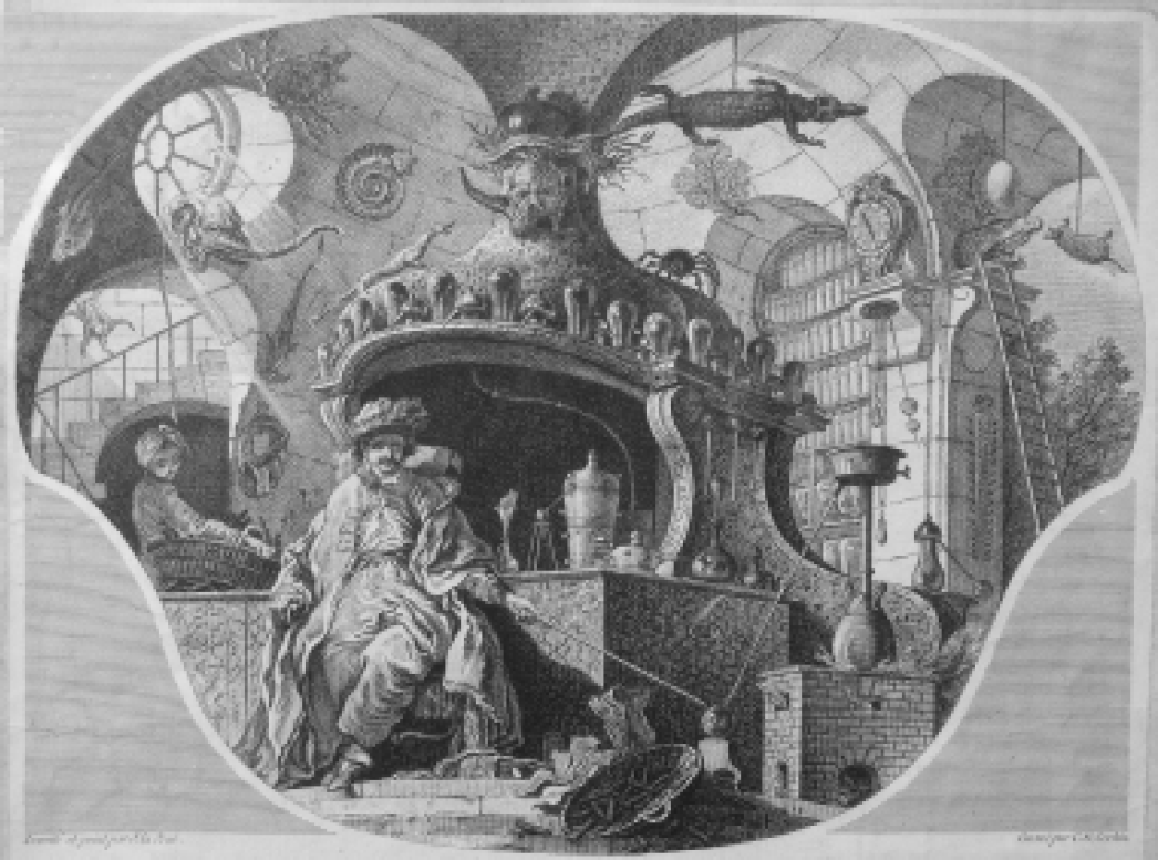
The laboratory of the Duke of Picquigny by J. La Joue. Engraving by Charles Nicolas Cochin (1715–1790).
Medieval anatomy at the School of Salerno was primarily based on the swine (1), while during the Renaissance, William Harvey tackled any number of species including dogs, hogs, sheep, ox, fish, serpents, mussels, oysters, sponges, snails, whelks, partridge, frogs, tortoises, bees, slugs, shrimps, crabs, crayfish, wasps, hornets and flies (6). The founder of experimental surgery, John Hunter (1728–1793), studied blood flow in the antlers of deer from Richmond Park when establishing his theory of collateral circulation. His student, Sir Astley Paston Cooper (1768–1841), ligated the common carotid artery of the dog, in order to further develop the aneurysm surgery developed by his teacher (1).
Most contemporary stroke experiments are performed on rats and mice, although rabbits, gerbils, cats, dogs and a variety of primates are also studied. Many modern strains of rat can trace their lineage to the Wistar institute in Philadelphia, with the first outbred commercial colony established early in the 20th century.
Future
Anatomy and physiology, once the primary drivers of experimentation, are ceding ground to proteomics, genomics and bioinformatics. Just as the invention of the compound microscope around 1600 resulted in a quantum leap in our understanding of the cerebrovasculature, now genetics and proteomics systems will increase the information content per experiment allowing us to understand quickly the broad physiological context of even the smallest perturbations of the body. Increasingly sophisticated computational models and multicell culture systems will help reduce in vivo experimentation.