
Abstract
Wound management in hidradenitis suppurativa (HS) patients is often challenging due to worsening infections and impaired circulation, which can lead to prolonged hospitalization and poorer quality of life. Traditionally, autografting—a leading option in skin grafting—exhibits risks, including donor site morbidity. However, xenograft products, such as KerecisTM, derived from North Atlantic Cod fish, offer a novel solution for conditions with chronic wounds by promoting natural tissue regeneration. This case report presents a 35-year-old woman with HS who presented with persistent abscesses and fistulas in bilateral axillary regions. Following multiple surgical incisions and drainage procedures, the patient was treated with intact fish skin grafting, which resulted in significant granulation tissue deposition and wound edge approximation. The successful outcome in this case suggests that intact fish skin grafting is a viable option for non-healing wounds, particularly HS wounds, and can be integrated into surgical and wound care practices.
Keywords
Key Takeaways
• Managing conditions with chronic wounds, particularly in hidradenitis suppurativa (HS), is challenging due to infection and impaired healing, often requiring surgical intervention. • An intact fish skin graft offers a promising alternative to traditional grafting methods, promoting wound healing through its anti-inflammatory properties and facilitating tissue regeneration via granulation. • This case demonstrates the successful use of intact fish skin grafting in a patient with non-healing HS wounds, offering a less invasive alternative and preventing complications associated with autografting or allografting.
Introduction
The field of wound care is rapidly evolving, with an increasing need for innovative treatment approaches. Conditions requiring chronic wound care can often be complicated by reduced circulation and persistent infection, leading to rehospitalization and diminished quality of life. 1 One such condition is hidradenitis suppurativa, a recurrent inflammatory skin disease presenting with painful abscesses and sinus tracts, affecting intertriginous areas most frequently. 2 The burden of chronic wounds on the health care system is substantial, impacting both the physical and mental well-being of patients. 3 Split-thickness autografting has long been the gold standard for chronic wound treatment; however, it poses the risk of an additional wound at the donor site, which may lead to complications such as pain, delayed healing, and scarring. 4
Xenografting, using biological materials obtained from other species, is an alternative approach. Intact fish skin xenografts (IFSGs) such as Kerecis TM, derived from the North Atlantic Cod fish in Ísafjörður, Iceland, are a decellularized skin matrix homologous to polyunsaturated fatty acids (PUFAs) and non-cellular proteins found in humans. These grafts are minimally manipulated to preserve a porous structure, allowing dermal and capillary ingrowth while maintaining fatty acids that reduce inflammation by modulating cytokine signaling during the healing process. 4 These grafts are used to treat various complex wounds, including traumatic, diabetic, and necrotic wounds. 5 Polyunsaturated fatty acids in these grafts exhibit anti-inflammatory properties and reduce bacterial colonization at the wound site. 4 Numerous studies have demonstrated a significant success in wound healing using IFSGs. 6 This study presents a case report of a 35-year-old woman with non-healing hidradenitis suppurativa (HS) who presented with persistent abscesses and fistulas in bilateral axillary regions.
Case Presentation
A 35-year-old South Asian woman with a history of Hurley stage III HS diagnosed with postpartum diabetes, gestational diabetes, and postpartum septicemia presented to the outpatient clinic with chronic, non-healing abscesses and fistulas in the axillae, groin, and lower back. Upon examination, the bilateral axillae showed tender erythematous dome-shaped nodules and indurated pits with purulent, foul-smelling discharge and hyperpigmented tissue. The patient was initially treated with doxycycline, clindamycin, and spironolactone, in addition to adalimumab (Humira®) injections in the outpatient setting. However, the abscesses worsened and progressed to recurrent axillary and vulvar wounds extending to the inguinal and buttock regions. Upon consultation with the dermatology department, additional therapy with systemic secukinumab (Cosentyx®) was initiated. Recurrent flare-ups significantly affected the patient’s quality of life, leading to severe depression and anxiety.
Six months after diagnosis, the patient presented to a community-level emergency department, reporting progressively worsening right axillary pain and inflammation over a 2-week period despite compliance with the treatment and wound care. Laboratory results indicated leukocytosis (12.1 × 10⁹ cells/mL) and elevated C-reactive protein (CRP; 8.4 mg/L). Cultures identified 2+ Streptococcus viridians group, 1+ diphtheroid-like organisms, and 1+ coagulase-negative Staphylococcus. Right axillary ultrasound (US) displayed a large, loculated abscess (7.6 × 2.6 cm). The patient was administered intravenous clindamycin and opted for surgical incision and drainage based on the US findings and a tender and fluctuant axillary abscess. The right axillary abscess was infiltrated with lidocaine before an incision was made, yielding 10 mL of purulent material. The incision was then extended to form a cruciate pattern over the abscess cavity. All loculations were disrupted, and the cavity was thoroughly irrigated with saline until clear effluent returned. Hemostasis was achieved, and the cavity was packed with iodoform gauze, followed by dressing with a sterile 4 × 4 cm gauze and tape. The patient tolerated the procedure well with no complications. She was discharged on the following day with a prescription of oral clindamycin at a dosage of 300 mg every 6 hours.
Thirty days after the initial incision and drainage procedure, the patient returned to the emergency department with severe pain in both axillae. On examination, the bilateral axillae showed purulent, foul-smelling discharge, as well as diffuse tenderness and erythema (Figure 1). Laboratory findings revealed leukocytosis (14.3 × 10⁹ cells/mL) and a significantly elevated CRP (14.4 mg/L). Computed tomography of bilateral axilla showed bilateral axillary abscesses with suspected gas and fistula formation throughout the bilateral axilla. After admission, intravenous vancomycin, cefepime, and metronidazole were administered. The patient again opted for surgical incision and drainage, as well as excision of the fistula tracts in the bilateral axilla. Left and Right Axilla Surgical Wound Prior to Grafting in a Patient With Hidradenitis Suppurativa (HS)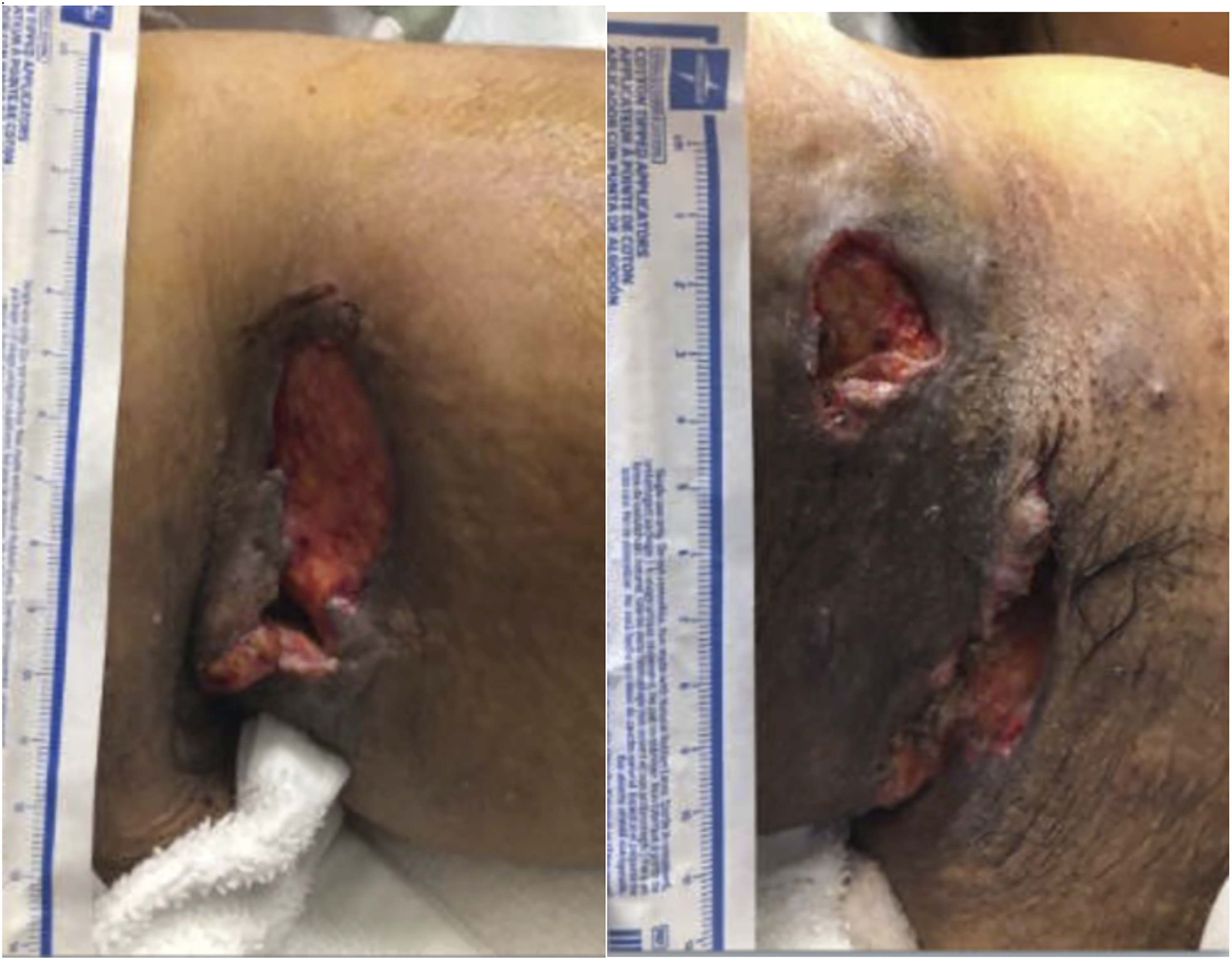
Two days after the admission, the patient was brought to the operating room under monitored anesthesia. Starting with the left axilla, a large fistula tract was opened using blunt electrocautery and completely drained, releasing copious amounts of purulent fluid. A similar fistula tract opening and drainage procedure was then completed in the right axilla. Following tract drainage, silver-impregnated dressings were inserted into both axillae, covered with betadine-soaked gauze abdominal pads, and secured with tape. Patient opted to avoid negative pressure wound therapy (NPWT) as anatomically it would have caused discomfort. The patient tolerated the procedure well and was subsequently admitted for intravenous antibiotic treatment and daily wound care.
Ten days postoperatively, the patient’s wound infection resolved, with significant reductions in erythema, purulent drainage, and pain upon palpation of the region. She opted to undergo IFSG because of the persistent, non-healing open wound and inadequate response to local wound care, aiming to promote wound closure by secondary intention. The patient was considered an appropriate candidate, as the wound site had visible healthy granulation tissue deposition, indicating potential for accelerated healing with IFSG.
The patient was returned to the operating room for wound graft placement under monitored anesthesia. Intraoperatively, wide excision around the demarcated area to the level of fascia was performed to ensure complete removal of diseased tissue with wound edges sharply debrided with to facilitate graft adherence. The fresh wound beds were cleaned with copious amounts of betadine solution and abraded with a scratch pad to promote back bleeding. A 6 × 8 cm intact fish skin biological graft was hydrated in saline for 1 min, trimmed to an appropriate shape, and secured with 3-0 absorbable suture and staples in the right axilla. The same procedure was repeated on the left axilla. The bilateral wounds displayed good graft overlap and healthy granulation tissue below the graft. An abdominal dressing was applied over iodoform gauze secured with foam tape without use of NPWT due to anticipated anatomical discomfort. The patient tolerated the procedure well and recovered from anesthesia without any issues. Intravenous antibiotics were administered.
On postoperative day 4, the dressing was removed to evaluate wound healing, which showed a significant improvement. Bilateral axillary wounds showed reduced inflammation surrounding the wound edges, absence of pus or drainage, a translucent covering from graft material breakdown covering most of the wound surface, healthy granulation tissue throughout the wound bed, adequate moisture, and progressive wound edge contraction—indicative of overall wound size reduction. On postoperative day 5, the patient was switched to oral amoxicillin-clavulanate (500 mg twice daily for 7 days) and doxycycline (100 mg twice daily for 7 days). Ten days after grafting, the patient was discharged with a prescription of oral antibiotics and instructions for home care, including ongoing wound dressing changes. On postoperative day 20, the wound sites exhibited well-approximated edges, no active infection, and progressively increased granulation tissue deposition (Figure 2). Left and Right Axilla 20 days Post-grafting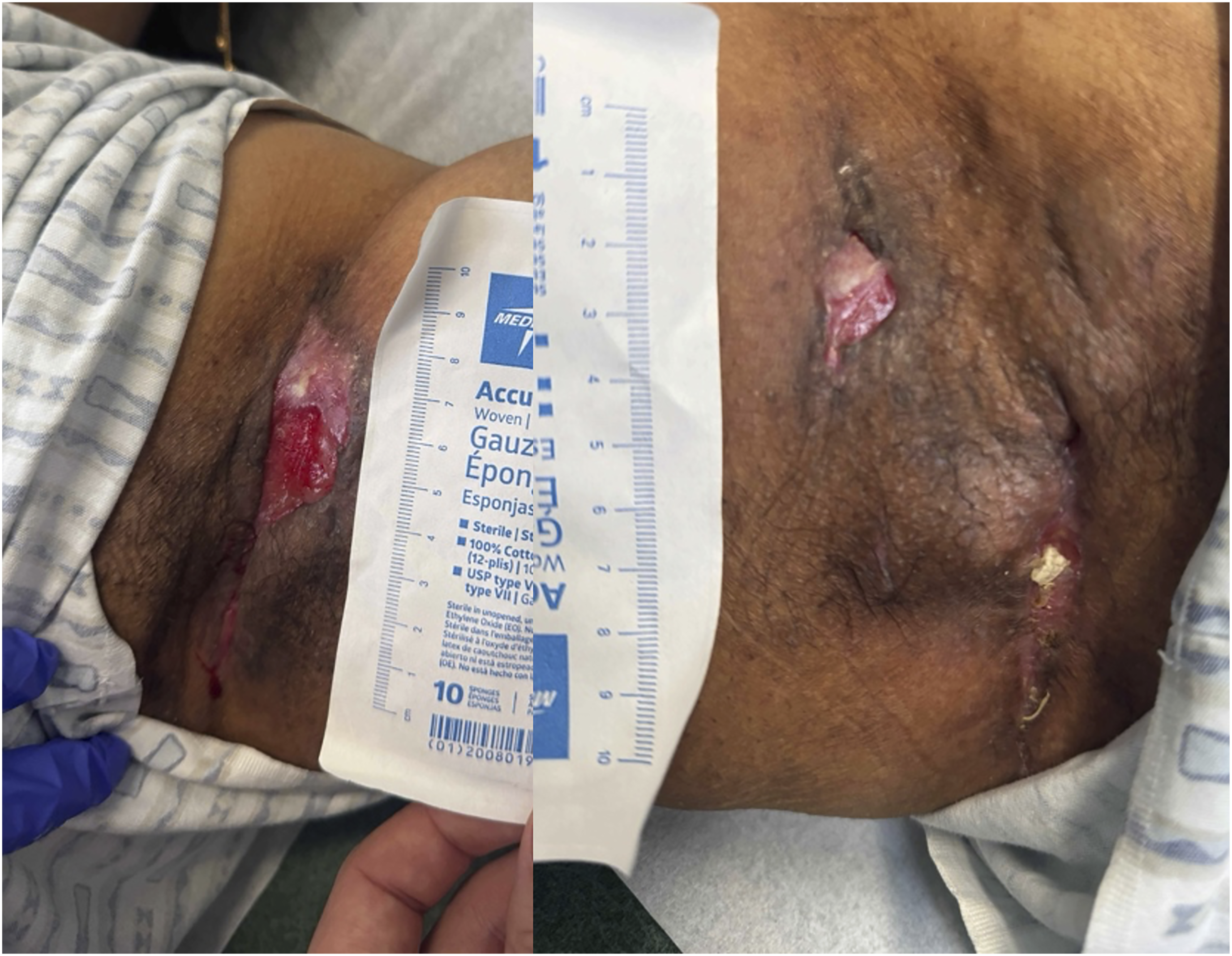
Discussion
Open abscesses and fistulas in HS can often be managed nonoperatively; however, surgical intervention may be required in more severe cases. One traditional approach is autografting, where a patient’s own tissue is transplanted from a healthy site to the wound site to aid healing. This requires a wide local surgical excision of diseased tissue and wound bed optimization proportionate to the extent of HS disease. Although autografts can completely close the wound, they often pose a risk for additional wound sites and infections. 6
An alternative is cadaveric allografting, which avoids the creation of an additional donor site wound. Furthermore, it ensures the viability of the wound bed for autografting and promotes wound contracture and coverage. However, allografts exhibit their own challenges, including limited availability and the need for specialized culturing facilities and skin banks for appropriate preservation and storage.3,7
More recently, biological xenografts, such as IFSGs, have emerged as a promising option. These grafts overcome many of the limitations of autografts and allografts by eliminating the need for tissue harvesting, thereby reducing the risk of additional wound sites and infection. Rather than mechanically closing the wound, IFSGs support autologous healing by secondary intention. Furthermore, the presence of PUFAs and other proteins within the graft decreases the risk of infection during the healing process. 5
Although IFSGs facilitate healing with decreased pain by modulating inflammatory processes, they have certain limitations, including accessibility, as they are not as widely available as autografts. 6 Additionally, the financial burden of such grafting can vary depending on its accessibility. Consequently, it may be more costly than allografting. 7 Careful evaluation of patient demographics, risk factors, and the size and location of the wound is critical for determining the candidacy for IFSGs.
Intact fish skin xenografts integrate directly into the patient’s natural granulation tissue, promoting cell proliferation and stimulating the physiological healing process. In a study investigating periocular anterior lamellar skin defects following Mohs surgery, IFSGs facilitated remarkable tissue regrowth in 6 patients with minimal postoperative complications. 8 Additionally, a study comparing IFSGs to bovine collagen grafts in patients with acute burns demonstrated that IFSGs exhibited a faster healing time. 9 Therefore, the use of IFSGs in uniquely challenging wounds, including HS wounds, could represent a significant advancement in wound management.
Conclusion
This case highlights the unique benefits of IFSGs for treating non-healing abscesses and wounds while minimizing the risks associated with other grafting methods. The successful use of IFSGs in this patient suggests their potential utility in future studies for managing similar patients. Integrating this grafting approach into general surgical practices may significantly improve wound care deficits in our health care system.
Footnotes
Ethical Considerations
This case report does not involve any experimental procedures or clinical trials, and as such, our institution does not require ethical approval for reporting individual cases or case series. The patient’s identity has been protected, and all information presented in this report is anonymized.
Consent for Publication
Written informed consent was obtained from the patient for publication and any accompanying images. A copy of the written consent is available for review by the Editor-in-Chief of this journal on request.
Author Contributions
(1) Roma Tarar—corresponding author. (2) Dr Christopher Engler—oversight and administration.
Funding
The authors received no financial support for the research, authorship, and/or publication of this article.
Declaration of Conflicting Interests
The authors declared no potential conflicts of interest with respect to the research, authorship, and/or publication of this article.
Permission to Reproduce Material from Other Sources
The test reproduced in this manuscript is in the public domain and does not require formal permission.