






Abstract
Objective:
Thyroglossal duct cysts (TGDCs) are relatively common congenital midline neck masses that are treated with surgical excision. Traditionally these are removed along with any associated tract and the central portion of the hyoid bone through an anterior neck incision. Some patients with TGDCs want to avoid an external neck scar.
Methods:
We describe the details of a transoral endoscopic vestibular excision of a TGDC and the associated hyoid bone in an adolescent patient.
Results:
This novel approach was successful and there were no complications.
Conclusion:
We propose that cervical TGDCs can be safely and completely removed with this approach in appropriately selected patients while avoiding a neck scar.
Introduction
Thyroglossal duct cysts (TGDCs) are congenital neck masses arising from the remnant of embryologic descent of the thyroid from the foramen cecum down to the final resting position in the anterior neck. 1 TGDCs are the most common congenital neck mass and typically first become apparent in childhood, but may present in adulthood. Surgical excision is recommended because of a high risk of infection and a small risk of developing a malignancy within the cyst. 2 TGDCs are traditionally removed through an anterior neck incision overlying the mass. A Sistrunk procedure is typically performed to remove the cyst, the central portion of the hyoid bone and any associated tract. 1 This technique reduces the risk of recurrence seen with simple cyst excision. 1 The resulting anterior neck scar may have an undesirable appearance.3,4 Regardless of its appearance, the scar may also negatively affect quality of life as has been seen in the thyroidectomy population.5,6 A transoral endoscopic vestibular technique is an alternative approach to a modified Sistrunk procedure and avoids an external neck scar.
Case Report
A 16-year-old healthy male presented after new onset of a tender neck mass in the setting of an upper respiratory illness. A prior ultrasound done at another institution’s emergency department suggested an infected TGDC, but did not evaluate the entire neck or thyroid. The mass decreased in size after a course of antibiotics but remained palpable and tender. On our physical exam a 1 cm palpable mass was present overlying the superior aspect of the left thyroid cartilage ala and left side of the hyoid. The overlying skin was normal. Magnetic resonance imaging (MRI) showed a 1.1×1.1×0.6 cm T2 intense irregular mass in the same area that was consistent with a TGDC (Figure 1). The thyroid gland was normal on physical exam and on MRI.
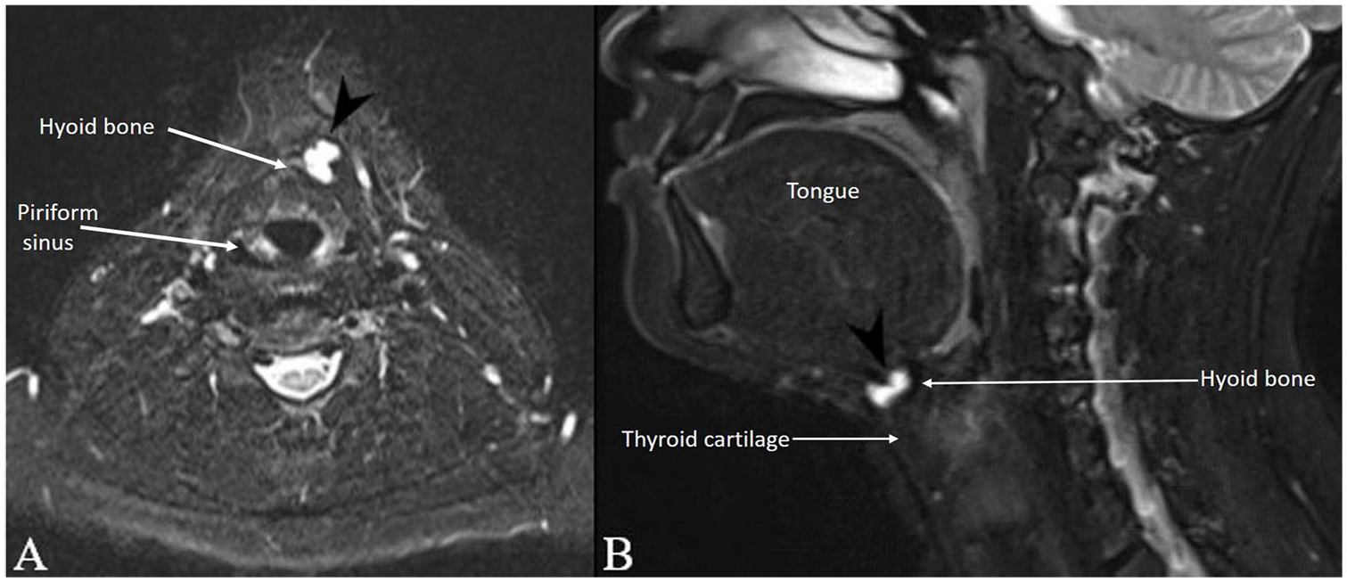
T2-weighted magnetic resonance images (MRI) with gadolinium contrast of the thyroglossal duct cyst (arrowheads) (A) axial and (B) sagittal.
Excision of the cyst as well as the central portion of the hyoid bone and any associated tract was recommended. The MRI was completed rather than a repeat ultrasound in order to also assess appropriateness for the transoral approach. The option of a transcervical versus transoral approach was discussed including all risks of both approaches. He and his family were interested in avoiding an external neck scar and elected for a transoral approach. Informed consent for publication of clinical information and images was provided by the patient and his legal guardian.
At the time of surgery, general anesthesia was induced and then he was orally intubated. He was positioned on a shoulder roll. He received intraoperative ampicillin-sulbactam. The proposed midline gingivobuccal and oral commissure incisions (Figure 2) were infiltrated with 1% lidocaine with 1:100 000 epinephrine and the soft tissue overlying the mentum was also infiltrated through the oral vestibule. The mucosal incisions were made with a 15 blade. While pulling up gently in a cephalad fashion a curved Crile and then a Kelly hemostat were used to open the subplatysmal submental space through the central incision. The tract was dilated with sequential Hegar dilators and then the central port with a 10 mm 0° endoscope was placed. Later during the dissection 30° and 45° scopes were used to improve visualization. The 5 mm ports were placed parallel to the skin to join the submental pocket. Silk sutures were placed through the skin in the vertical midline overlying the thyroid cartilage for upward retraction to open the subplatysmal pocket.
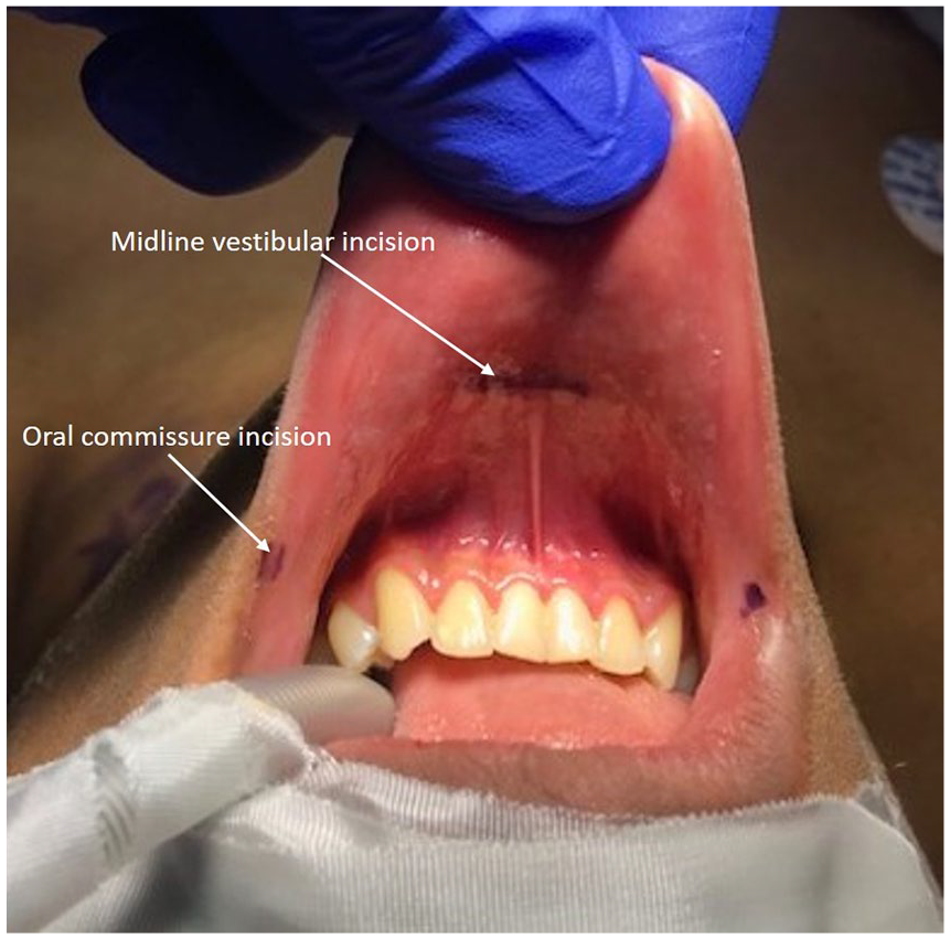
Proposed incisions at oral commissures and vestibule for transoral approach to Sistrunk procedure.
The subplatysmal pocket was insufflated with carbon dioxide (CO2) to 5 to 7 mmHg. A Maryland dissector and Harmonic shears (Ethicon US, LLC, Cincinnati, OH, USA) were used to identify and define the thyroid notch. The thyrohyoid membrane and hyoid were identified and defined. The cyst was identified and dissected from the surrounding sternohyoid muscle while keeping it attached to the hyoid (Figure 3A). The muscle attachments to the central portion of the hyoid were divided and then lateral cuts were made with the Sonopet ultrasonic aspirator (Stryker, Kalamazoo, MI) to release the central third of the hyoid (Figure 3B). Soft tissue inferior and superior to the central portion of the hyoid was dissected but remained attached to the central hyoid. The soft tissue and suspected embryologic tract deep to the hyoid extending toward the tongue base was ligated with the ultrasonic shears. The entire specimen including the cyst, soft tissue and central portion of the hyoid was removed in an endocatch bag (Medtronic, Minneapolis, MN, USA) through the central incision. The wound was irrigated and the oral vestibule incisions closed with single layers of running 4.0 chromic suture. A pressure dressing with jaw support was placed. There were no intraoperative complications.
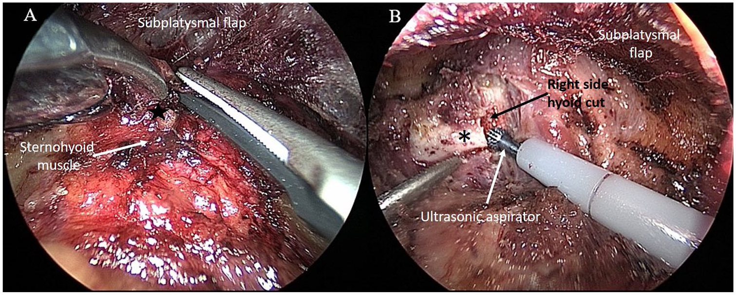
Intraoperative endoscopic image of (A) dissecting cyst (star) from the sternohyoid muscle while keeping it attached to the hyoid and (B) dividing central portion of hyoid bone (asterix) with ultrasonic aspirator.
Postoperatively he was monitored as an inpatient for 24 hours. Then the pressure dressing was removed and he was discharged home with 5 days of prophylactic amoxicillin-clavulanate, 1 week of soft diet, saline mouth rinses after all meals and at bedtime and instructions for neck range of motion exercises to prevent neck stiffness. Pathology confirmed a TGDC. He had no postoperative complications. At follow-up 2 weeks after surgery he had mild mental and submental fullness and tenderness. He was tolerating a regular diet; he had normal tongue movement, intact chin sensation, and full neck range of motion. His intraoral incisions were healed. He was seen again 3 months after surgery and the submental fullness and tenderness had resolved. There was no evidence of recurrent mass and future follow-up will be as needed.
Discussion
A transoral endoscopic vestibular approach to a modified Sistrunk procedure for a TGDC can be considered when an external scar is a deterrent to undergoing surgery. This can be considered in patients with a personal history or increased risk of hypertrophic scarring or keloid formation. The transoral approach to a TGDC excision through a different approach with a frenotomy incision has been previously reported.7-9 A transoral approach with a single vestibular incision without insufflation has also been reported. 10 This is the first published report of a novel three-port transoral vestibular Sistrunk operation for a TGDC. Our technique is adapted from that of a transoral endoscopic thyroidectomy vestibular approach (TOETVA), which has been well described and also used for parathyroidectomy and central neck dissections.11-13
The indications and contraindications for a transoral endoscopic vestibular approach to a TGDC are similar to those described for TOETVA. The indication is a history of hypertrophic scarring or strong motivation to avoid a neck scar. 14 Contraindications include a history of neck surgery, neck irradiation, oral abscesses, other oral pathology, or morbid obesity.14,15 Surgeons should be mindful that the cervicofacial angle of candidate patients may make the case more or less challenging.
A preoperative computed tomography (CT) scan or MRI is recommended to evaluate the TGDC and thyroid gland as well as the surrounding anatomy for appropriateness of a transoral approach, including the depth of tongue base involvement. A CT or MRI can be done instead of the more typical ultrasound, which may not provide adequate anatomic detail for this approach.
The additional risks with the transoral approach and potential need to convert to a transcervical approach should be thoroughly discussed preoperatively. The patient and family should be counseled that temporary or permanent mental nerve injury can occur and result in chin or lower lip numbness. The risk of TGDC recurrence with both approaches and the unknown difference in risk between the two approaches should also be discussed before surgery.
As with all laparoscopic surgery there is a small but accepted risk of a CO2 embolus from insufflation. It has a reported occurrence of 0.001% in laparoscopic surgeries. 16 There has been one published case report of CO2 embolus during transoral endoscopic vestibular thyroid surgery resulting from anterior jugular vein laceration. 17 Two-way open communication between the anesthesia and surgical teams is especially important should there be unexpected hemodynamic swings, arrhythmias, or rapid changes in end tidal CO2 that may indicate a CO2 embolus. If suspected, several key steps should follow including the cessation of insufflation, application of manual pressure to the neck, positioning the patient in Trendelenburg to trap the embolus in the apex of the right ventricle, discontinuing anesthetic agents, and hyperventilation with 100% oxygen. Due its rarity and broad range in presentation, open communication regarding risk of CO2 embolism is key to timely management.
Intraoperative ampicillin-sulbactam and a 5-day postoperative course of amoxicillin-clavulanate (or clindamycin with metronidazole if penicillin allergic) are used because of contamination of the submental space with mixed oral microbes. There may be an increased risk of infection with the clean-contaminated transoral approach compared to the clean wound of the transcervical approach. The patient may be exposed to additional side effects from this brief postoperative antibiotic course. The need for postoperative antibiotics remains unclear, however.
The 24 hours of pressure dressing minimizes the risk of hematoma and seroma. Additionally, a small drain could be placed through the cervical skin flap or transorally 10 to prevent any fluid accumulation. A drain was not required in this case since hemostasis was easily achieved and he had no risk factors for bleeding. The ultrasonic aspirator also maintained excellent hemostasis at the cut ends of the hyoid bone.
This carefully selected patient did not experience any complications and had excellent anatomy for a transoral approach. He had a normal mandible and adequate lip laxity that allowed for ease of port placement. His relatively long neck and his hyoid position made soft tissue and bony dissection feasible and efficient. At first this transoral approach may take longer from incision to closure than a transcervical approach and that additional risk and cost should be considered. 18 However, as with all new techniques, efficiency will be gained with experience and this has already been shown for TOETVA.18,19
Conclusion
A TGDC can be safely removed with modified Sistrunk operation through a transoral endoscopic vestibular approach in the appropriate pediatric or adult patient who is motivated to avoid an external neck scar.
Footnotes
Declaration of Conflicting Interests
The author(s) declared no potential conflicts of interest with respect to the research, authorship, and/or publication of this article.
Funding
The author(s) received no financial support for the research, authorship, and/or publication of this article.
