
Abstract
In this work, a microwave assisted method was developed for synthesis of red fluorescent copper nanoclusters (NCs) using trypsin as a template (trypsin–Cu). The as-synthesized trypsin–Cu NCs are stable and water soluble, exhibiting fluorescence emission at 657 nm when excited at 490 nm. The as-prepared red-emitting trypsin–Cu NCs were characterized by using several analytical techniques such as ultraviolet–visible (UV–Vis) and fluorescence, fluorescence lifetime, Fourier transform infrared, and X-ray photoelectron spectroscopic techniques. Red-emitting trypsin–Cu NCs acted as a nanosensor for sensing both Pb2+ and Hg2+ ions through fluorescence quenching. Using this approach, good linearities are observed in the range of 0.1–25 and of 0.001–1 μM with the lower limit of detection of 14.63 and 56.81 nM for Pb2+ and Hg2+ ions, respectively. Trypsin–Cu NCs-based fluorescence assay was successfully applied to detect both Hg2+ and Pb2+ ions in water and tobacco samples.
Keywords
Introduction
Noble metal clusters, for example, Au, Cu, and Ag nanoclusters (NCs), are recognized as a new type of nanomaterial with a particle size of less than 2 nm, consisting of between a few to about 100 atoms.1,2 The metal NCs are a new class of fluorescent nanomaterials because of their optimistic fluorescence and molecular properties. 3 Metal NCs have many advantages, including ultrasmall size, high water solubility, low toxicity, and biocompatibility, just like other fluorescent nanomaterials, such as organic dyes, quantum dots, carbon dots, and lanthanides. Therefore, it has been found that fluorescent metal NCs are found to be excellent candidates for bio-labeling, biomedical targeting, and sensing applications.4–7 Several ligands such as enzymes, 8 thiols,9,10 proteins,11–13 and DNA 14 have been explored as ligands for fabrication of metal NCs with selective sensing applications. Therefore, a simple and green synthetic approach is developed for fabrication of highly water-soluble fluorescent Cu NCs using trypsin as a ligand.
Transition and heavy metals are important for the maintenance of different biological processes; however, they are highly bioaccumulative nature, causing a serious threat to the human health and environment. 15 For centuries, lead (Pb2+) has been used in various fields, including building, plumbing, the military, painting, and sculpture. 15 Although it is widely used in various sectors, Pb2+ ions are recognized as a toxic element. The central and peripheral nervous systems are compromised by excess levels of Pb2+ >10 μg/dL in human blood, causing behavioral disorders, learning difficulties, and hearing and speech deficiencies. 16 The European Food Safety Authority (EFSA) has given a warning that even a a low Pb2+ concentration (<2.1 μg/L) has an adverse effect on childhood intelligence. 17 Higher Pb2+ levels > 70 μg/dL are responsible for devastating health issues, including seizures, coma, and eventually death. 18 In modern times, Pb2+ exposure is primarily caused by fuel, paint, toys, burning of gas, air pollution, and batteries. 19 Pb2+ leaches from these sources and can enter bodies of water and become a human exposure source. 20 It is difficult to detect Pb2+ in environmental bodies, since Pb2+ is colorless and odorless.
Mercury (Hg2+) is one of the most toxic components in the environment. There is a heavy metal ion Hg2+, which is not biodegradable, present in air, soil, food, water, and other natural environments. By eating foods containing Hg2+ or its derivatives, these elements can enter living systems and accumulate in the body, posing serious health risks. An excessive accumulation of Hg2+ in the kidney, central nervous system, and digestive system can result in acrodynia, Minamata disease, and memory and movement problems.21–24 In view of this, CuO nanoparticles were successfully integrated with resonance Rayleigh scattering technique for assaying Hg2+ ions.
25
Several reports have been demonstrated on the fabrication of Cu NCs with different capping agents such as bovine serum albumin (BSA),
26
metallothionein (MT),
27
and
Various types of analytical techniques, such as inductively coupled plasma–mass spectrometry, 29 atomic absorption spectroscopy, 30 fluorescence, 31 and ultraviolet–visible (UV–Vis) 32 spectrophotometric tools have been examined to evaluate the amounts of Pb2+ and Hg2+ ions in biological and environmental samples. 33 Although these techniques can detect metal ions with a lower limit of detection (LOD), they exhibited some limitations such as expensive devices and complex protocols.34–36 An easy and fast analysis method is therefore highly desirable for the detection of trace level metal ions (Pb2+ and Hg2+) at once and simultaneously. Fluorescent metal NC-based sensors were particularly used as effective analytical protocols for the determination of trace metal ion levels because of their remarkable features such as rapid response, efficient selectivity, and high sensitivity.37,38 Among fluorescent metal NCs, Cu NCs have shown promising advances as compared with other metal NCs (Au, Mo, and Ag). Importantly, Cu is significantly inexpensive and extensively used in industries due to its high conductivity and similar characteristics to Au and Ag. Moreover, precursors used for the preparation of Cu NCs are comparatively abundant, cheap, and easily available from commercial sources as compared to other metal NCs precursors. It was also observed that Cu NCs displayed strong fluorescence properties, good water solubility, low toxicity, and contain multifunctional groups on its surface, allowing them to act as nanosensor for selective and sensitive detection of various heavy metal ions, biomarkers, and drugs. In this work, a low-cost and simple microwave synthetic approach was introduced for the preparation of red-emitting Cu NCs using trypsin as a surface ligand. When excited at 490 nm, trypsin–Cu NCs showed fluorescence emission at 657 nm. Trypsin–Cu NCs showed high ability for the selective and sensitive detection of Pb2+ and Hg2+ with fluorescence using a “turn-off” sensing mechanism. The addition of Pb2+ and Hg2+ ions separately to a trypsin–Cu NCs solution results in complete quenching of the emission peak of trypsin–Cu NCs CNPs, resulting in a drastic change in the fluorescence of trypsin–Cu NCs from red to non-fluorescence under UV light. This probe exhibited the LODs are 14.63 nM and 56.81 nM for Pb2+ and Hg2+ ions, respectively. Furthermore, 2,2′,2′′,2′′′-(ethane-1,2-diyldinitrilo)tetraacetic acid (EDTA) and ascorbic acid (AA) were used as masking agents for Hg2+ and Pb2+ ions, favoring the assay of both ions simultaneously. By taking advantage of the developed probe, the method was successfully demonstrated to assay both ions in tobacco and water samples.
Experimental
Materials and Methods
Enzyme Bioscience Pvt. Ltd., India, sent trypsin as a gift sample. Copper sulfate (CuSO4), NaOH, hydrazine hydrate, and metal salts were obtained from Sigma-Aldrich. A Milli-Q water purification system was used throughout the experiment. In the whole analysis, only analytical grade chemicals were used.
Instrumentation
An optical spectrophotometer (Ocean Insight, U.S.A.) was used to record the absorption spectrum of trypsin–Cu NCs. A fluorescence spectrophotometer from Cary Eclipse (Agilent Technologies, U.S.A.) was used to measure the excitation and emission spectrum of trypsin–Cu NCs. Functional groups of trypsin–Cu NCs were determined using a Fourier transform infrared (FT-IR) spectrometer (II, Bruker, Germany). The morphology of trypsin–Cu NCs was studied using high-resolution transmission electron microscopy (HR-TEM) (JEM 2100, JEOL, Japan).
Trypsin–Cu Nanocluster Synthesis
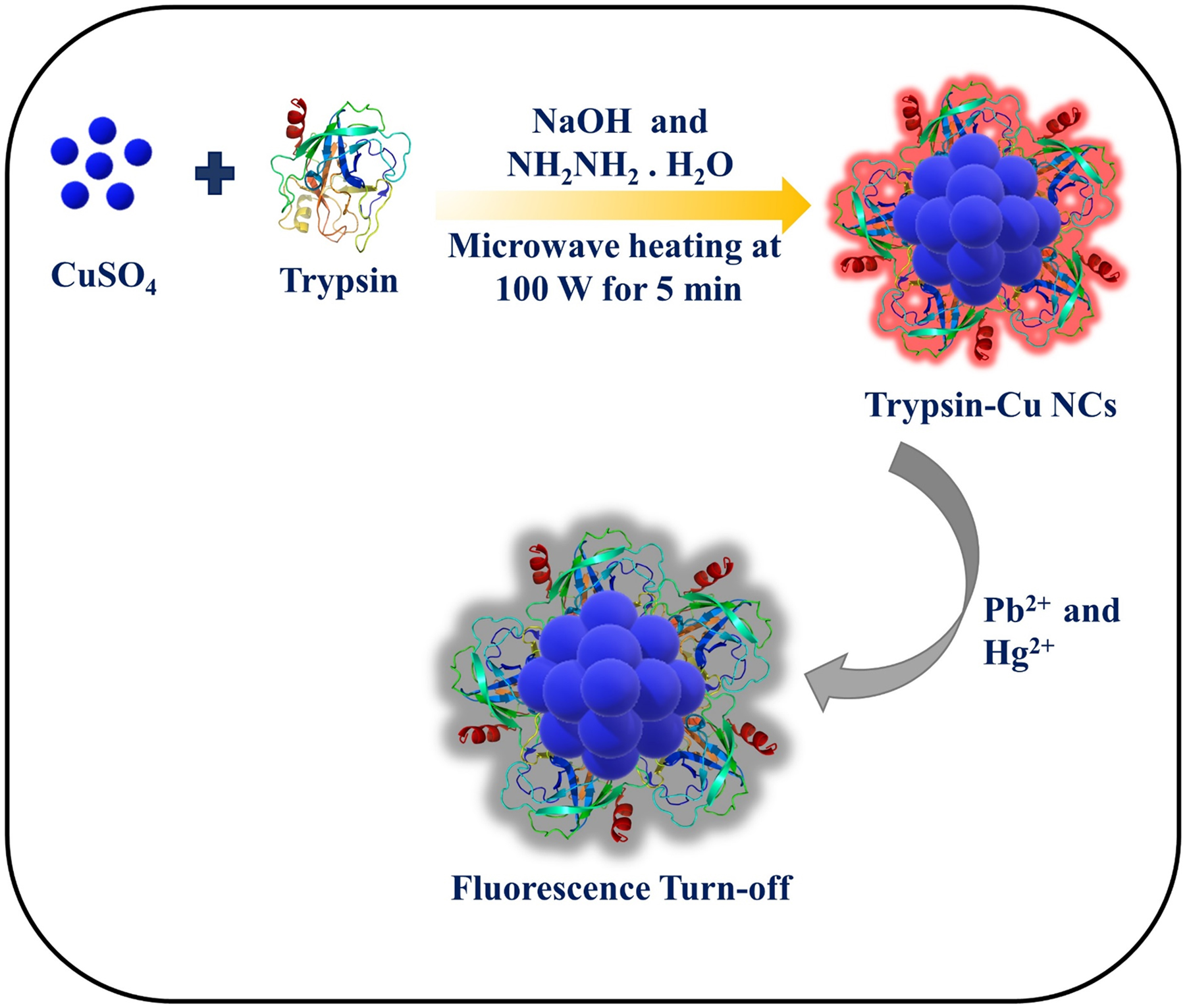
Fluorescent Cu NCs were prepared by using CuSO4, trypsin, and hydrazine hydrate as precursors. Briefly, CuSO4 (10 mM, 5 mL) and trypsin solution (125 mg/mL, 5 mL) were added into a beaker under vigorously stirring conditions. After 5 min of stirring, 500 μL of 1 M NaOH was added into the above reaction mixture to adjust the solution pH 8 and then 100 μL of hydrazine hydrate was added to the above solution and stirred for 10 min. The solution color changed from light blue to brown. The solution was irradiated with microwaves for 5 min at 100 W. It was observed that the color of solution was gradually changed from brown to dark yellow, which indicates the formation of trypsin–Cu NCs. Once the microwave reaction was completed, the solution must be allowed to cool down to room temperature and then centrifuged for 10 min at 6000 rpm. The collect supernatant liquid displayed red fluorescence, signifying the formation of trypsin–Cu NCs (Scheme 1). Schematic representation for microwave-assisted synthesis of fluorescent trypsin–Cu NCs for assaying of Pb2+ and Hg2+ ions using the fluorescence “turn-off” mechanism.
Detection of Pb2+ and Hg2+ Ions
For the detection study, 1 mM of stock solutions of different metal ions (i.e., Pb2+, Hg2+, Al3+, Cr3+, Zn2+, Fe3+, Cd2+, and Co2+) were prepared, and then 0.5 mL of each metal ion solution separately into 1 mL of trypsin–Cu NCs solution was added and vortexed for 1 min and allowed to stand for 2 min. The emission spectra of trypsin–Cu NCs metal ion solutions were studied by measuring the emission peak at 657 nm. It was noticed that only Pb2+ and Hg2+ ions selectively quenched the emission peak of trypsin–Cu NCs, illustrating that trypsin–Cu NCs acted as a fluorescent probe for sensing of Pb2+ and Hg2+ ions.
Quantum Yield Measurement
The quantum yield (QY) of trypsin–Cu NCs was calculated using rhodamine6G as a reference. The absorbance of the standard and trypsin–Cu NCs samples was determined at the excitation wavelengths and the fluorescence emission spectra, respectively. The following equation was used to estimate the QY of trypsin–Cu NCs.

Results and Discussion
Optimization and Characterization of Trypsin–Cu Nanoclusters
Trypsin–Cu NCs were synthesized by a reduction of CuSO4 in which CuSO4 acts as a precursor, hydrazine hydrate acts as a reducing agent, and trypsin acts as a ligand. Trypsin contains various amino acids like cysteine, histidine, and tyrosine, allowing it to be an effective encapsulating agent for trapping Cu2+ ions in basic medium that favors controlled reduction of Cu2+ ion to form red emissive Cu NCs. Therefore, in this case trypsin acts as an encapsulating agent during the fabrication of trypsin–Cu NCs. The UV–Vis spectra of trypsin–Cu NCs, trypsin, and hydrazine hydrate are shown in Figure S1 (Supplemental Material). To investigate the role of trypsin concentration on the formation of Cu NCs, the fluorescent trypsin–Cu NCs were synthesized using various concentrations of trypsin (50–150 mg/mL) and 10 mM of CuSO4 (Figure S2a, Supplemental Material). The maximum emission intensity of trypsin–Cu NCs was noticed at 657 nm, demonstrating 125 mg/mL of trypsin is optimal concentration for preparing fluorescent trypsin–Cu NCs. Further, the emission spectra of trypsin–Cu NCs were also investigated by synthesizing Cu NCs at different pH levels from 2 to 12 (Figure S2b, Supplemental Material). It was revealed that trypsin–Cu NCs exhibited the highest emission peak at 657 nm at pH 8.0, suggesting the pH of the solution plays a key role in fabricating red-emitting Cu NCs and the preparation of Cu NCs is pH-dependent. The fabrication of red emitting Cu NCs is in situ reduction of Cu2+ ions, followed by the encapsulation of Cu2+ ions by trypsin where NaOH plays crucial role in controlled reduction of Cu2+ ion. The intensity of emission peak at 657 nm was very weak in acidic pHs; however, the emission intensity was maximum at pH 8.0. These results revealed that histidine and aromatic amino acids can effectively encapsulate Cu2+ ions, favoring the formation of Cu NCs with precise number of Cu atoms in Cu NCs, which confirms that trypsin has high ability to encapsulate Cu2+ ion in basic medium via coordination covalent bonds. Furthermore, the addition of NaOH will also avoid the formation of large nanoparticles with irregular shapes and sizes.
8
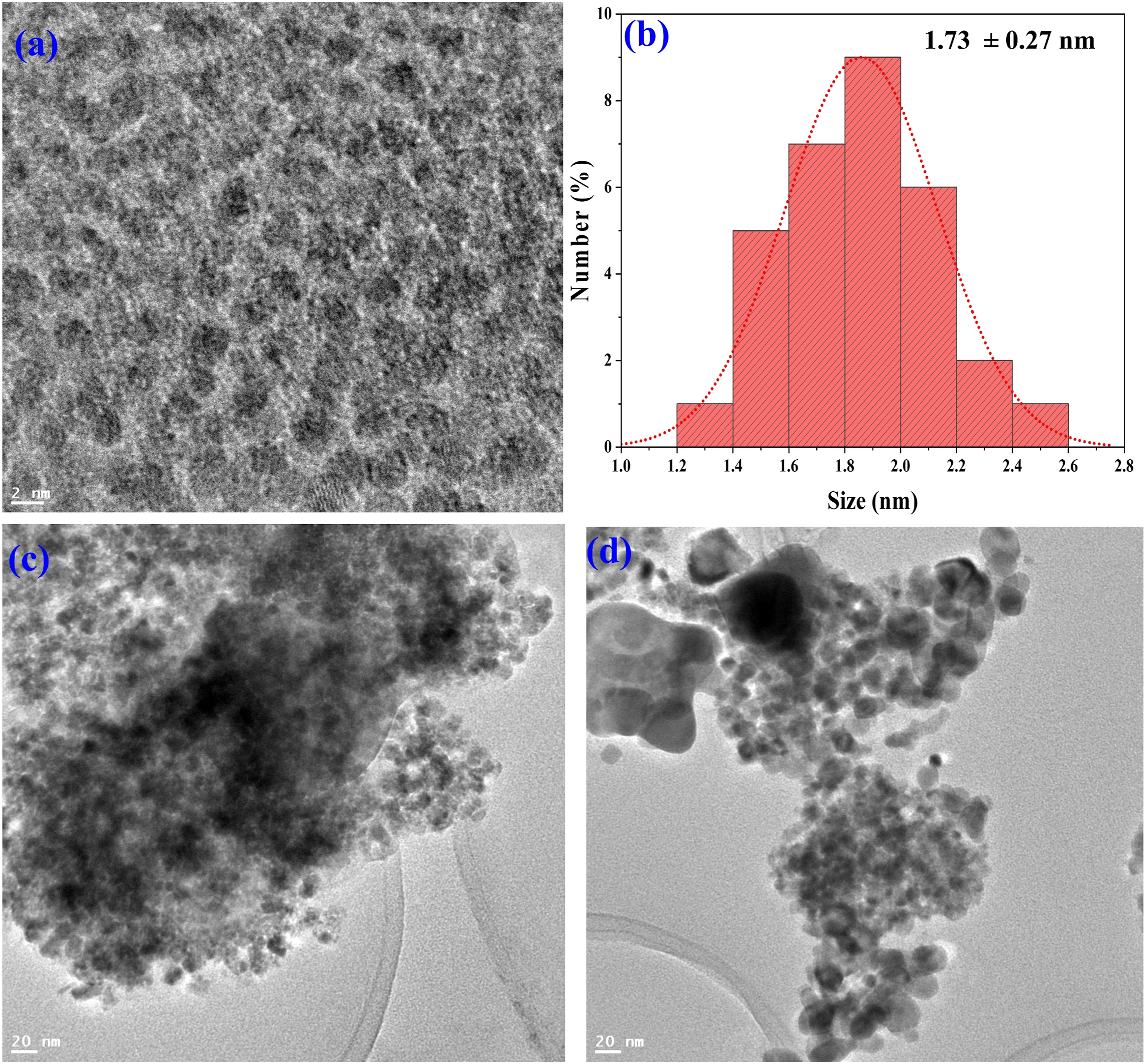
Thus, the addition of NaOH plays a key role in preparing red-emissive trypsin–Cu NCs. After that we have also performed the excitation dependent emission spectra of trypsin–Cu NCs at different wavelengths ranging from 410 nm to 500 nm in which the maximum fluorescence intensity was noticed at 657 nm upon excitation at 490 nm (Figure S3, Supplemental Material). Similarly, UV–Vis and fluorescence spectra data illustrated that trypsin–Cu NCs showed a notable emission peak at 657 nm upon the excitation at 490 nm and absorption peak at 335 nm. Trypsin–Cu NCs display a yellow color in daylight, while it shows red fluorescence under UV lamp at 365 nm (Fig. 1). In order to confirm the size and shape of as-synthesized trypsin–Cu NCs, we have studied the HR-TEM analysis of trypsin–Cu NCs, revealing the as-prepared trypsin–Cu NCs are spherical in shape with an average size of 1.73 ± 0.27 nm (Figs. 2a and 2b). The as-fabricated trypsin–Cu NCs exhibited excellent fluorescence properties and showed high QY as compared to other reported Cu NCs (Table S1, Supplemental Material). Spectral characteristics (UV–Vis, fluorescence excitation and emission spectra) of trypsin–Cu NCs. (Inset: Photographs of trypsin–Cu NCs in daylight and under UV lamp). HR-TEM images of (a) trypsin–Cu NCs at 2 nm scale bars and (b) histogram plotted at 2 nm scale bar, trypsin–Cu NCs with (c) Pb2+ ion and (d) Hg2+ ion.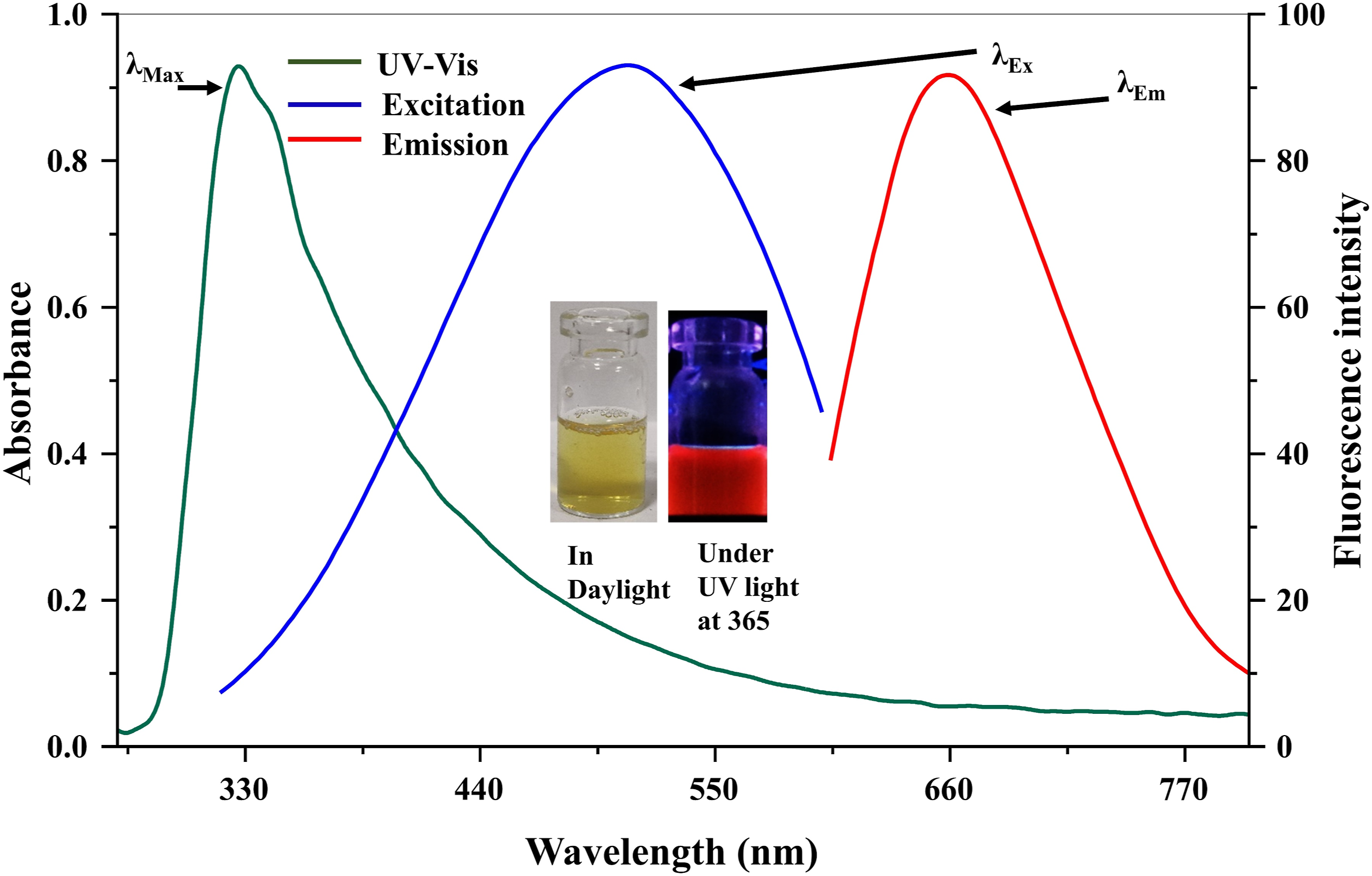
To know the presence of different functional groups, we have performed FT-IR characterization of trypsin–Cu NCs (Figure S4, Supplemental Material). The infrared spectra of trypsin–Cu NCs showed broad peak at 3394 cm−1 due to –OH stretching and the peaks at 2963 and 2932 cm−1 correspond to –CH– stretching. The peaks at 1654, 1539, 1399, and 1115 cm−1 are ascribed to –C=O stretching, –NH bending, and –C–O stretching, respectively. Infrared spectra of pure trypsin are displayed in Figure S4b (Supplemental Material). The broad peak at 3292 cm−1 was due to –OH stretching. It was noticed that –CH, –C–O stretching, and =C–H bending were observed at 2932, 1648, 1452, 1242, and 669 cm−1, respectively. 39 These spectra results confirmed that slight changes in the characteristic peak are strongly supported the formation of trypsin–Cu NCs. Moreover, copper NCs chemical composition as well as valence electrons were determined by using X-ray photoelectron spectroscopy or XPS (Figure S5, Supplemental Material). Binding energies of C, N, O, Na, Cl, and S elements appeared at 285.5, 400.8, 531.85, 1071.82, 198.44, and 164.06 eV, respectively, in the XPS data of trypsin–Cu NCs (Figure S5a, Supplemental Material). The peaks at 954 and 935.45 eV belonged to the distinct binding energies from Cu2p3/2 and Cu2p1/2, signifying both Cu0 and Cu+ in the as-synthesized trypsin–Cu NCs. Importantly, no peak was observed at 942 eV, which means that Cu2+ is not present in trypsin–Cu NCs40,41 (Figure S5b, Supplemental Material). Trypsin–Cu NCs spectrum of C1s peak fitting curves illustrates the three characteristic peaks at 284.7, 286.1, and 287.8 eV, which confirms the presence of C=C, C–N/C–O, and C=O groups 42 (Figure S5c of Supplemental Material). These results confirmed the use of trypsin as a ligand led to form red fluorescent Cu NCs, exhibiting several functional groups –OH, –COOH, and –NH2 on the surface of trypsin–Cu NCs, which improves their solubility in water and sensing ability. The powder X-ray diffraction (XRD) pattern of trypsin–Cu NCs showed a broad peak around 20°, indicating there was no significant population of crystallized Cu nanoparticles in trypsin–Cu NCs 43 (Figure S6, Supplemental Material). The fluorescence decay time data are shown in Figure S7 (Supplemental Material). The fluorescence lifetime results showed that trypsin–Cu NCs (2.98 ns) exhibited higher lifetime decay value than the trypsin–Cu NCs with Pb2+ (1.57 ns) and Hg2+ (0.099 ns) ions. It was found that the trypsin–Cu NCs were stable up to 15 days, showed stable fluorescence intensity from days one to seven, and then decreased from days 10–15 (Figure S8, Supplemental Material). This finding showed that as synthesized trypsin–Cu NCs exhibited good stability, which could be enough to use them as a fluorescence sensor in analytical sciences.
Trypsin–Cu Nanoclusters as a Nanosensor for the Detection of Metal Ions
In order to demonstrate how trypsin–Cu NCs worked as sensors, the different metal ions were added to trypsin–Cu NCs independently and examined for fluorescence characteristics of trypsin–Cu NCs with different metal ions. Briefly, 1000 μL of trypsin–Cu NCs solution was added individually to different vials containing 500 μL of various metal ions (Pb2+, Hg2+, Al3+, Cr3+, Zn2+, Fe3+, Cd2+, and Co2+, 1 mM). The vials were vortexed for 1 min and stood for 2 min. The emission spectral characteristics of the above solutions at 657 nm were examined upon λEx at 490 nm (Fig. 3). Interestingly, trypsin–Cu NCs exhibited a remarkable recognition capability towards Pb2+ and Hg2+ ions than the other metal ions, as demonstrated by fluorescence quenching of trypsin–Cu NCs by both ions. Only Pb2+ and Hg2+ considerably quenched the fluorescence emission at 657 nm, whereas other metal ions had no or negligible effect on the emission peak of trypsin–Cu NCs. These results revealed that trypsin–Cu NCs might be used as a probe for Pb2+ and Hg2+ assaying using the fluorescence “turn-off” mechanism. Fluorescence spectra of trypsin–Cu NCs after addition of various metal ions (Pb2+, Hg2+, Al3+, Cr3+, Zn2+, Fe3+, Cd2+, and Co2+, 1 mM). (Inset) Photograph of trypsin–Cu NCs with above metal ions under UV lamp at 365 nm.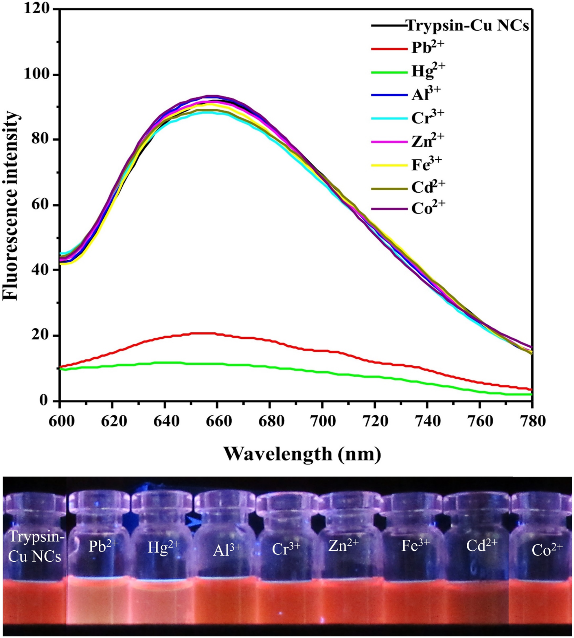
Effect of pH
Before examining the effects of phosphate-buffered saline (PBS) pH on the sensing ability of trypsin–Cu NCs, the fluorescence emission spectra of trypsin–Cu NCs were investigated with the addition of various metal ions (Pb2+, Hg2+, Al3+, Cr3+, Zn2+, Fe3+, Cd2+, and Co2+, 1 mM) in the presence of the PBS (Figure S9, Supplemental Material). This data revealed that the emission intensity of trypsin–Cu NCs was quenched only with Pb2+ and Hg2+ ions, indicating trypsin–Cu NCs show a sensing ability toward both ions even in a PBS medium. The effect of pH on trypsin–Cu NCs with Pb2+ and Hg2+ were further examined in this study. First, the fluorescence emission spectra of trypsin–Cu NCs was measured without Pb2+ and Hg2+ in PBS pH (2.0–12.0), revealing there is no effect on the emission intensity of trypsin–Cu NCs (Figure S10, Supplemental Material). The fluorescence emission spectra of trypsin–Cu NCs were studied after addition of Pb2+ and Hg2+ in various PBS pH (2.0–12) in which the emission intensity of trypsin–Cu NCs were quenched at all PBS pH values (Figures S11a,b, Supplemental Material). Interestingly, the emission intensity of trypsin–Cu NCs was greatly quenched with Pb2+ and Hg2+ without PBS pH, suggesting the fluorescence assay of Pb2+ and Hg2+ was performed by using trypsin–Cu NCs as a probe without adjustment of the solution pH.
Sensitivity Study
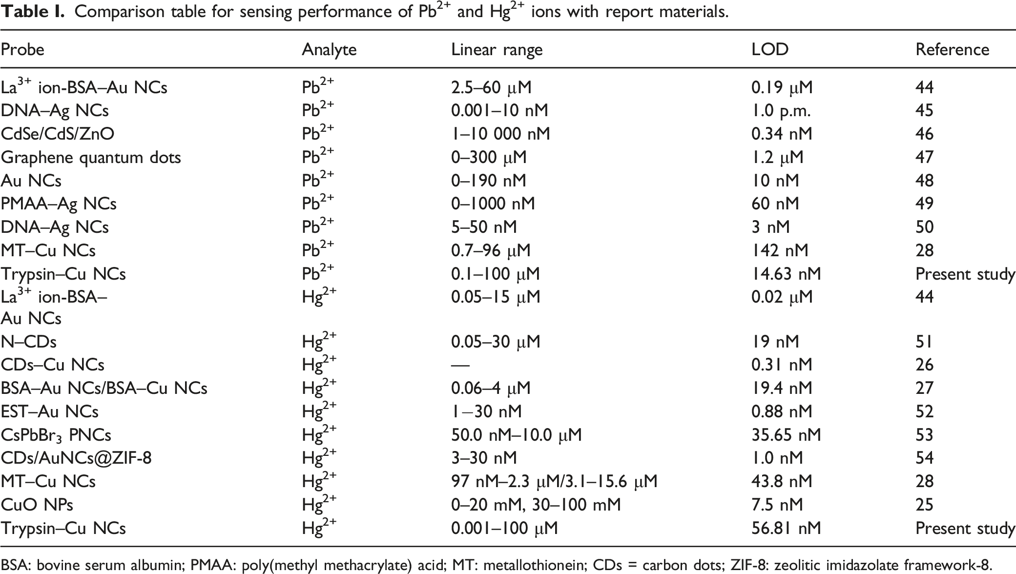
Additionally, the sensitivity of synthesized trypsin–Cu NCs was examined for the quantification of Pb2+ and Hg2+. With the addition of Pb2+ and Hg2+ ions, the emission intensity of trypsin–Cu NCs was quenched, resulting in the establishment of calibration graphs by examining the emission spectra of trypsin–Cu NCs with the addition of different concentrations of Pb2+ (0.1–100 μM) and Hg2+ (0.001–100 μM) ions (Figs. 4a and 4b). These results indicated that the fluorescence emission intensity of trypsin–Cu NCs was linearly quenched with increasing amounts of both ions, exhibiting good linear response within the concentration ranges of 0.1–25 μM and of 0.001–1.0 μM for Pb2+ and Hg2+ ions, respectively. Noticeably, Pb2+ and Hg2+ ions effectively quenched the fluorescence spectra of trypsin–Cu NCs, even at concentrations of 0.1 and 0.001 μM. As a result, the calibration graphs were plotted against the fluorescence quenching intensity of trypsin–Cu NCs versus various concentrations of Pb2+ and Hg2+ (Figure S12, Supplemental Material). The LOD was estimated by using Eq. 2: Fluorescence spectra of (a) trypsin–Cu NCs with various concentrations of Pb2+ (0.01–100 μM) and (b) of Hg2+ (0.01–25 μM). (Inset) Photographic image of above solutions under UV lamp at 365 nm. Comparison table for sensing performance of Pb2+ and Hg2+ ions with report materials. BSA: bovine serum albumin; PMAA: poly(methyl methacrylate) acid; MT: metallothionein; CDs = carbon dots; ZIF-8: zeolitic imidazolate framework-8.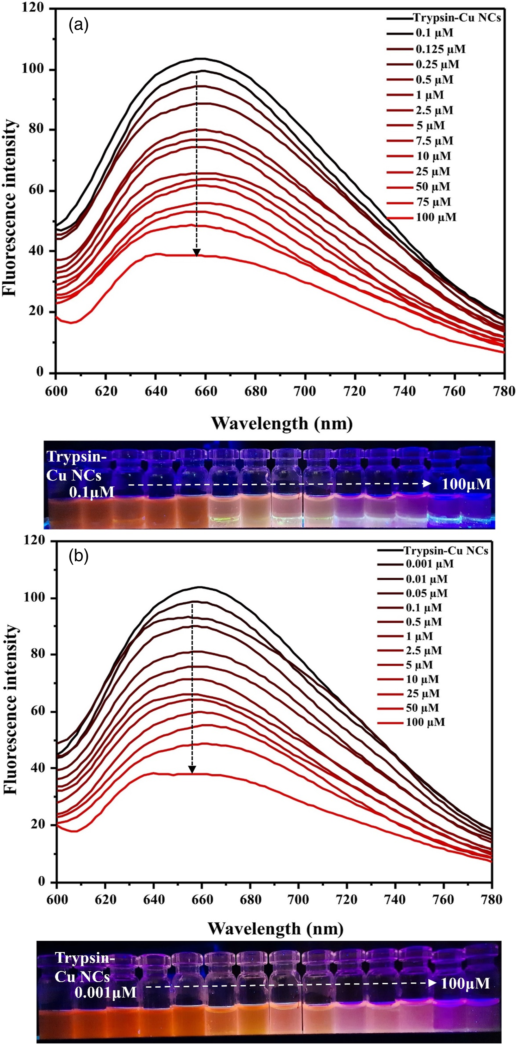

Sensing Mechanism
Functional groups such as–NH2, –COOH, and –OH present on the surface of trypsin–Cu NCs are responsible for binding with Pb2+ and Hg2+ ions. The interaction between functional groups and metal ions destroyed the fluorescence characteristics of trypsin–Cu NCs, which results in fluorescence quenching. In order to understand possible sensing mechanism of trypsin–Cu NCs, HR-TEM analysis was conducted in the presence of Pb2+ and Hg2+ ions (Figs. 2c and 2d). When Pb2+ and Hg2+ ions were added to trypsin–Cu NCs, the size of trypsin–Cu NCs was increased, causing the aggregation of trypsin–Cu NCs by the addition of both ions, which induced the fluorescence quenching of trypsin–Cu NCs. To further investigate, the fluorescence lifetimes of trypsin–Cu NCs with and without Pb2+ and Hg2+ ions were examined; the spectral data are shown in Figure S7 (Supplemental Material). A typical lifetime decay for trypsin–Cu NCs was noticed at 2.98 ns, but when Pb2+ and Hg2+ ions were added the lifetimes decreased to 1.57 and 0.099 ns, respectively. These results also indicated that the dynamic quenching of Cu NCs with both ions are due to significant changes in the lifetime of trypsin–Cu NCs after adding Pb2+ and Hg2+ ions. Based on the life-time spectra, it was found that a complex was formed between trypsin–Cu NCs and target analytes, which results to form Pb2+–trypsin–Cu NCs and Hg2+–trypsin–Cu NCs aggregates.
Selectivity Study
Interference of different chemical species on the fluorescence “turn-off” sensing performance of trypsin–Cu NCs for both Pb2+ and Hg2+ ions were also studied (Figs. 5a and 5b). Figs. 5a and 5b display the changes of emission spectra of trypsin–Cu NCs with and without both ions (Pb2+ and Hg2+ ions) in the presence of various different cations (Cu2+, Fe3+, Cr3+, Ni2+, Zn2+, Mg2+, and Mn2+, 500 μM), mixture of anions (SO42–, S2–, F−, PO43–, Cl−, Br– CH3COO−, and NO3−, 500 µM), and a mixture of pesticides (hexythiazox, lambda cyhalothrin, metalaxyl, hexaclonazole, and deltamethrin, 500 µM), respectively. The spectral data illustrated no interference was noticed for the above interfering chemical species, and other metal ions did not affect the selectivity of trypsin–Cu NCs toward both ions (Pb2+ and Hg2+ ions), signifying the trypsin–Cu NCs acted as selective fluorescent nanosensor for sensing of Pb2+ and Hg2+ ion. Investigation of interference from mixture of cations (Cu2+, Fe3+, Cr3+, Ni2+, Zn2+, Mg2+, and Mn2+, 500 μM), mixture of anions (SO42–, S2–, F−, PO43–, Cl−, Br– CH3COO−, and NO3−, 500 µM) and mixture of pesticides (hexythiazox, lambda cyhalothrin, metalaxyl, hexaclonazole, and deltamethrin, 500 µM) for the detection of (a) Pb2+ and (b) Hg2+ ions. (Insets) Photographic image of the above solution under UV lamp at 365 nm.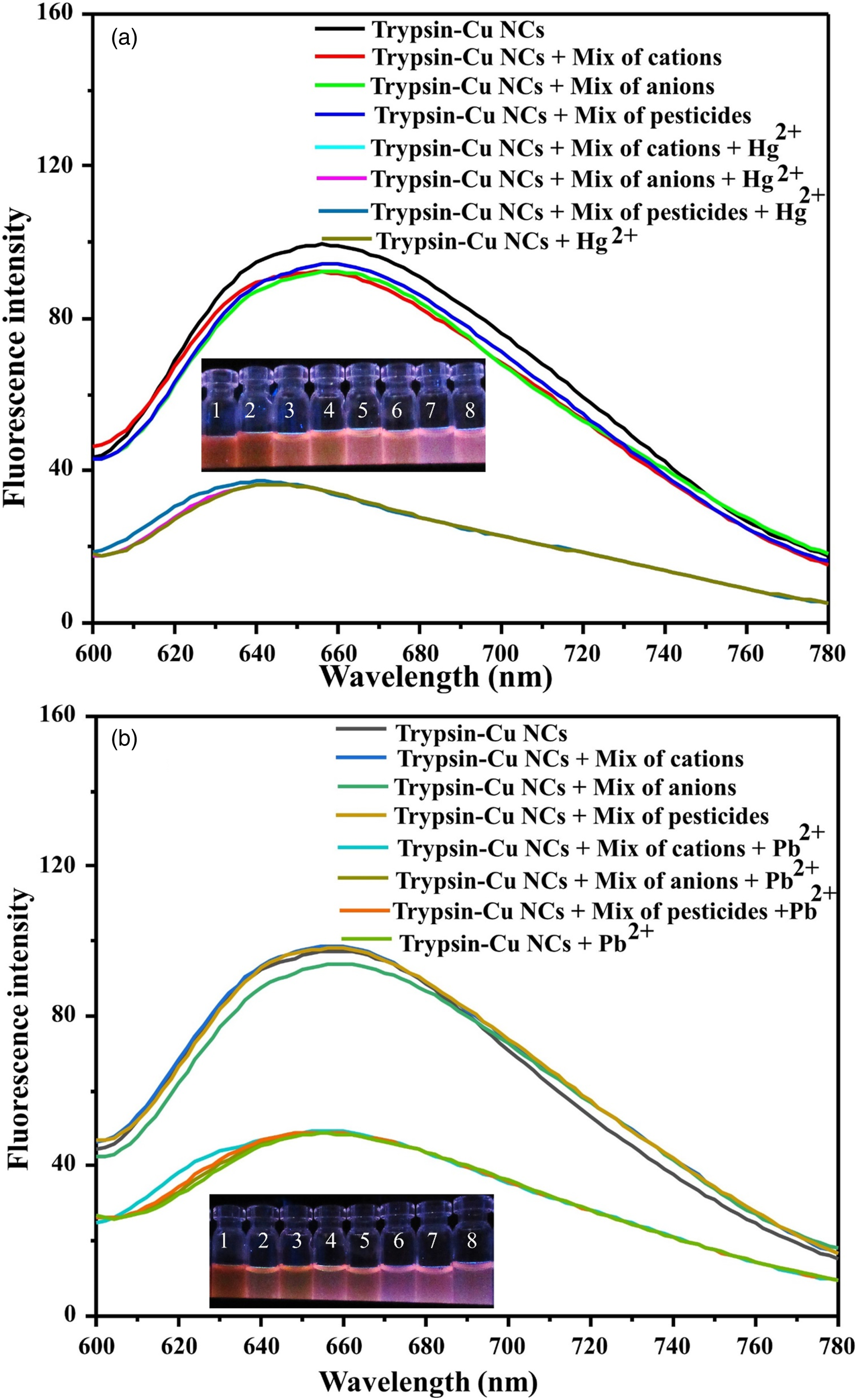
Masking Study
In order to tune the selectivity of trypsin–Cu NCs for sensing each target ion in the presence of other target ions, EDTA and AA were used as masking agents to ensure that the interference of each target analyte would completely mask during the sensing of each target metal ion in the presence of other target ion (Fig. 6). For Pb2+ detection, AA was used as a masking agent to suppress the interference from Hg2+ ion, thereby facilitating the quenching o the fluorescence of trypsin–Cu NCs by only Pb2+ ions, even in the presence of Hg2+ ion. Furthermore, EDTA was used as masking agent to mask Pb2+ ion interference during the sensing of Hg2+ ions, allowing the formation of complexes with only Pb2+ ions. Therefore, trypsin–Cu NCs-based fluorescent sensors showed high selectivity towards one metal ion in the presence of another sensing metal ion, demonstrating the developed method could be used as a promising nanosensor for the simultaneous detection of Pb2+ and Hg2+ ions. Fluorescence spectra of trypsin–Cu NCs for selective detection of (a) Pb2+ ion in the presence of Hg2+ ion using EDTA as a masking agent. (b) Emission spectra of trypsin–Cu NCs for selective detection of Hg2+ ion in the presence of Pb2+ ion using AA as a masking agent.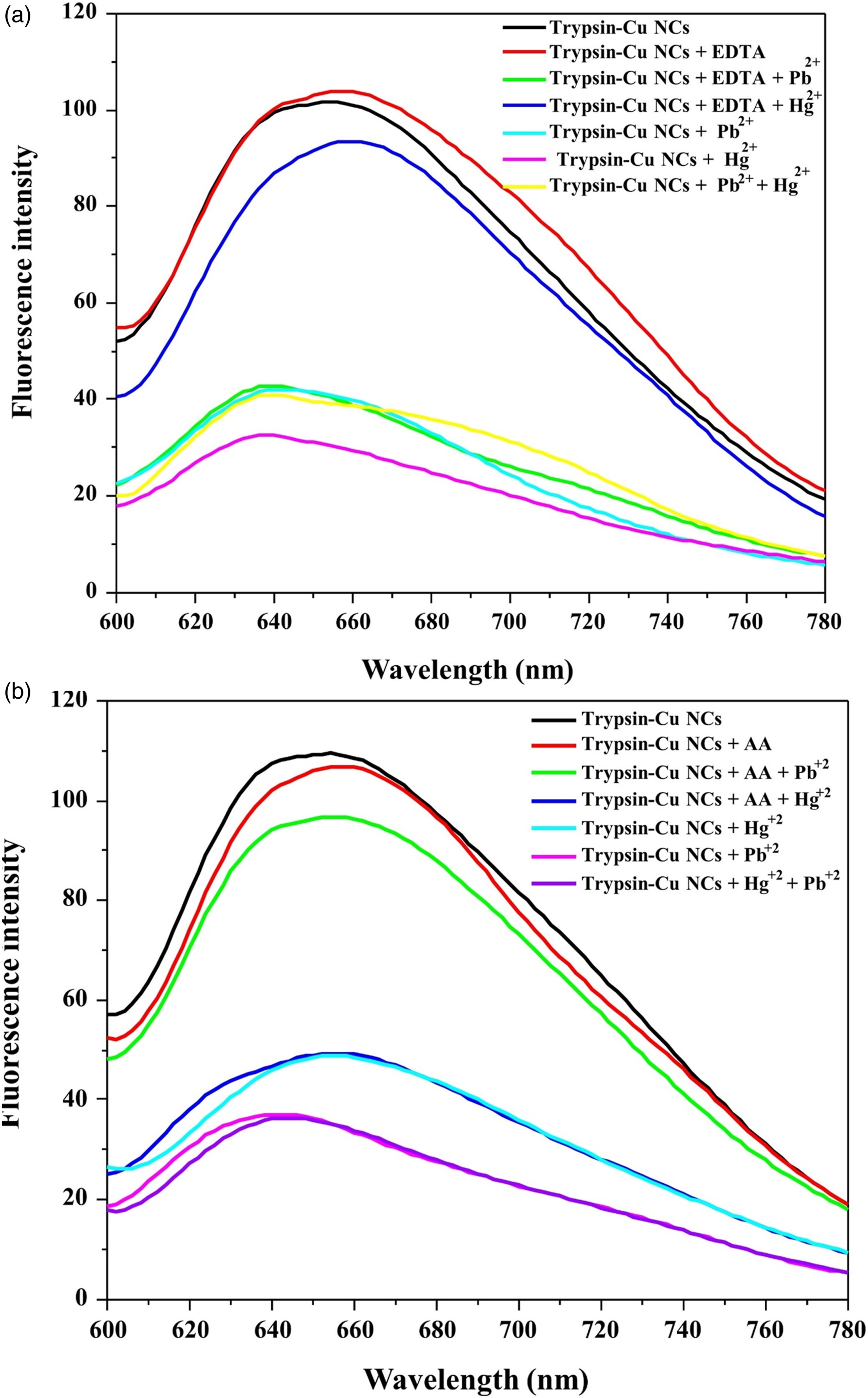
Real Sample Analysis
To demonstrate the efficiency of trypsin–Cu NCs as a probe, the developed probe was used to analyze Pb2+ and Hg2+ ions in tobacco samples, that is, F1 (feedstock), TM1 (low concentration tuibur), NC1 (nut-C solution), and T9 (high-concentration tuibur), and water samples (textile industry and tap). Tobacco samples are prepared using the following procedure. Briefly, tuibur is a peculiar form of "liquid" tobacco product, that is, tobacco-smoke aqueous condensate which is commonly used by smokers as well as the smokeless-tobacco-consuming populace of Mizoram, India. This unusual way of tobacco consumption as a liquid has been culturally practiced by the Mizo people for many generations. Tuibur is sipped several (5–30) times a day, retained in the mouth for a short period of time (10–15 sec) and then is spat out. 55 Stream water is normally used in the tuibur preparation as it is converted into an alkaline solution (pH ∼ 8–11) when treated with plant ash/bamboo ash and this alkaline solution rich in potash and potassium nitrate is known as feedstock. By virtue of the higher alkalinity, the feedstock solution can extract higher concentrations of an “unprotonated” form of nicotine from gas-phase tobacco smoke. Subsequently, the passage of tobacco smoke, produced by slowly smoldering tobacco stalk (mid-rib, petiole, and tobacco plant stem) rather than tobacco leaves, goes through the feedstock solution until the solution turns cognac in color and has an astringent and sharp nicotine smell. This tobacco smoke aqueous condensate is known as “crude” tuibur. This crude tuibur solution is then “filtered” by passing the crude tuibur solution through spent tobacco stalk (tobacco ash) to remove suspended particles and the non-polar component droplets of tobacco smoke. The filtered clear, highly alkaline solution is called tuibur, which will be consumed by the user as such. Nut-C is prepared by keeping two mature dry tobacco leaves immersed in the boiling water kept in an open container (mostly plastic tubs) and allowed to cool overnight (∼8–10 h). Subsequently, the remaining tobacco leaf matter is then removed from the solution in the morning. This tobacco solution is then mixed with lime (calcium hydroxide) paste and this alkaline tobacco extract solution is known as nut-c. The nut-c solution is normally consumed (∼1–1.5 mL) along with betel leaves and fresh mature betel nuts.
In the present study, lime-mixed nut-c, feedstock, and filtered tuibur solution were analyzed for heavy element concentration levels by the above-described procedure. Furthermore, trypsin–Cu NCs were utilized as a basic optical probe to investigate Pb2+ and Hg2+ ions in textile industrial wastewater obtained from fabric production in Surat, India, as well as different tobacco samples collected from Mizoram. The collected textile industrial wastewater and tobacco samples were first filtered through Whatman filter paper to remove microsize particles. Next, water and tobacco samples were spiked with different concentrations of Pb2+ (0.1, 0.25, and 0.5 μM) and Hg2+ (0.005, 0.01, and 0.05 μM) ions, and the concentrations of both ions were calculated using the aforementioned procedure (Table S2, Supplemental Material). The obtained results demonstrated that the suggested approach had good recoveries for Pb2+ and Hg2+ ions in diverse water and tobacco samples in the ranges of 94–99% and 93–99% with relative standard deviation (RSD) values of >2%. As a result, the current approach for alternate fluorescence detection of Hg2+ and Pb2+ ions in water and tobacco samples have remarkable precision and accuracy. The analytical performance of the trypsin–Cu NCs-based fluorescence approach possesses remarkable advantages including simplicity, rapidity, selectivity, and sensitivity toward the assaying of Pb2+ and Hg2+ ions, which suggests trypsin–Cu NCs can be potentially used for simultaneous sensing of trace level Pb2+ and Hg2+ ions in real samples.
Conclusion
In summary, a microwave-assisted synthetic approach was introduced for the preparation of red-emissive Cu NCs using trypsin as a ligand. When excited at 490 nm, the fluorescent trypsin–Cu NCs emitted an emission peak at 657 nm, exhibiting QY of 19.94%. Among the metal ions, two metal ions, that is, Pb2+ and Hg2+ ions strongly interacted with the surface of trypsin–Cu NCs, leading to diminishment of the fluorescence of trypsin–Cu NCs, which favors the development of a fluorescence “turn-off” mechanism for the assaying of both metal ions simultaneously. The probe exhibited good linearities in the ranges of 0.1–25 μM and of 0.001–1 μM for Pb2+ and Hg2+ ions with LODs of 14.63 nM and 56.81 nM, respectively. In addition, trypsin–Cu NCs acted as a potential probe for simultaneous assay of Pb2+ and Hg2+ ions by using AA and EDTA as masking agents for the Pb2+ and Hg2+ ions. Furthermore, it was demonstrated that the trypsin–Cu NCs based fluorescence method was successfully utilized to assay Hg2+ and Pb2+ ions in water and tobacco samples with good recoveries to illustrate this method could open-up new analytical pathways for the monitoring of Pb2+ and Hg2+ ions in real samples.
Supplemental Material
Supplemental Material - Microwave-Assisted Synthesis of Red Emitting Copper Nanoclusters Using Trypsin as a Ligand for Sensing of Pb2+ And Hg2+ Ions in Water and Tobacco Samples
Supplemental Material for Microwave-Assisted Synthesis of Red Emitting Copper Nanoclusters Using Trypsin as a Ligand for Sensing of Pb2+ And Hg2+ Ions in Water and Tobacco Samples by Dharaben J. Joshi, Lalrinhlupuii, Naved I. Malek, Rajendra Bose Muthukumaran, and Suresh Kumar Kailasa in Applied Spectroscopy.
Footnotes
Acknowledgments
DJJ sincerely acknowledges the Director, SVNIT, Surat, India for providing the doctoral fellowship to carry this work.
Declaration of Conflicting Interests
The author(s) declared no potential conflicts of interest with respect to the research, authorship, and/or publication of this article.
Funding
The author(s) disclosed receipt of the following financial support for the research, authorship, and/or publication of this article: This work was financially supported by the Department of Science and Technology, Government of India (EMR/2016/002621/IPC).
Supplemental Material
All supplemental material mentioned in the text is available in the online version of the journal.
References
Supplementary Material
Please find the following supplemental material available below.
For Open Access articles published under a Creative Commons License, all supplemental material carries the same license as the article it is associated with.
For non-Open Access articles published, all supplemental material carries a non-exclusive license, and permission requests for re-use of supplemental material or any part of supplemental material shall be sent directly to the copyright owner as specified in the copyright notice associated with the article.