
Abstract
Umbilical vein varix (UVV) is a rare vascular anomaly of the umbilical cord, most commonly identified in the fetal intra-abdominal segment during prenatal ultrasound. Extra-abdominal UVVs are exceedingly uncommon and may be missed on routine obstetric imaging, with diagnosis often made postnatally. We report an isolated extra-abdominal UVV in a term newborn that was not detected antenatally and was recognized immediately after delivery on gross examination of the cord. Postnatal ultrasound with Doppler demonstrated no intra-abdominal extension and no evidence of abdominal wall herniation. Placental pathology confirmed a 3-vessel cord with focal dilation of the umbilical vein consistent with an extra-abdominal UVV. The infant remained clinically stable, required no intervention, and was discharged in good condition. This report adds to the limited literature suggesting that isolated extra-abdominal UVV can follow a benign course, and it highlights the value of careful postnatal cord inspection to prompt targeted evaluation when prenatal imaging is unrevealing.
Educational Objectives
Recognize the clinical presentation and diagnostic challenges of extra-abdominal umbilical vein varix (UVV), a rare vascular anomaly often missed on routine prenatal imaging.
Differentiate intra-abdominal UVV from extra-abdominal UVV with respect to diagnostic approaches and clinical implications.
Describe the role of targeted postnatal imaging (ultrasound with Doppler) to confirm the diagnosis and exclude intra-abdominal extension, thrombosis, or other associated findings.
Appreciate that isolated extra-abdominal UVV may be benign and that reporting normal outcomes helps reduce publication bias in rare cord anomalies.
Background
Umbilical vein varix (UVV) refers to focal dilation of the umbilical vein and is most often described in the fetal intra-abdominal segment near the abdominal wall insertion.1,2 Extra-abdominal UVV occurs in the intra-amniotic portion of the cord and is much rarer.3,4 Extra-abdominal lesions may be missed on routine prenatal ultrasound because systematic evaluation of the entire free cord is not universally performed at the mid-trimester scan, and routine third-trimester screening ultrasound practices vary by setting. 5 Historically, UVV has been linked to adverse outcomes in cases with associated anomalies or when complicated by thrombosis; however, contemporary evidence suggests that isolated intra-abdominal UVV is often associated with favorable outcomes, while the evidence base for isolated extra-abdominal UVV remains limited to case reports and small series.6-8
Case Presentation
A 20-year-old G1P0 Hispanic woman delivered at 40 weeks and 5 days. A fetal anatomic survey at 20 weeks and 5 days noted a left choroid plexus cyst and moderate tricuspid regurgitation; the remainder of the fetal anatomy was reported as normal. A subsequent growth ultrasound at 33 weeks and 3 days documented that the placental cord insertion was suboptimally visualized (limited visualization), without reported concern regarding cord appearance. Prenatal laboratory studies were unremarkable. Maternal blood type was O negative with a negative antibody screen; Rho(D) immune globulin was administered during pregnancy. Group B Streptococcus screening was positive and treated with penicillin. The patient denied alcohol, tobacco, or drug use. The mother was admitted with spontaneous rupture of membranes. Labor was briefly augmented with oxytocin. Due to a non-reassuring fetal heart rate tracing, she underwent cesarean delivery after labor onset under spinal anesthesia. Membranes had been ruptured for 9 hours and 46 minutes with meconium-stained fluid. At delivery, the umbilical cord was immediately noted to have a visibly abnormal segment proximal to the infant’s abdomen. Delayed cord clamping for 30 seconds was performed, and the cord was clamped and cut distally, leaving approximately 10 cm of normal-appearing cord distal to the abnormal segment. The infant transitioned well on room air with Apgar scores of 8 and 9 at 1 and 5 minutes, respectively, and was transferred to the neonatal intensive care unit (NICU) for evaluation of the cord abnormality. On inspection, an 18-cm segment of cord originating from the infant’s abdomen appeared dark red and opaque. (Image 1) The remaining cord appeared normal, and 3 vessels were identified along the cord length. Due to the markedly dilated segment, abdominal and abdominal wall ultrasound with Doppler was performed to exclude herniation of bowel/abdominal contents and to assess for intra-abdominal extension. Ultrasound showed no evidence of abdominal wall defect or herniated contents. Doppler evaluation of the liver and portal/hepatic venous system showed no intra-abdominal varix or abnormal dilation. Placental pathology demonstrated a 3-vessel umbilical cord with focal vascular dilatation consistent with an extra-abdominal UVV; the placental disk was otherwise unremarkable. (Image 2) The cord insertion type was not specified in the pathology report. Postnatal cardiovascular examination was reassuring; echocardiography was not deemed necessary. The umbilical stump was subsequently clamped and trimmed without complication. The stump remained dry without bleeding, inflammation, or discharge. The infant received routine newborn care, initiated breastfeeding, remained clinically stable, and was discharged on day of life 3. Outpatient follow-up on day of life 7 showed a dry stump without inflammation. Per maternal report, the stump separated on day of life 10, and the infant continued to do well.
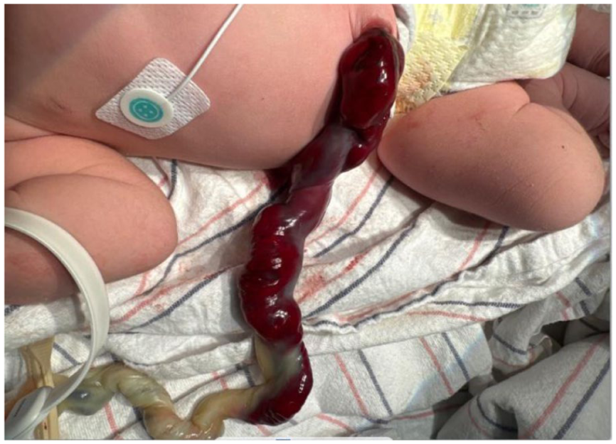
Photo of extra-abdominal umbilical vein varix immediately after birth.
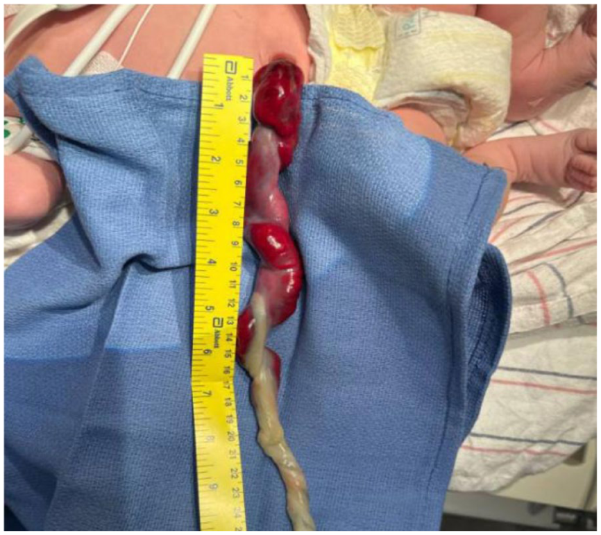
Umbilical cord varix segment measurement.
Discussion
The UVV comprises focal dilation of the umbilical vein and is most commonly described in the fetal intra-abdominal segment, whereas extra-abdominal UVV occurs in the intra-amniotic portion of the cord and is far less frequently reported. 2
Intra-abdominal UVV has traditionally been defined sonographically as an umbilical vein diameter ≥ 9 mm or as a focal dilation measuring at least 50% wider than the adjacent non-dilated segment. 2 Nomogram-based approaches have also been used; early work demonstrated that the normal fetal intra-abdominal umbilical vein increases with gestation (approximately 3 mm at 15 weeks to ~8 mm at term), enabling interpretation relative to gestational age. 2 Earlier reports, particularly those describing cases accompanied by additional anomalies or diagnosed earlier in gestation, described significant morbidity; however, contemporary evidence indicates that isolated intra-abdominal UVV generally has a favorable prognosis, with some studies suggesting a possible association with fetal growth restriction.7,8
Extra-abdominal UVV is rare and may be missed on routine prenatal ultrasound because systematic examination of the umbilical cord is not universally performed. 5 When visualized prenatally, it can mimic a cystic lesion; Doppler confirmation of venous flow and continuity with the umbilical vein are helpful.3,4 Unlike intra-abdominal UVV, standardized size criteria for extra-abdominal UVV remain limited. Reference work describing measurements along the umbilical vein may support future standardization and counseling. 5 Published reports describe variable outcomes; while some neonates have an uncomplicated course after postnatal diagnosis, others have experienced severe complications including thrombosis, fetal anemia, or fetal demise, particularly when thrombosis is present or monitoring is limited.4,9,10
There is no evidence-based consensus defining an optimal antenatal surveillance regimen for UVV, particularly for extra-abdominal lesions. 6 If UVV is diagnosed prenatally, assessment is typically individualized and may include targeted ultrasound with Doppler to confirm location and evaluate for thrombus or turbulent flow, alongside fetal growth assessment. Additional fetal testing is generally tailored to gestational age and any concerning sonographic features. 6
This report contributes additional evidence of a benign outcome in isolated extra-abdominal UVV, helping offset publication bias toward adverse cases.3,4 It also underscores that careful postnatal cord inspection can identify rare anomalies not detected antenatally and can appropriately prompt targeted imaging to exclude clinically important complications.3,4
Early recognition, whether antenatal or postnatal, combined with targeted imaging and multidisciplinary evaluation, is essential to optimizing outcomes. As prenatal imaging technology continues to evolve, future research should aim to refine diagnostic criteria and surveillance strategies for both intra-abdominal and extra-abdominal UVVs. Establishing clearer guidelines will help clinicians balance vigilance with appropriate intervention, ultimately improving perinatal outcomes in these rare yet clinically significant cases.
Author Contributions
P. Jairath and E. Rosier conceptualized and prepared the case report, contributed to the clinical assessment and management of the patient, and drafted the initial manuscript.
P. Jairath reviewed the current literature, critically reviewed and revised the manuscript for important intellectual content.
All authors approved the final manuscript as submitted and agree to be accountable for all aspects of the work in ensuring that questions related to the accuracy or integrity of any part of the work are appropriately investigated and resolved.
This case report complies with ethical standards and patient consent was obtained for publication.
Footnotes
Acknowledgements
The authors would like to thank the NICU and NBN staff of the York Hospital Women and Children’s Service Line for helping take care of this patient and their dedication in providing excellent health care to the infants of our community.
Ethical Considerations
IRB submission and approval are not required for case reports.
Informed Consent
Maternal consent was obtained and is attached.
Funding
The authors received no financial support for the research, authorship, and/or publication of this article.
Declaration of Conflicting Interests
The authors declared no potential conflicts of interest with respect to the research, authorship, and/or publication of this article.