
Abstract
Green composites of waste silk short fibers/cellulose were prepared by dissolving cellulose in (7%NaOH/12%Urea) aqueous solution pre-cooled at −12°C. The effect of fiber loading on the tensile, optical, thermal degradation, and cell viability was studied. The interfacial bonding between the fibers and matrix was assessed using the fractograph.
Introduction
Due to their outstanding properties like lightweight, higher strength-to-weight ratio, ease of manufacturing, low cost, etc. polymers in general and polymer composites in particular are finding many applications in every field. Unfortunately, because of their nondegradable nature, the environment problems posed by them are also simultaneously increasing. 1 In order to overcome these problems, the trend is now shifting toward developing environment friendly green composites using natural materials. In this direction, many natural matrix materials like soy protein,2,3 wheat protein, 4 polybutylene succinate, 5 etc. were tried for making green composites. As these matrix films are brittle, they were reinforced with natural fibers/fabrics like hemp, 6 – 8 sisal, 9 Alfa, 10 kenaf, 11 Hildegardia populifolia, 12 etc. However, these matrices are expensive and some of them are food materials. Nowadays, research is going on to try nonedible, biodegradable, renewable, and low cost matrix materials. Cellulose is the most abundant natural, renewable, and biodegradable low-cost source. Hence, the authors selected cellulose as matrix. As cellulose degrades thermally before going to its melting point, the only way of converting it into film form is by solution casting and subsequent regeneration. Unfortunately, dissolution of cellulose is complicated and time-consuming process. The conventional solvents of celluloses like CS2 have low-boiling points and are toxic. Efforts were made to use ionic liquids as green solvents. But their usage is limited due to their high cost and energy consumption. Recently, a new solvent of 7 wt% NaOH/12 wt% urea aqueous solution pre-cooled to −12°C was successfully used to dissolve cellulose rapidly. 13 – 16 The authors chose this method for the dissolution of cellulose as it is a green process. Both China and India are major producing countries of silk fabrics. Annually thousands of tons of waste silk fibers (WSF) are generated in silk industries in these two countries. The authors chose WSFs as reinforcement in order to add value to the waste generated. Silk is the strongest natural fiber is another reason for its selection as reinforcement in this study. The authors studied the effect of fiber loading on the tensile, optical clarity, thermal degradation, and cell viability of WSF/cellulose composite films. The interfacial bonding between the fibers and matrix was investigated using scanning electron microscopic technique.
Materials and methods
Materials
Cotton linter pulp supplied by Hubei Chemical Fiber Co. Ltd. (Xiangfan, China) was used as received. The degree of polymerization provided by the manufacturer was ca. 620. NaOH and urea were supplied by Shanghai Chemical Reagent Co. Ltd., China, and they were used without further purification. The WSFs were obtained from silk industries in Dharamavaram town of Anantapur district, Andhra Pradesh state, India.
Dissolution of cellulose
An aqueous solution containing 7 wt% NaOH and 12 wt% urea was prepared and cooled to −12°C as described elsewhere. 13 – 16 Cellulose was added (4 wt%) to the pre-cooled solvent and stirred vigorously at room temperature. A clear solution of cellulose was obtained within 2 min of stirring. This solution was centrifuged at 5°C at a speed of 7200 rmp for 15 min to separate any undissolved cellulose and impurities. This stock solution was stored at 5°C, till it was used.
Preparation of WSF/cellulose composite films
The WSFs were treated with 5% aq.NaOH solution for 1 h to remove the sericin. The alkali treated WSFs were washed thoroughly using detergent to remove grease and dirt, and then rinsed in distilled water several times and dried to constant weight. The dried fibers were cut to small pieces of ∼1 mm length and stored in desiccators until they were used. The fiber content was varied from 1 to 5 wt% in cellulose and stirred well. The WSF/cellulose solutions were degassed to remove any air bubbles. The films of thickness ∼0.1 mm were made by evenly spreading the respective solutions on a glass plate using a spreader and subsequently regenerated in 5% dil.H2SO4 acid bath for 5 min. The regenerated films were washed thoroughly in distilled water and dried.
Tensile test
The tensile strength, modulus, and %elongation at break of the films were determined using an Instron 3369 Universal testing machine at a cross head speed of 5 mm/min at 23°C and relative humidity of 50%. The composite films were cut to 100 mm length and 10 mm width and used as specimens. Ten specimens were used in each case and their average values are reported.
Optical clarity
In order to assess the optical clarity of the composite films, their UV–visible spectra were recorded on a TU-1901 spectrophotometer.
Thermal degradation
To assess the thermal stability of the composite films, their thermograms were recorded on a Perkin Elmer TGA-7 instrument in a nitrogen atmosphere at a heating rate of 10°C/min.
Cell viability
To assess the biocompability of the composite films, their cell viability was examined using 3-(4,5-simwrhylthiazol-2-yl)-2,5-diphenyl tetrazolium bromide (MTT) cell viability test. For this, two types of cells – Hela cancer cells and COS7 normal cells were employed. The procedure as described elsewhere17,18 was adopted. In brief, the composite films were cut to 1.2 × 1.2 cm2 size, UV sterilized, and placed in 24 well plates. On each film sample, 60,000 cells treated with MTT dye were introduced and kept for 48 h. The living cells containing succinate dehydrogenase will deoxidize MTT to blue crystals whereas the dead cells fail to form them. At the end of the test, the blue crystals formed were dissolved in dimethyl sulfoxide and the cell concentration was determined colorimetrically using pre-calibrated curves.
Microscopic analysis
Scanning electron micrographs of the brittle fractured specimens were recorded on a JEOL JSM 6700 F microscope. An accelerating voltage of 5 kV and current of 10 µA were used for operating the scanning electron microscope.
Results and discussion
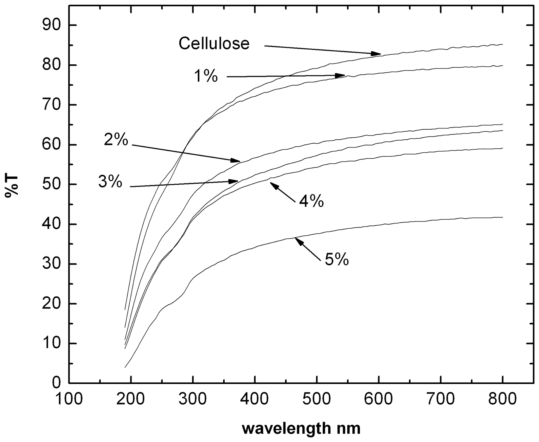
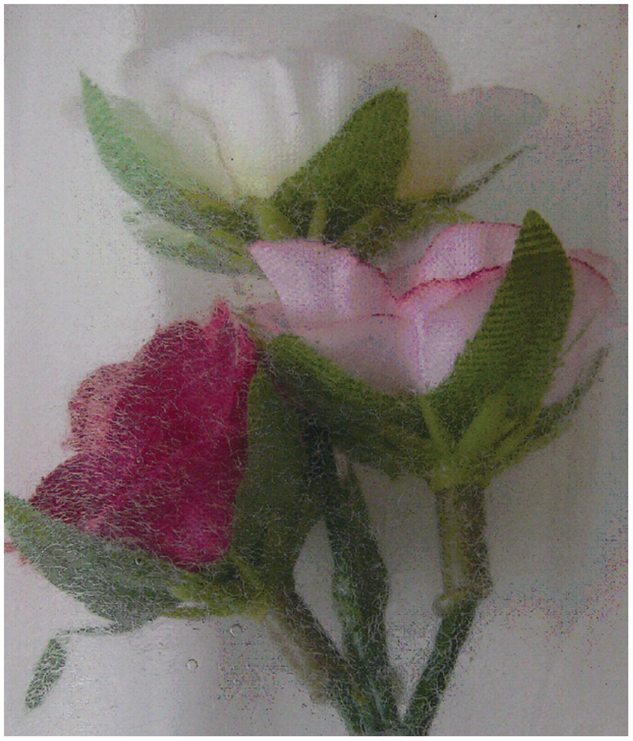
A photograph of the composite film with 5 wt% WSF is presented in Figure 1. From Figure 1, it is evident that the fibers are uniformly distributed in the matrix. In order to assess the optical clarity of the composite films under study, their UV–visible spectra are presented in Figure 2. From Figure 2, it is evident that the composite films were less transparent than the pure cellulose films and the transparency of the films was decreasing with increasing fiber content. The opaqueness of silk fibers and the difference in refractive indices of the matrix and silk fibers might be responsible for this decreasing trend. A photograph of the flowers wrapped with 5 wt% WSF/cellulose films is presented in Figure 3. From the figure, it is evident that the films had sufficient optical clarity for usage as package films.
Photograph of 5 wt% WSF/cellulose composite film. UV–visible spectra of cellulose and WSF/cellulose composite films. Photograph of the flowers wrapped with 5 wt% WSF/cellulose film.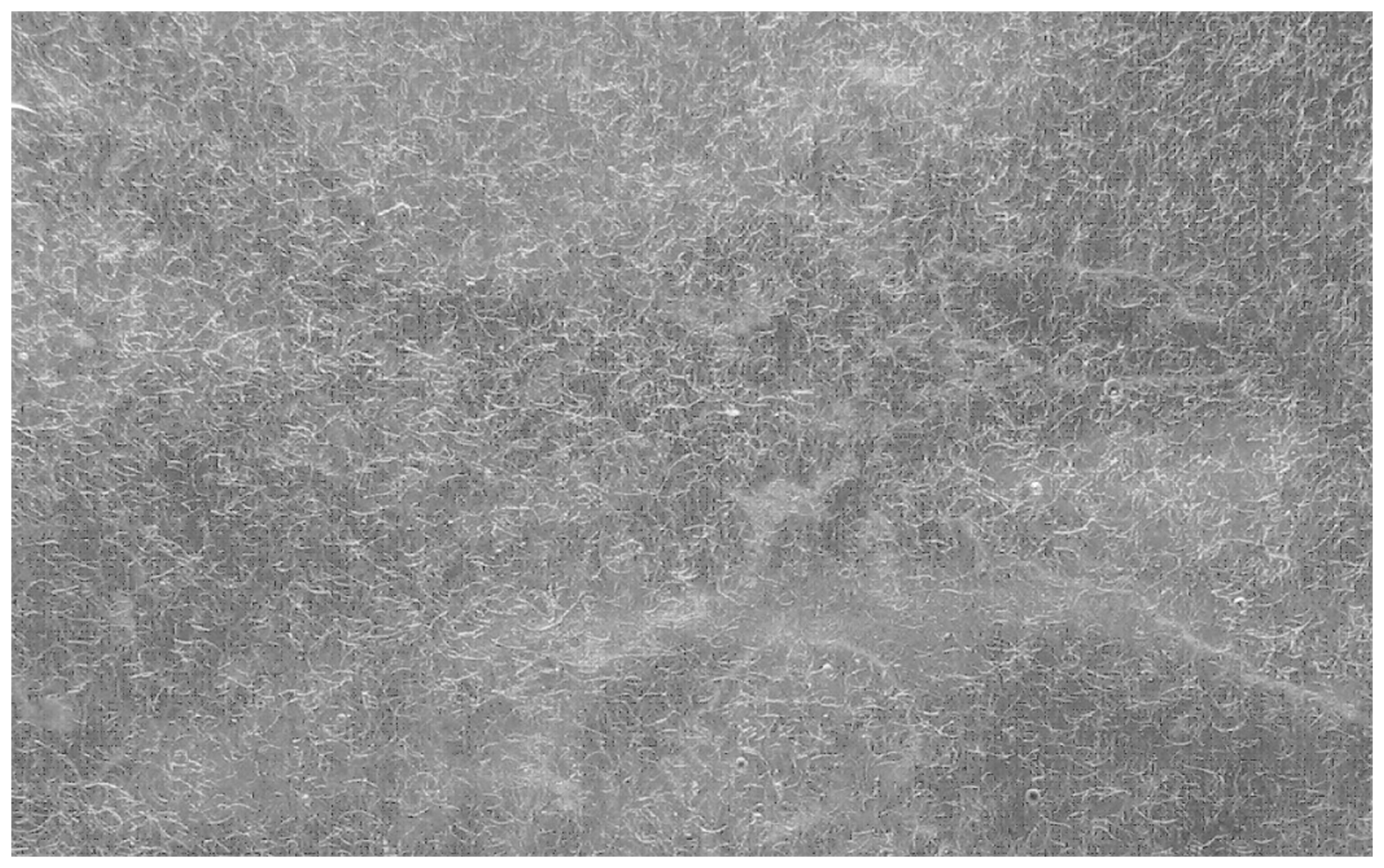
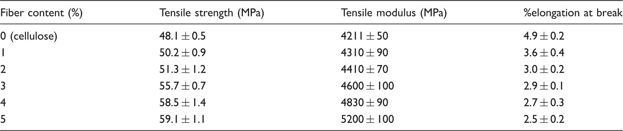
The tensile properties of the composite films are presented in Table 1. From this table, it is evident that the tensile strength and modulus of the composite films were slightly higher than those of the matrix film. Further, these properties are increasing with increasing fiber content. Unfortunately, the authors could not load the fibers beyond 5 wt% due to dispersion problems. Though silk fibers are the strongest among the natural fibers, only moderate increase in the tensile strength and modulus was achieved in the composites under study. The random orientation of the fibers may be responsible for such a behavior. The %elongation at break was found to be marginally lower for the composites when compared to the matrix. Generally, commercial package films are treated with glycerol. In the present case also, the %elongation at break can be increased with glycerol. The scanning electron micrograph of the brittle fractured surface of 5 wt% WSF/cellulose film is shown in Figure 4. From the micrograph, it is evident that predominantly fiber breakage rather than fiber pull out was observed for the composites under study. In a recent study on silk fiber/fibroin composites,
19
though both fiber and matrix were of the same nature, the interfacial bonding between them was poor. In the present case, as the fibers were treated with aq.NaOH solution, the resulted surface roughening might have facilitated moderate adhesion by mechanical bonding. The marginal improvement of the tensile strength and modulus with fiber content further supported this view.
Scanning electron micrograph of the fractured surface of 5 wt% WSF/cellulose composite film. Tensile properties of cellulose and WSF/cellulose composite films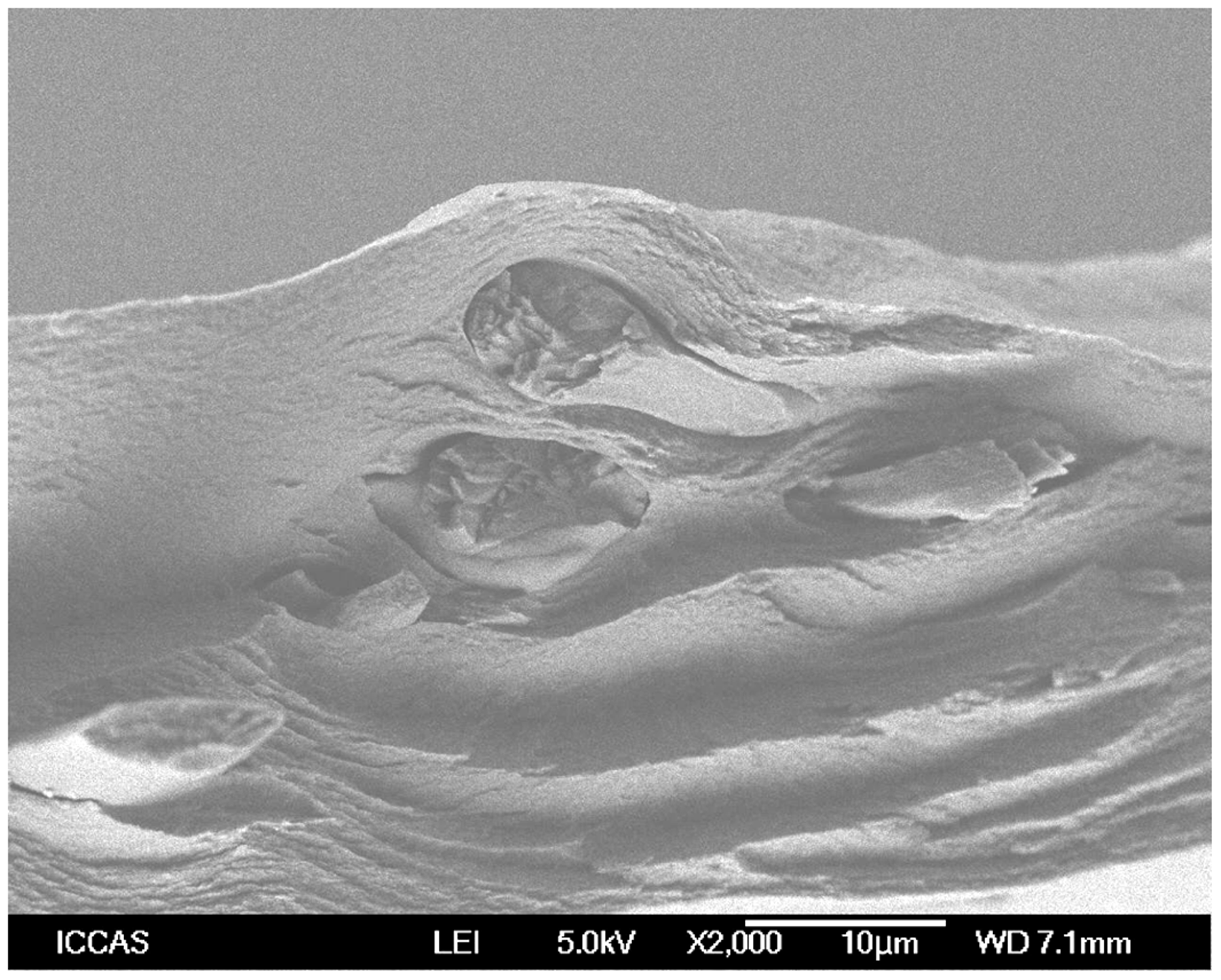
In order to investigate the biocompatibility of the composites under study, their cell viability is presented in Figure 5. From this figure, it is evident that in most cases, the cell viability for normal cells was good (>80%), whereas the growth of the cancerous cells decreased both on matrix and composite films. This observation clearly indicates the biocompatibility of the films under study.
Normal and cancerous (Tesla) cell viability of cellulose and WSF/cellulose composite films.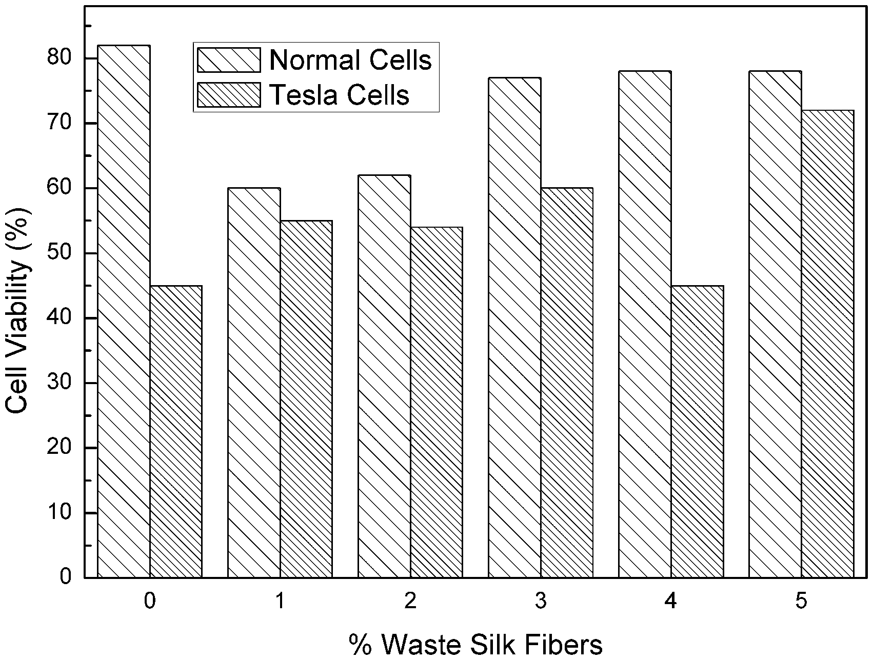
In order to assess the thermal stability of the composite films under study, their primary thermograms are presented in Figure 6. For comparison, the thermogram for cellulose matrix is also presented in the same figure. From this figure, it is evident that both cellulose and the composite films were stable up to 350°C. However, the weight percentage of the composite films after 350°C was lower than that of the matrix and decreased with the fiber content in the composites.
Primary thermograms of cellulose and WSF/composite films with different fiber content.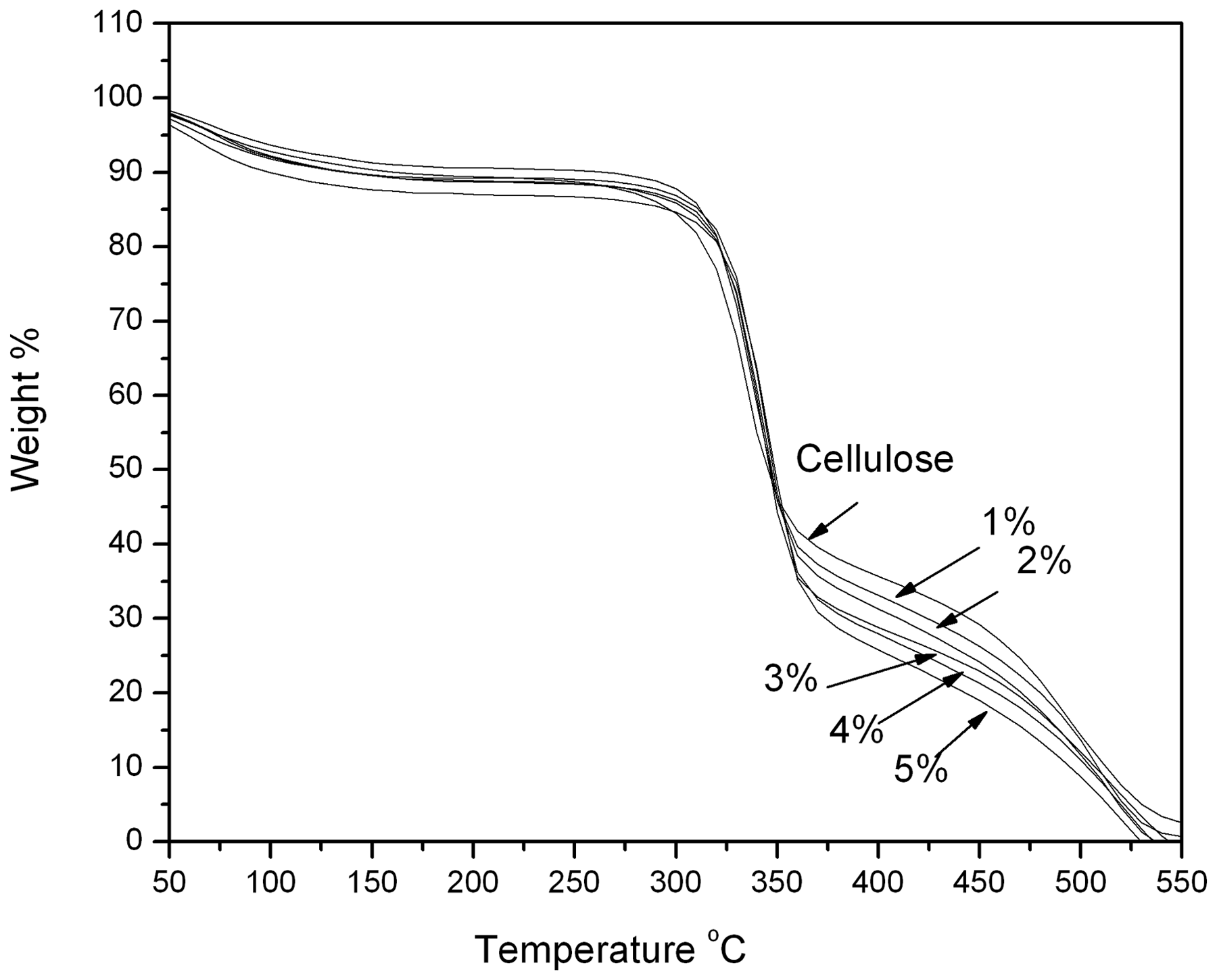
Conclusions
WSF/cellulose green composite films were prepared first by dissolving cellulose in a aq.(7 wt% NaOH/12 wt% urea) pre-cooled to −12°C, reinforcing them with different amounts (1–5%) of short fibers and subsequently regenerating the spread films in aq.5 wt% H2SO4 bath. The effect of fiber loading on the optical, tensile, cell viability, and thermal stability was studied. The tensile and cell viability were found to increase, whereas the optical clarity and thermal stability decrease for the composite films with increasing fiber content. The interfacial bonding between the fibers and cellulose was found to be moderate.
Footnotes
Acknowledgments
The author (AVR) thanks University Grants Commission of India for the award of Emeritus Fellowship [F.6-74/10 (SA-II) dt:22-12-2010] under which this study was carried out. The author also thanks the authorities of Wuhan University for providing a visiting fellowship to carry out part of this study at Wuhan.