
Abstract
Tooth caries is a prevalent chronic oral disease with microorganisms as the initiating factor and carbohydrates as the key environmental factor. Clinical antimicrobial therapies rely mainly on broad-spectrum antibiotics, which usually increase the risk of bacterial resistance. Recently, phototherapy has shown powerful antibacterial effects, although it cannot effectively eliminate cariogenic microenvironments and the antibacterial effect is not sustained after the light is removed. Here, we developed novel bioheterojunctions (bio-HJs) comprising MXene/Ag3PO4 (MX/AgP) and glucose oxidase (GOx), denoted MX/AgP-GOx, aiming at both the chemical and biological components of dental plaque biofilm. The bio-HJs decomposed the glucose rich in the cariogenic environment through GOx while providing abundant H2O2 for subsequent Fenton reaction. Under near-infrared (NIR) light, the bio-HJs produced hyperthermia and generated large amounts of reactive oxygen species based on the above H2O2, exerting powerful phototherapy properties (log reduction: 1.45 log 10 CFU/mL). It is worth noting that MX/AgP-GOx still exerted antibacterial effects in the dark via Ag+ bactericidal effects, Ag0 NPs catalytic activity, and GOx-mediated glucose depletion (log reduction: 0.39 log 10 CFU/mL), ensuring a sustained anticaries effect after the removal of NIR light. In addition, the rat caries model revealed that MX/AgP-GOx significantly reduced enamel mineral loss and had good biocompatibility. This study constructed efficacy-cascade bio-HJs targeting the sugar-rich cariogenic microenvironment, which promotes subsequent photodynamic therapy and combines photothermal and metal ion synergistic antibacterial means to continuously and effectively eliminate biofilm and prevent the occurrence and development of tooth caries.
Introduction
Tooth caries is a multifactorial, sugar-dependent disease driven by microorganisms, representing a global public health challenge (Peres et al. 2019; Wen et al. 2022). Specifically, Streptococcus mutans metabolizes dietary sucrose into glucose and forms dextran polymer, known as exopolysaccharides (EPSs), which protect and nourish microorganisms while contributing to antimicrobial resistance (Bertolini et al. 2022). Therefore, targeting the microbial etiology in sugar-rich cariogenic microenvironments is the key to preventing caries initiation and progression. However, conventional broad-spectrum antibiotics in clinical practice show limited efficacy due to poor penetration of low-permeability plaque biofilms and disrupt the natural diversity of the oral microbiome (Chatzigiannidou et al. 2020).
Photothermal therapy (PTT) has emerged as a promising alternative to antibiotics by destroying pathogens’ integrity through photothermal conversion processes (Qi et al. 2023). However, the temperatures required to eliminate biofilm would exceed the thermal tolerance of healthy cells (Huo et al. 2021). To enhance antibacterial efficiency with less light energy, researchers have explored synergistic strategies that combine PTT with other technologies, such as photodynamic therapy (PDT) (Hu et al. 2022). Yet, the PTT/PDT strongly depends on the external energy supply, whose effect disappears with the withdrawal of light, allowing biofilm formation to occur. Continuous biofilm inhibition after light withdrawal remains a challenge. In addition, blocking EPS formation is also important given its central role in plaque formation and bacterial defense, which is beyond the scope of phototherapy. Accordingly, a comprehensive strategy incorporating chemical and biological perspectives is essential for effective biofilm elimination.
Biologically compatible heterojunctions (bio-HJs) comprise 2 functional semiconductors with different bandgaps and energy levels, which is a superior platform for biological systems by integrating PTT, PDT, chemodynamic therapy (CDT), and metal ion therapy (MIT) through structural and compositional modulation (Low et al. 2017; Geng et al. 2024). Here, we proposed cascade anti-biofilm bio-HJs constructed of functionalizing MXene-Ag3PO4 HJs with glucose oxidase (GOx) through polydopamine (pDA) linkage, designated as MXene/Ag3PO4-GOx (MX/AgP-GOx). MXene is a category of 2-dimensional layered materials with broad specific surfaces, atomically thin thickness, tunable bandgaps, and facile surface functionalization (Naguib et al. 2014; Hao et al. 2022). Ag3PO4 serves as a photocatalyst with a narrow bandgap and high visible light responsiveness, releasing Ag+ to destroy bacterial membrane proteins and forming catalytically active Ag0 nanoparticles (NPs) reduced by pDA (Ma et al. 2022; Yang et al. 2022). GOx catalyzes glucose in cariogenic biofilms to generate hydrogen peroxide (H2O2) (Fu et al. 2018), providing substrates for chemodynamic and photodynamic processes. Under near-infrared (NIR) radiation, the bio-HJs demonstrate photothermal efficiency and generate reactive oxygen species (ROS) via Fenton-like reactions, chemical reactions that generate ROS through catalytic H2O2 decomposition (Tang et al. 2021). Furthermore, bio-HJs are sustainedly anticaries in darkness through the Ag+ bactericidal effects, Ag0 NPs catalytic activity, and GOx-mediated glucose depletion, overcoming the light-dependency limitation of conventional phototherapy. The MX/AgP-GOx bio-HJs effectively eliminate sugar-rich cariogenic environments while enhancing ROS generation, achieving cascading anticaries effects in dark and NIR environments.
Materials and Methods
Synthesis of Bio-HJ
MXene was synthesized through liquid-phase exfoliation of Ti3AlC2 using a 1:1 mixture of LiF and HCl at 45 °C for 12 h (Wang et al. 2022). The product was washed, centrifuged at 4,000 rpm, and lyophilized. MX/AgP was constructed via electrostatic self-assembly by adding silver nitrate (AgNO3, 0.1 M) to dispersed MXene, dialyzed overnight, and tested with sodium phosphate. Aqueous sodium phosphate solution (Na2HPO4, 0.05 M) was then applied to the dialysate and vigorously agitated. The MX/AgP composite was modified with pDA (2 mg mL−1) and GOx (2 mg mL−1) through sequential addition and incubation to obtain MX/AgP-GOx.
Sample morphology and microstructure were characterized using scanning electron microscopy (SEM) equipped with energy dispersive spectrometer (EDS) mapping (JSM-7500-F, JEOL), transmission electron microscopy (TEM; Tecnai F20), and X-ray photoelectron spectroscopy (XPS; ESCALAB 250 instrument, Thermo).
Photothermal and Chemophotodynamic Performance
The photothermal performance of MX/AgP-GOx was analyzed under different powers of 808 nm NIR (0.5, 1.0, and 1.5 W cm−2). The temperature was monitored using an infrared radiant thermal camera (E6, FLIR) at 30-s intervals. The photothermal stability of MX/AgP-GOx was assessed under 1.5 W cm−2 NIR illumination through 3 heating–cooling cycles (10-min heating, 15-min natural cooling) (He et al. 2022). Photodynamic performance was evaluated by detecting hydroxyl radicals (·OH), superoxide radicals (·O2−), and singlet oxygen (1O2) using methylene blue (MB) spectrophotometry (Huang et al. 2023) and 1,3-diphenylisobenzofuran (DPBF) assays (Shu et al. 2023). Glutathione (GSH) depletion was assessed through the Ellman assay (Deng et al. 2022).
Antibacterial Activity
S. mutans (UA159) was cultured anaerobically in brain heart infusion broth (Oxoid). Bacterial suspensions of S. mutans were treated with different samples under NIR irradiation or in darkness. Antibacterial activity was evaluated using the plate spread method, and bacterial morphology was observed by SEM. In addition, antibiofilm performance was assessed using crystal violet (CV) staining.
The log reduction was calculated by

where AE is the average CFU of the experimental groups and AC is the average CFU of the control group.
Cytocompatibility
Human oral keratinocytes (HOK) cells were cultured in high-glucose DMEM with samples under standard conditions. Cell morphology was observed using SEM and confocal laser scanning microscope (CLSM, Nikon) after fluorescent staining. Cell proliferation was evaluated using a cell counting kit (CCK-8) assay.
Anticaries Evaluation In Vivo
Twelve male Sprague–Dawley rats (4 wk old) were used to establish a caries model following ARRIVE guidelines and permitted by the Animal Care and Experiment Committee of West China Stomatology Hospital of Sichuan University. After streptomycin pretreatment for 4 d, oral swabs were taken from the rats’ mouths for the plate spread method to confirm no S. mutans infection (Tao et al. 2022). Then, the rats’ mouths were infected with S. mutans suspension (108 CFU/mL) for 3 d and fed with a cariogenic diet (Diet 2000, Trophic Animal Feed) and sucrose aqueous solution (10% w/v). Sterile oral swabs and plate spread method were used again to confirm the successful infection of S. mutans. The rats were randomly divided into 4 groups (n = 3): phosphate-buffered saline (PBS) (NIR−), MX/AgP-GOx (NIR−), PBS (NIR+), and MX/AgP-GOx (NIR+). Treatments were applied twice daily with or without NIR irradiation (808 nm, 1.5 W cm−2) for 4 wk.
Caries progression was evaluated using micro–computed tomography (micro-CT; Quantum GX system, PerkinElmer) and Keyes scoring after ammonium purpurate staining (Wu et al. 2018). Biocompatibility was assessed through hematoxylin and eosin (H&E) staining of oral mucosa and major organs.
Statistical Analysis
The results are presented as the mean ± standard deviation from at least 3 independent experiments. Statistical significance was determined using one-way analysis of variance or t test (SPSS 24.0), with P < 0.05 considered significant.
Further details are included in the Appendix.
Results
Characterization of MX/AgP-GOx Hjs
SEM and TEM were employed to observe the micromorphology and structure of the samples. As the zoomed-out image SEM in the upper right corner of Figure 1B shows, MXene presented a typical accordion-like multilayer nanostructure (Venkateshalu and Grace 2020). After stratification, the SEM (Fig. 1B) and TEM (Fig. 1C) images of MXene exhibited single-layer nanostructures with larger relative surface areas. In Figure 1D, MX/AgP were evenly coated with particles (ø = 10~200 nm). Corresponding EDS results (Fig. 1F) reflect that Ag, C, O, and Ti elements were uniformly distributed in MX/AgP.
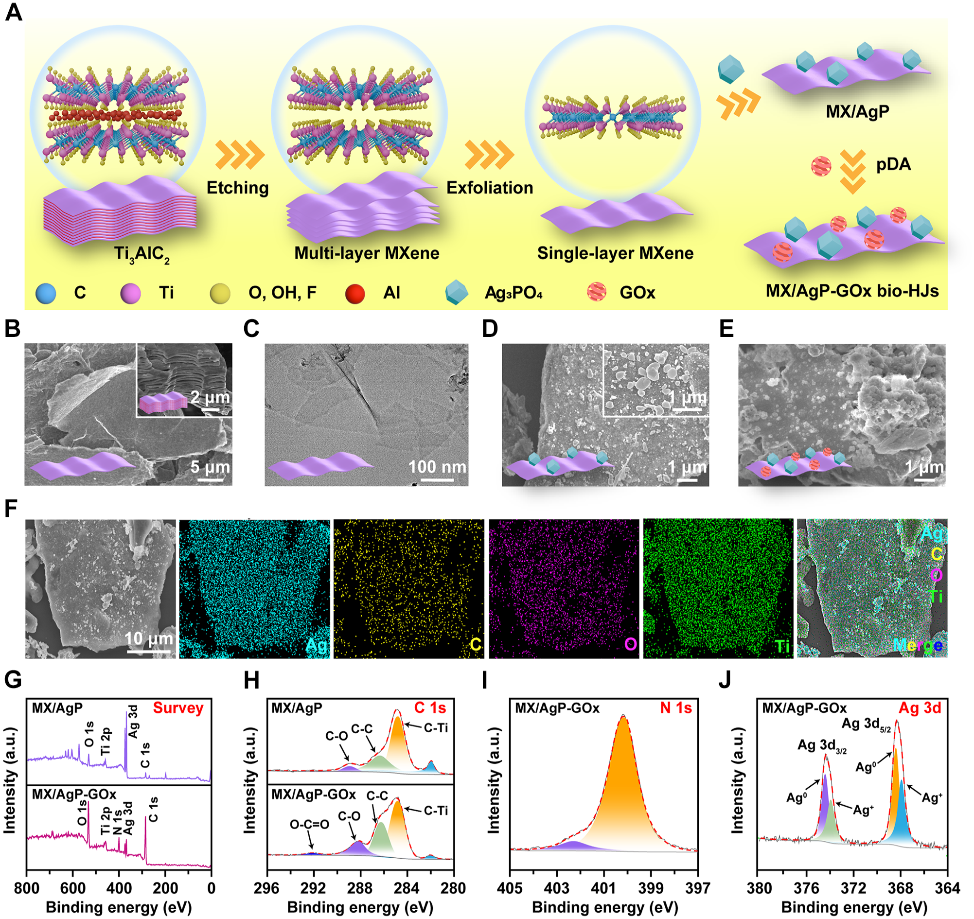
Bioheterojunctions (bio-HJs) assessment characterization. (
The surface of the MX/AgP-GOx (Fig. 1E) shows an incorporation of spherical particles. The chemical valence and elemental composition of MX/AgP and MX/AgP-GOx were detected by XPS spectra (Fig. 1G). The MX/AgP-GOx shows a distinct O-C = O bond (Fig. 1H) and N 1s peak (Fig. 1I), confirming the incorporation of GOx (C6H12O6) and pDA (C8H11NO2) (Guiomar et al. 1999). In addition, it exhibits peaks of Ag 3d5/2 and Ag 3d3/2 (Fig. 1J), which further split into signals for Ag+ in Ag3PO4 and Ag0 NPs reduced by pDA (Duan et al. 2023).
Photothermal Performance of MX/AgP-GOx Hjs
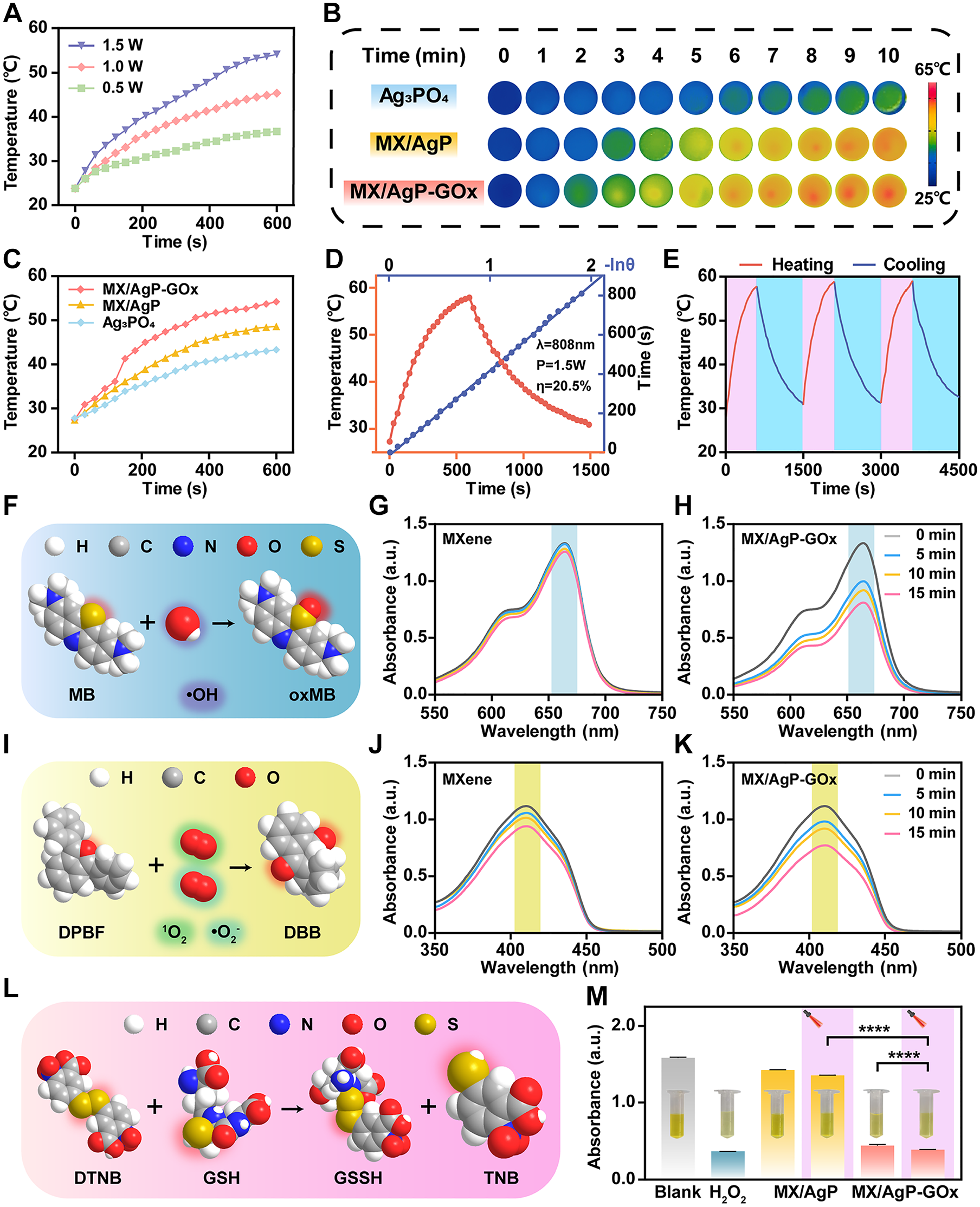
The photothermal effect was studied by monitoring temperature changes under NIR light. As shown in Figure 2A, the temperature of MX/AgP-GOx increased to 36.7, 45.4, and 54.2 °C after 10-min 808 nm NIR illumination at powers of 0.5, 1.0, and 1.5 W cm−2. The heating images and curves (Fig. 2B and C) show the temperature of MX/AgP-GOx significantly increased to 54.2 °C, higher than those of Ag3PO4 and MX/AgP. The photothermal conversion efficiency of MX/AgP-GOx attained 20.5% under 1.5 W cm−2 NIR irradiation (Fig. 2D), and it had no significant damping in the heating curves during 3 heating–natural cooling cycles (Fig. 2E).
Photothermal and chemophotodynamic performance. (
Chemophotodynamic Performance of MX/AgP-GOx Hjs
The production of OH was detected by MB, which caused a peak decline at about 664 nm, whose principle is illustrated in Figure 2F. After a 15-min NIR irradiation, the MB absorbance of MXene decreased very slightly (Fig. 2G). However, the MB absorption intensity of MX/AgP-GOx decreased obviously after 5-min NIR irradiation and continued to decline with the increase in irradiation time (Fig. 2H). The ·O2− and 1O2 was captured by DPBF, which caused a peak decline at about 410 nm, whose principle is illustrated in Figure 2I. Under NIR irradiation, the DPBF absorbance of MXene decreased slightly (Fig. 2J), while that of MX/AgP-GOx decreased more distinctly (Fig. 2K).
The GSH consumption capacity (Fig. 2L) was determined by the sulfhydryl reaction of 5,5′-dithiolide (2-nitrobenzoic acid) (DTNB) with glutathione to form yellow 5-thiole-2-nitrobenzoic acid (TNB) (Rahman et al. 2006). Compared with the blank group, the absorbance of MX/AgP (NIR−) and MX/AgP (NIR+) slightly reduced. Notably, MX/AgP-GOx consumed a significant amount of GSH in the dark, and the GSH consumption under NIR radiation was close to the H2O2 group (Fig. 2M).
Antibacterial Properties In Vitro
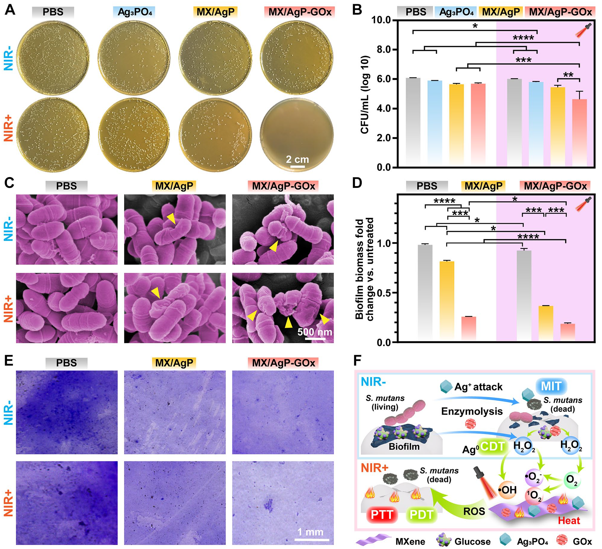
The spread plate method (Fig. 3A and B) shows that in the absence of NIR light, the log reduction of the MX/AgP and MX/AgP-GOx groups was 0.43 and 0.39 log 10 CFU/mL, respectively. Under NIR irradiation, the log reduction of MX/AgP-GOx reached 1.45 log 10 CFU/mL. Under 2.0 W cm−2 and 3.0 W cm−2 NIR irradiation, the log reduction of MX/AgP-GOx further reached 2.53 and 3.02 log 10 CFU/mL, respectively, in just 5 min (Appendix Fig. 1A and B).
Antibacterial properties in vitro. (
Furthermore, the morphology and the cell membrane integrity of S. mutans were observed by SEM. As Figure 3C shows, S. mutans in the PBS group appeared to have a typical oval shape with a smooth surface. In contrast, bacterial cytoplasm contraction was observed in the MX/AgP (NIR−) group, while the contraction was more pronounced in the MX/AgP (NIR+) and MX/AgP-GOx (NIR−) groups. Notably, S. mutans in the MX/AgP-GOx (NIR+) group exhibited more severe morphological shrinkage and cell membrane rupture.
The influence of different materials on S. mutans biofilm was investigated by CV staining (Fig. 3E). Mature S. mutans biofilm in PBS groups was stained purple. In the darkness, the MX/AgP-GOx group (≈75%) exhibited much higher biofilm clearance than the MX/AgP group (≈20%) (Fig. 3D). Under NIR irradiation, the residual amount of biofilm in the MX/AgP-GOx group was less, and the biofilm clearance rate reached greater than 80%.
Cytocompatibility In Vitro
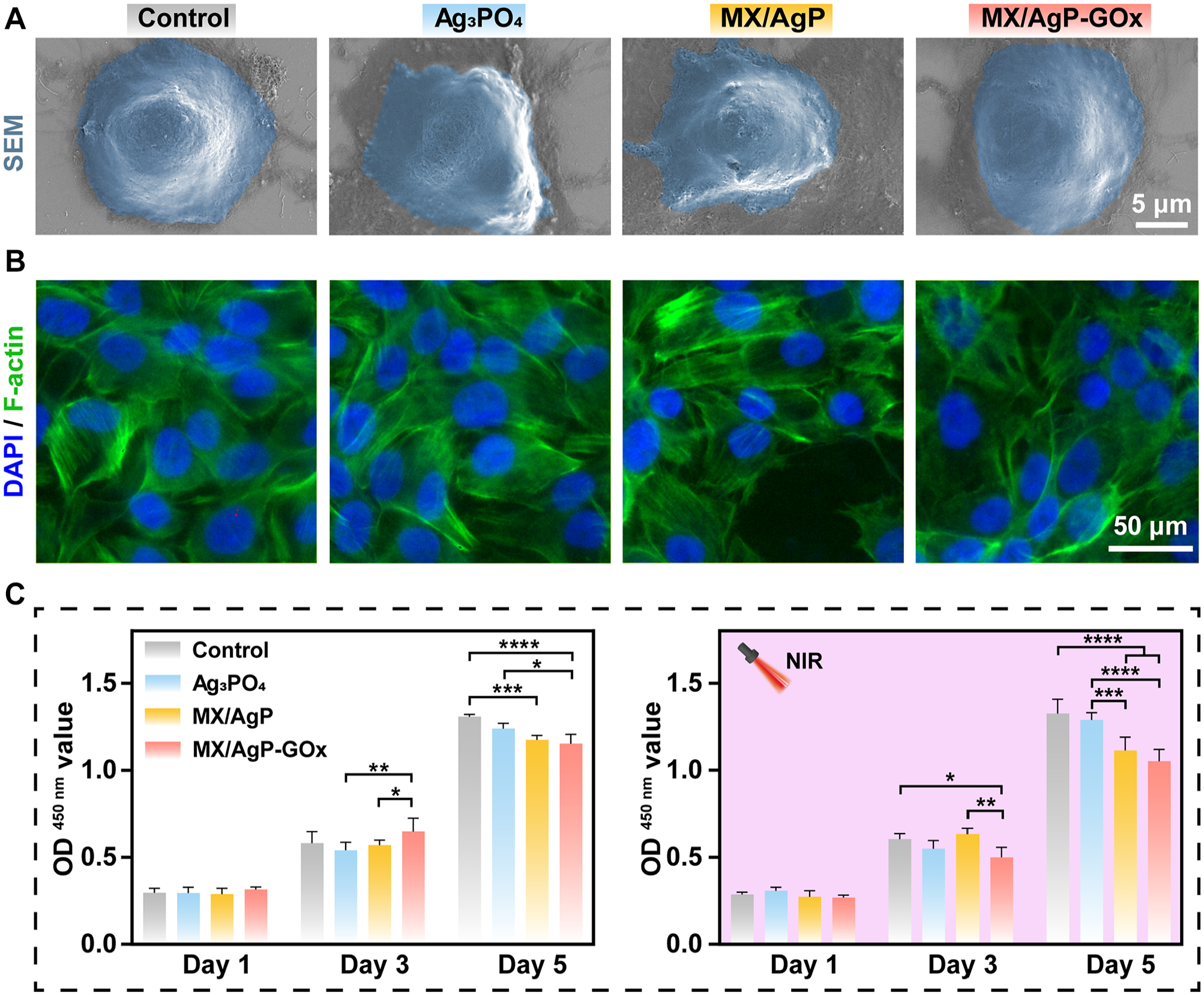
The HOK cells in the MX/AgP and MX/AgP-GOx groups presented a round and smooth morphology similar to that in the control group (Fig. 4A). All the HOK cells exhibited clear, complete nuclei and an extended cytoskeleton (Fig. 4B). The CCK-8 results (Fig. 4C) show that the cell number in all groups was close to that of the control group after 1 d of culture. The cell proliferation rate in the MX/AgP-GOx groups under dark and NIR light was 88% and 79% of the control group.
Cytocompatibility evaluation. (
Anticaries Property In Vivo
Given the excellent antibacterial activity and good biocompatibility of MX/AgP-GOx in vitro, we established a rat caries model to verify its anticaries performance in vivo (Fig. 5A).
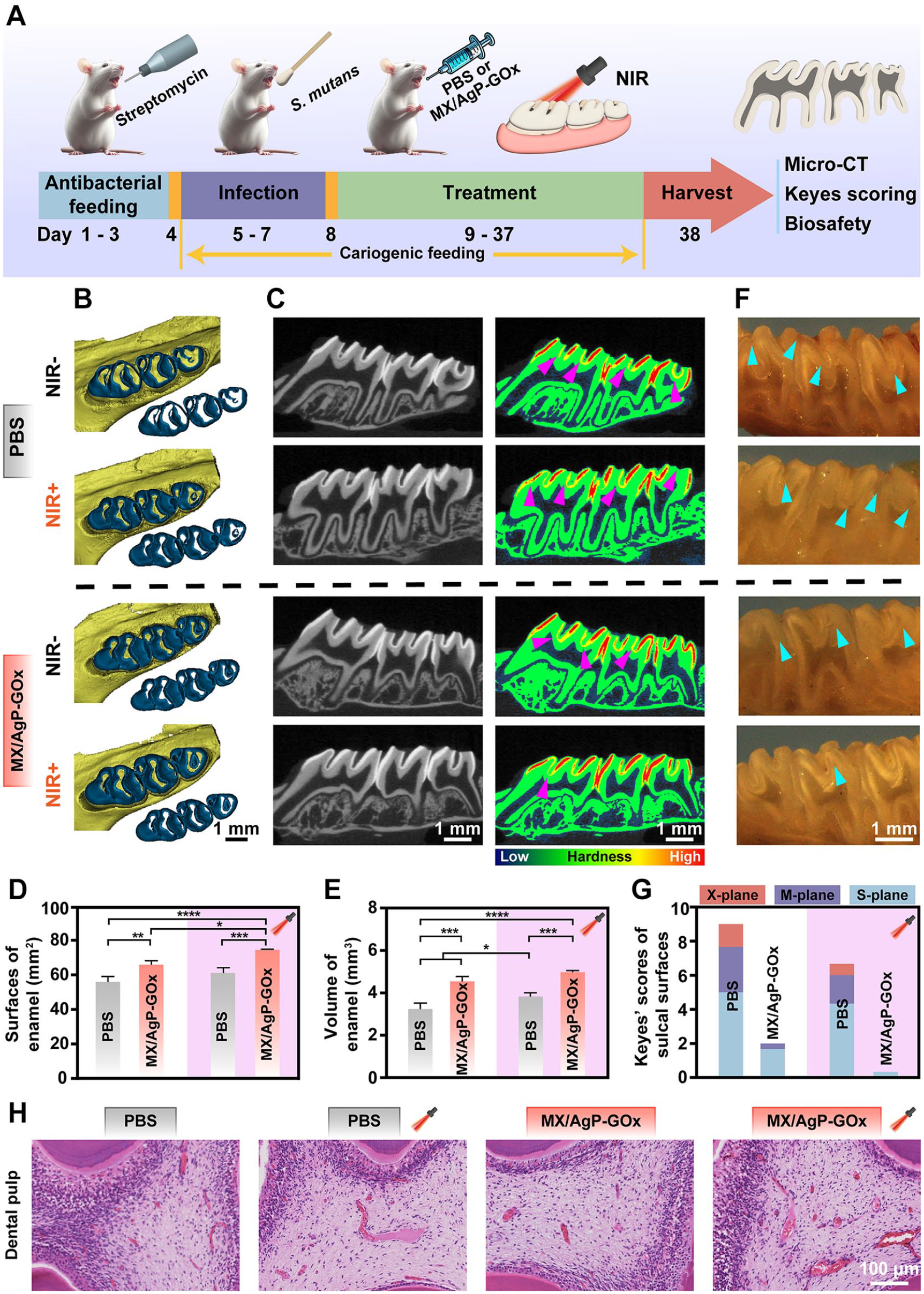
Anticaries performance in vivo. (
From the top view of micro-CT (Fig. 5B), the PBS (NIR−) group involves a large area of enamel loss in the tooth cusps and fossa, while the MX/AgP-GOx (NIR+) group involves only a small amount of enamel wear in the cusps. The tomography images (Fig. 5C) show a density reduction shadow at the bottom of the fossa and groove of the teeth in the PBS (NIR−) group, which reflects tooth demineralization, whereas there was no significant density reduction shadow in the MX/AgP-GOx (NIR+) group. The enamel surface area (Fig. 5D) and volume (Fig. 5E) of the MX/AgP-GOx (NIR+) group and MX/AgP-GOx (NIR−) group were higher than that of the PBS (NIR−) group.
After the noninvasive CT scan, we adopted the modified Keyes scoring, one of the most commonly used methods to assess the biological course of caries, which evaluates the lesions from 3 planes of depth and width and provides more morphological details. The images of stained teeth sections (Fig. 5F) show the PBS (NIR−) group had extensive caries (stained red) that invaded the dentin, while barely red-stained lesions were observed in the MX/AgP-GOx (NIR+) group. The score (Fig. 5G) of the MX/AgP-GOx (NIR−) group decreased significantly compared with the PBS (NIR−) group. The score of the MX/AgP-GOx (NIR+) group (0.3 ± 0.6) further decreased with only slight plane.
Histopathological Analysis
Hematoxylin and eosin (H&E) staining was performed on sections of oral tissues and vital organs to assess biosafety in vivo. The H&E staining images of the dental pulp (Fig. 5H) and oral mucosa (Appendix Fig. 2) of all groups showed no obvious inflammatory infiltration. In addition, there was no obvious damage or inflammation in the H&E staining images of the vital organ sections (Appendix Fig. 3).
Discussion
Dental caries is a biofilm-mediated disease in which S. mutans exploits sugar-rich microenvironments, metabolizing carbohydrates into glucose. This process leads to EPSs formation, nourishing microorganisms, and inducing antibiotic resistance (Du et al. 2020; Bertolini et al. 2022; Ciofu et al. 2022). While phototherapy is a promising alternative antibiotic strategy due to its deep penetration and high bactericidal efficiency, its reliance on NIR allows for potential bacterial growth following the withdrawal of light. To address this limitation, we developed MX/AgP-GOx bio-HJs that target the cariogenic microenvironment to consume glucose while generating H2O2 to enhance subsequent CDT and phototherapeutic efficacy. After removing NIR, the system maintains antibacterial activity through the MIT of Ag+, the chemodynamic property of Ag0 NPs, and the high-sugar environment elimination of GOx, continuously blocking the caries process.
The construction of MX/AgP-GOx (Fig. 1A) starts with extracting single-layer MXene (Ti3C2Tx), which provides abundant nucleation sites (Fig. 1C) for Ag3PO4. Via an electrostatically driven self-assembly strategy, Ag3PO4 was uniformly bound to MXene (Fig. 1F) and formed HJs due to different bandgaps (Yang et al. 2022). Eventually, by the excellent adhesion property of pDA, GOx was attached to the MX/AgP HJs surface and obtained MX/AgP-GOx bio-HJs. With redox-active catechol groups, pDA can reduce some Ag+ to Ag0 NPs, which has catalytic activity due to the large specific surface area and quantum effects (Yang et al. 2023).
The photothermal and photodynamic properties are critical determinants of bactericidal efficiency. The photothermal curves (Fig. 2A) show that the photothermal performance of MX/AgP-GOx increases proportionally with NIR power intensity. Under 1.5 W cm−2 irradiation, the final temperature exceeded 50 °C, surpassing the bacterial thermal ablation threshold (Huo et al. 2021), so it was chosen for subsequent experiments. MX/AgP-GOx exhibited superior heating efficiency compared with AgP and MX/AgP under NIR irradiation (Fig. 2B and C), indicating its enhanced capacity to convert NIR light energy into thermal energy for PTT. Besides, it possesses good photothermal stability (Fig. 2E).
The photodynamic efficiency correlates with ROS production. MB and DPBF assays revealed that MX/AgP-GOx HJs function as photosensitizers to produce various ROS under NIR irradiation, including ·OH, ·O2−, and 1O2. The outstanding ROS generation efficiency of MX/AgP-GOx is due to the establishment of HJs between MXene and Ag3PO4, and the energy generated by hot electrons during the surface plasmon resonance decay of MXene promotes the isolation of electron-hole pairs (Chan et al. 2021). Under NIR light excitation, the holes will remain and extend to the surface of MXene, reacting with the surrounding O2 to form 1O2, while the hot electrons will be reverse transferred to Ag3PO4, reacting with H2O to form ·OH.
GSH is a nonenzymatic antioxidant that can oxidize organic thiols (R-SH) to disulfides (R-S-S-R) and mediate ROS neutralization (Zhou et al. 2022). Extracellular GSH uptake is essential for the antioxidant stress responses and protein function regulation in S. mutans (Cheng et al. 2024). MX/AgP-GOx consumed large amounts of GSH in both darkness and NIR irradiation, reflecting its sustaining chemodynamic and photodynamic ROS generation functionality. The chemodynamic process is attributed to the ROS generation from H2O2 catalyzed by the reduced Ag0 NPs, which makes up for the dependence of the photodynamic ROS generation on NIR.
The above photothermal, photodynamic, and chemodynamic efficiency of MX/AgP-GOx indicates its sustained antimicrobial potential under NIR and dark conditions. Considering S. mutans is recognized as the predominant cariogenic bacteria in caries initiation and progression (Zhang et al. 2021), we chose it for antibacterial evaluation. In darkness, MX/AgP and MX/AgP-GOx exhibited a certain antibacterial ability, attributed to the direct bactericidal action of Ag+ and ROS produced by the chemodynamic process. Under NIR irradiation, the antibacterial ability of MX/AgP-GOx was significantly enhanced, demonstrating its potent photothermal and photodynamic bactericidal effect. Bacterial morphological analysis revealed that when MX/AgP-GOx was exposed to NIR light, it destroyed the bacterial cell membrane integrity under the synergistic effects of Ag+, heating, and ROS, thereby significantly inhibiting bacterial growth and proliferation.
Furthermore, dental plaque biofilm is the initiating factor of caries (Jakubovics et al. 2021), making biofilm inhibition efficacy critical in evaluating potential anticaries materials. The biofilm biomass of S. mutans in the MX/AgP-GOx (NIR−) group was significantly reduced compared with the MX/AgP (NIR−) group, demonstrating GOx’s capacity for EPSs degradation. Under NIR irradiation, MX/AgP-GOx suppressed biofilm formation more obviously, owing to the EPSs degradation producing H2O2, which enhanced ROS generation. Eventually, MX/AgP-GOx effectively inhibited biofilm formation through synergistic therapy of MIT, PTT, CDT, and PDT, as depicted in the proposed antibiofilm mechanism (Fig. 3F).
This study successfully developed MX/AgP-GOx bio-HJs that catalyze glucose and produce H2O2 in cariogenic conditions, triggering cascade antibacterial actions. Under NIR irradiation, MX/AgP-GOx bio-HJs effectively clear biofilms through the combination of MIT, PTT, and enhanced CDT/PDT. In addition, due to the Ag+ bactericidal effects, Ag0 NPs’ catalytic activity, and GOx-mediated glucose depletion, MX/AgP-GOx continues to work in the dark, making up for the dependence on external irradiation. In the rat caries model, MX/AgP-GOx bio-HJs reduced mineral loss of the teeth in the dark and further reduced mineral loss under NIR light. In addition, MX/AgP-GOx showed good biocompatibility both in vitro and in vivo. The MX/AgP-GOx bio-HJs provide a multidirectional template for blocking biofilm initiation diseases not only limited to tooth caries.
The current study has some limitations. Although MX/AgP-GOx showed effective antibacterial activity at 1.5 W cm−2 NIR irradiation for 10 min, its antibacterial efficiency can be further improved by enhancing NIR intensity and shortening the duration of irradiation in the future. While our study focused on S. mutans to evaluate the antibacterial efficacy of MX/AgP-GOx bio-HJs and the ability to target primary cariogenic factors, we recognize that the oral cavity hosts a diverse microbial community. This includes beneficial symbiotic bacteria that promote oral health by competing with cariogenic bacteria and maintaining ecological balance. To minimize the risk of microbial dysbiosis, we designed the MX/AgP-GOx bio-HJs to amplify their antibacterial effect specifically in cariogenic high-sugar environments. However, more assessments of the impact of MX/AgP-GOx bio-HJs on oral microbial diversity and community composition are needed in further research.
Author Contributions
J. Zhu, contributed to conception, design, data acquisition and interpretation, drafted and critically revised the manuscript; L. He, contributed to conception, design, data analysis and interpretation, critically revised the manuscript; X. Xu, H. Wu, and J. Li, contributed to data analysis and interpretation, critically revised the manuscript; B. Yan, Y. Deng, contributed to data acquisition, critically revised the manuscript; Y. Gao, K. Liang, contributed to conception and design, critically revised the manuscript. All authors gave final approval and agree to be accountable for all aspects of the work.
Supplemental Material
sj-docx-1-jdr-10.1177_00220345251329334 – Supplemental material for Bioheterojunctions Prevent Tooth Caries via Cascade Antibacterial Strategy
Supplemental material, sj-docx-1-jdr-10.1177_00220345251329334 for Bioheterojunctions Prevent Tooth Caries via Cascade Antibacterial Strategy by J. Zhu, L. He, X. Xu, H. Wu, J. Li, B. Yan, Y. Deng, Y. Gao and K. Liang in Journal of Dental Research
Footnotes
A supplemental appendix to this article is available online.
Declaration of Conflicting Interests
The authors declared no potential conflicts of interest with respect to the research, authorship, and/or publication of this article.
Funding
The authors disclosed receipt of the following financial support for the research, authorship, and/or publication of this article: This work was supported by the National Natural Science Foundation of China (82270970, 82470967), Natural Science Foundation of Sichuan Province (2023NSFSC1504), and Sichuan Science and Technology Program (2023YFQ0014).
References
Supplementary Material
Please find the following supplemental material available below.
For Open Access articles published under a Creative Commons License, all supplemental material carries the same license as the article it is associated with.
For non-Open Access articles published, all supplemental material carries a non-exclusive license, and permission requests for re-use of supplemental material or any part of supplemental material shall be sent directly to the copyright owner as specified in the copyright notice associated with the article.