






Abstract
Uterine inversion is a life-threatening medical emergency, which can result in fatal shock and sudden unexpected death during and after delivery. Most cases of uterine inversion occur due to mismanagement of the third stage of labour. We report a case of sudden death occurring post full-term vaginal delivery conducted by a traditional birth attendant (TBA) popularly known as dai. Autopsy revealed signs of haemorrhage and shock due to an inverted uterus. This emergency medical condition, though frequently fatal, can be treated if prompt action is taken by a trained and skilled obstetrician, preventing morbidity and mortality. We discuss the incidence, aetiology, symptomatology, complications and the management of uterine inversion. We also review the capability and eligibility of TBAs in handling such obstetrical emergencies, along with their culpability, if any, in such cases of negligence as per the applicable statutes.
Keywords
Introduction
Uterine inversion is a rare complication of vaginal delivery, defined as the uterus turning inside out, usually following childbirth. The reported incidence of puerperal inversion varies widely depending upon the country and the obstetric approach, from approximately 1 in 25001,2 to 1 in 20,000 normal deliveries.2,3 Maternal mortality is as high as 15%, due to life-threatening blood loss and shock.4,5 Though these cases are fatal if left untreated, successful management of this condition have been reported in many cases.6–8
In the developing world, facing constraints of health resources, traditional birth attendants (TBAs) are providing maternal and child health-care services in many regions, especially rural. TBAs have been a part of the Indian health-care system since antiquity and are socially accepted. Various cadres of health-care providers exist in India, including professional doctors, nurses or midwives, trained TBA and untrained local TBA or dais. Two different views of the traditional dai exists, one in favour due to the lack of resources in providing institutional or skilled care in every region of the country, and the other against due to their assumed lack of skill in handling deliveries effectively, especially emergencies. It is pertinent to say that given the scenario, TBAs cannot be ignored, and their role will continue as health-care providers in areas where professional services are not easily accessible. Whether uterine inversion is inherently fatal or it is treatable and what skills are required for its successful management has been briefly discussed in this case report.
Case report
A 26-year-old woman had delivered a full-term baby at her residence on the outskirts of Delhi, under the assistance of an unskilled TBA or dai. Soon after the delivery, she complained of severe abdominal pain and intractable vaginal bleeding. She was brought to the emergency department in an unconscious state and was declared dead, despite resuscitative efforts. There was a magisterial inquest, as the death had occurred within seven years of marriage, under the prevalent criminal procedure codes, that is, 17 c CrPC, and a post-mortem examination was requested.
At autopsy, the body was of an adult woman of average build and nourishment. The face, body and conjunctivae of the deceased appeared strikingly pale (Figure 1). The abdomen appeared flabby, with signs of striae gravidarum on the front and sides. Signs of recent delivery such as blood stains over the vulva and the inner aspects of the thighs with blood clots protruding from the vaginal orifice were found. An irregular perineal tear was present, extending from the posterior commissure on the right side measuring 3 cm × 1 cm × 0.5 cm.
Conjunctiva showing severe pallor.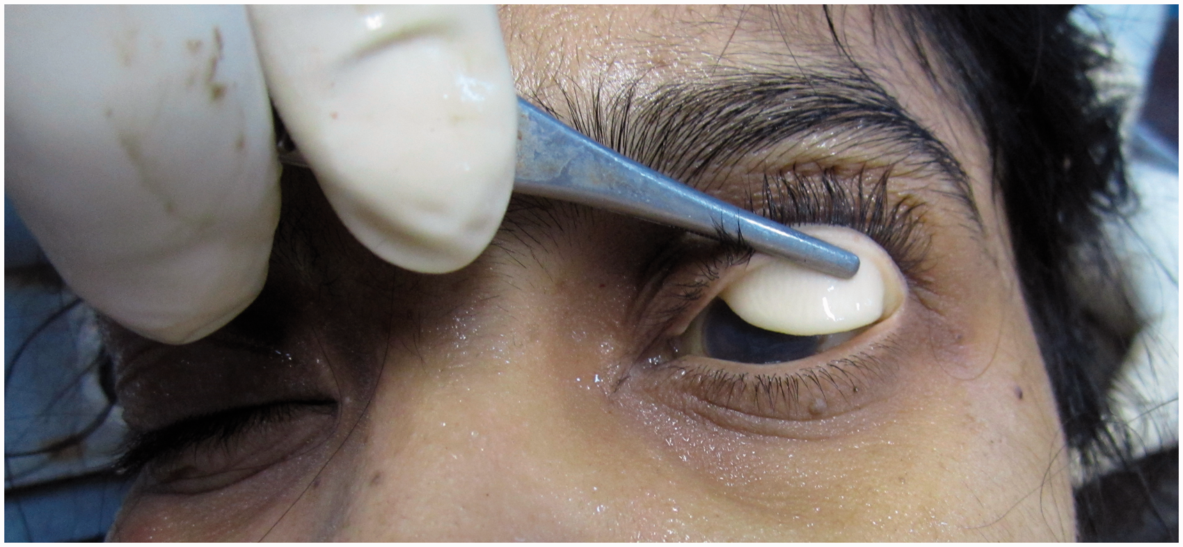
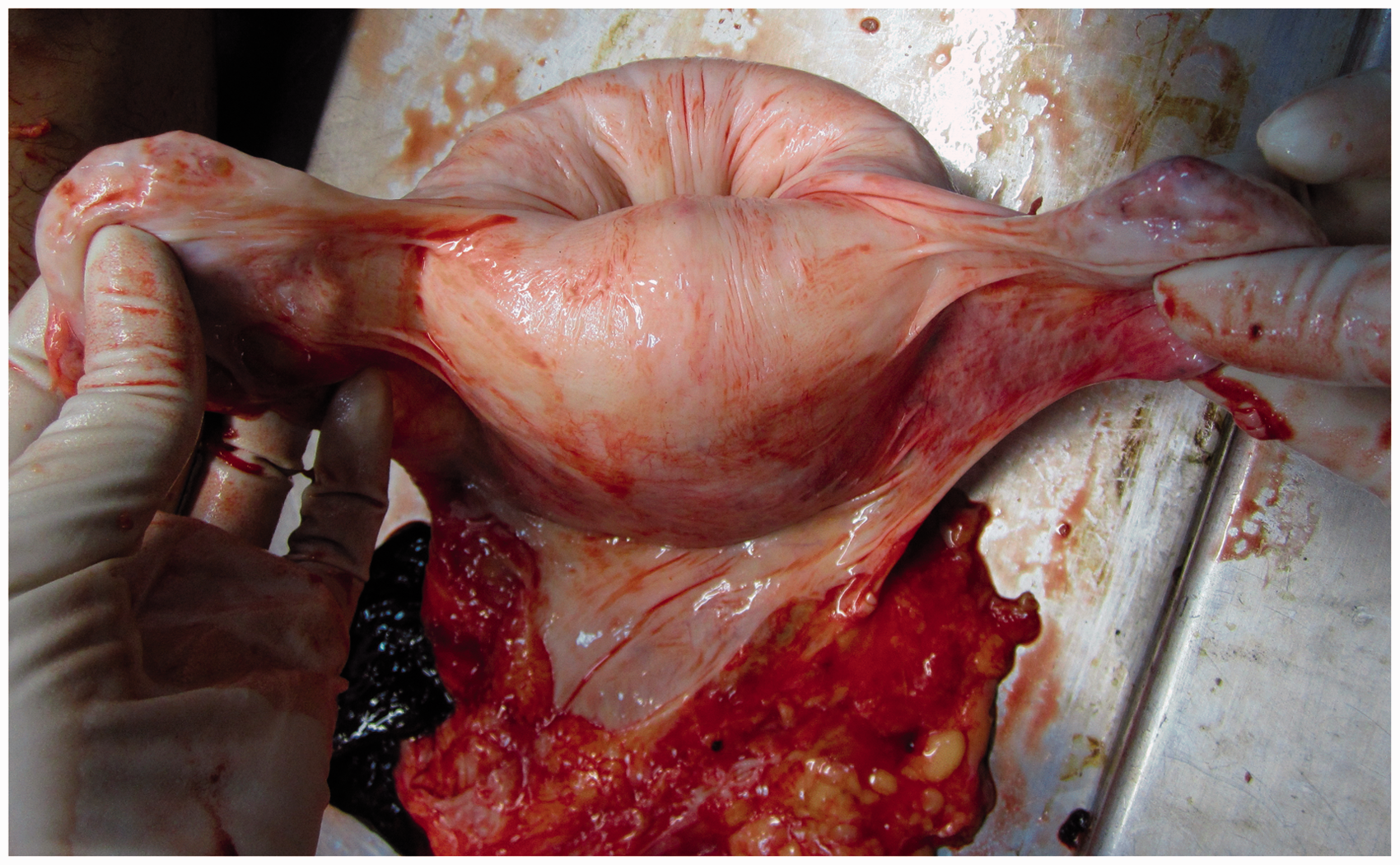
On internal examination, the organs, including the lungs, liver, brain, kidneys and stomach mucosa, showed severe pallor. The abdominal cavity contained an enlarged and globular uterus. The fundus of the uterus was unusually inverted, with the serosal surface dipping inwards, forming a cup-like depression (Figures 2 and 3). The uterus in its inverted state measured 17.5 cm × 12.5 cm × 7.5 cm, with the inverted fundus measuring 9.5 cm. On reverting the uterus, the dimensions were 23 cm × 12.5 cm × 7.5 cm. On opening the uterine cavity, there were about 250 mL of retained blood clots found adherent to the fundal region of the endometrium, which were easily separated on manual removal. Raw areas of denudation were present on the fundo-anterior wall of the endometrium, suggestive of placental attachment (Figure 4). The internal and external os of the cervix was found to be open and dilated. The uterus weighed 818 g.
Image of uterus showing dipping of the fundus into the cavity, along with the tubes and ovaries. Uterus with its adnexa showing a ‘cup-like depression’. Cut section of uterine cavity showing raw areas of denudation on the fundo-anterior wall.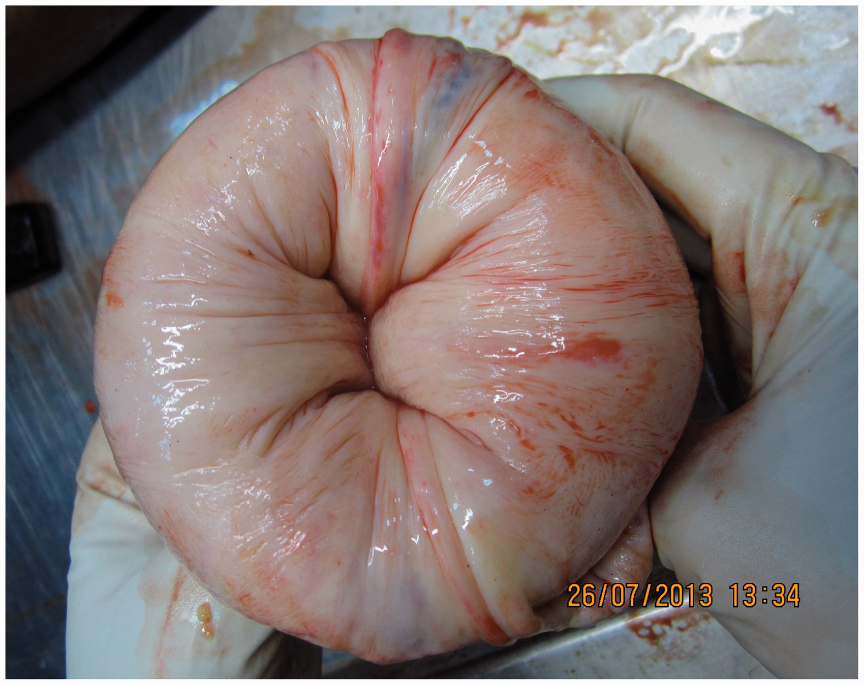

Death was attributed to shock and haemorrhage due to post-partum bleeding from an inverted uterus and incomplete evacuation of retained clots occurring in the third stage of labour. The case was referred to the State Medical Council (SMC) for further action. (As per the guidelines of the SMC, all cases of alleged medical negligence are referred to the SMC where a medical board is formed constituting doctors from concerned specialties for the final opinion regarding negligence. Therefore, the authors referred the case to the SMC).
Discussion
Puerperal uterine inversion is classified into acute, subacute and chronic inversion. The acute inversions occur immediately or within 24 hours of the delivery; subacute inversion occurs after 24 hours and before four weeks of the delivery; and chronic inversion occurs more than four weeks after the delivery. 9 The prevalence of each class of inversion (i.e. acute, subacute and chronic) is 83.4%, 2.62% and 13.9%, respectively.5,9,10 It is classified into four stages where, in the first stage, the uterine base is in the uterine cavity and does not cross the cervix of the uterus. In the second stage, the uterine base crosses the cervix and is passed into the vagina. The third stage involves the exteriorisation of the uterine base at the vulva, and in the fourth stage, the vaginal walls participates to the inversion. The present case was an acute inversion of the first stage.
The aetiology of uterine inversion has been attributed to many factors such as oxytocic arrest after a prolonged labour, premature umbilical cord traction or abdominal expression before placental separation, particularly in fundal attachment.11,12 Intrinsic risk factors such as primiparity, pauciparity, uterine hypotonia secondary to twin pregnancy, betamimetic, fundic or accreta placenta, fundic myoma and short umbilical cord5,9,13,14 has also been reported. Idiopathic inversion rarely occurs. 8
In this case, the deceased was a primigravida with a single uncomplicated pregnancy of normal duration of labour having fundal attachment of the placenta. Administration of oxytocics, placental accreta or uterine myoma were ruled out at history and autopsy. Hence, premature umbilical cord traction before complete placental separation which was fundally attached was the most reasonable explanation for the cause of uterine inversion.
The diagnosis of uterine inversion is usually based on clinical signs and symptoms such as severe lower abdominal pain in the third stage of labour, haemorrhage (present in 94% of cases) and severe shock. 15 In complete inversion, the inverted fundus is palpable at the cervical os or vaginal introitus, while in incomplete inversion, the fundus is palpable in the lower uterine segment. 13 Though haemorrhage and shock are the cause of death in most cases, sometimes it can occur due to air embolism during repositioning. 16
The management of uterine inversion requires immediate treatment for circulatory shock, uterine repositioning and antibiotic therapy. The nonsurgical methods for reverting the uterus are manual repositioning (Johnson’s manoeuver) and hydrostatic reduction (O’Sullivan); surgical methods include the incision of the constriction ring vaginally (Spinelli), upward traction on the round ligaments with the assistant applying upward pressure from the vagina (Huntington) and incising the constriction ring posteriorly at laparotomy (Haultain). 17 The newer methods include laparoscopic reduction, 18 the use of obstetric ventouse at laparotomy 19 and application of cephalad traction on the deepest visible part of the posterior uterus. 20
Recognition of such obstetrical emergencies and application of such manoeuvers require expertise and skill attainable only through formal training. In this case, the delivery was conducted by an untrained TBA who failed to appreciate the danger signals. Many successful reversions of this condition have been conducted by using the manual repositioning method which is easy, effective and can be initiated promptly.13,21–23 However, it is usually aided by tocolytics or halogenated anaesthetic agents. 13 Therefore, it would be fair to state that even though TBAs can be trained to recognise and infer such clinical clues and be trained to adopt the manual method for repositioning, bearing in mind their training, the circumstances they work under and the facilities available to them, it is not practical for them to administer tocolytics or general anaesthetics.
In India, approximately 80% of deliveries are conducted by untrained birth attendants who have always been outside the formal health-care system. 8 The nursing council of India recognises the courses of diplomas and degrees in nursing, including that of midwifery, 24 but the validity, recognition and an appropriate registration body for TBAs is ambiguous. However, TBAs are recognised under various government policies such as the Janani Suraksha Yojana (JSY) under the National Rural Health Mission. This Yojana identifies ASHA, the accredited social health activist, as an effective link between the government and the pregnant women in low performing states. However, even TBAs in the JSY facilities have low competence and lack the skills needed to provide appropriate care for emergency obstetric complications. 25 Several studies are of the consensus that TBAs are generally not able to handle most potentially fatal complications.26–28 Prompt diagnosis along with timely, eventful and aggressive management of potentially life-threatening complications during labour or a quick transportation to referral-level care, all of which require considerable skill, are critical to the success of life-saving interventions. 29 The 5th Millennium Development Goal aims to improve maternal health by providing every woman free or cashless skilled institutional delivery. However, the financial and infrastructural constraints faced by developing countries make this goal a difficult one to achieve in the near future. The indispensable role played by TBAs in maternal and child health cannot be ignored, as the rural women will continue using their services until skilled health workers reach them. The only solution in improving maternal and child health in rural areas is by strengthening TBAs’ abilities through training, proper supervision under skilled workers and developing referral pathways for every stage of pregnancy and labour in case of complications and unmanageable situations. However, TBAs’ skills and expertise should be clearly defined to prevent them from acting beyond their scope of expertise and making them accountable for any negligence. (As per Indian law, section 304 A of the Indian Penal Code is the common law duty of care applicable in any case of negligence by ordinary people or by any professional). It becomes mandatory to regulate the functions of TBAs. The loopholes in the health-care system and the accountability of every health-care provider should be fixed in order to prevent such avoidable deaths.
Footnotes
Funding
This research received no specific grant from any funding agency in the public, commercial or not-for-profit sectors.