






Abstract
Age estimation is one of the essential criteria in the identification process. The method of age estimation employed depends on the availability of skeletal material brought for forensic examination. McKern and Stewart's method constitutes one of the principal approaches towards pubic symphyseal age estimation. The method entails evaluating morphological changes within the pubic symphysis and subsequently allotting a specific score corresponding to the observed changes. Based on the obtained scores, an age range is then assigned to the remains presenting for examination. The present systematic review was undertaken to ascertain the applicability of the McKern-Stewart method for age estimation. Studies pertaining to the use of the McKern-Stewart method for age estimation in skeletal remains were retrieved by keying in a combination of MeSH terms and other free terms from four databases. The retrieved articles were subjected to a stringent inclusion and exclusion criteria, following which the risk of bias was assessed and the overall quality of evidence was established. Once the final tally of relevant articles was obtained, data specific to the mean age corresponding to each score was extracted. Non-parametric tests and boxplots were employed to compare the mean ages reported across multiple studies. The present systematic review concludes that the McKern-Stewart method can be applied for the purpose of age estimation in skeletal remains. Broader age cohorts for higher scores, as well as, overlapping values for age ranges in relation to the cumulative scores, however, can be considered a limitation for its applicability in forensic case work.
Introduction
Age estimation occupies a prominent niche in the identification process. 1 The role of age estimation in legal and medico-legal investigations includes, and is not limited to, cases dealing with child labour, pedopornography, 2 juvenile sentencing, and at other times, missing persons and illegal immigrants. 3 When skeletal remains present for examination forensic anthropologists rely on various age markers distributed throughout the bony framework. 4 Morphological changes ensuing within bony surfaces, and/or ossification and fusion of bones have often been employed to ascertain age-at-death of individuals.5–12 The pelvis is of particular importance as it exhibits significant age-dependent developmental, as well as, degradative changes, allowing for age estimation across a broader age cohort.5,9,11,13–18 Within the pelvis, more commonly researched parameters are the pubic symphysis, and the auricular surface of the ilium.19,20
The pubic symphysis displays high levels of preservation owing to the robusticity and taphonomic resilience conferred to this region within the pelvis. As an added advantage, transpiring pubic symphyseal changes encompass a broad age range, allowing for age estimation in sub-adults, as well as, adults. 5 The first documented age estimation method using the pubic symphysis was laid down in early 1900s. Todd in 1920, proposed a ten phase pubic symphyseal age estimation method. 21 Until the middle of the 20th century, Todd's method remained the primary method employed for pubic symphyseal age estimation. 22 Subsequently, multiple methods for age estimation through the evaluation of morphological changes within the pubic symphysis were developed.5,18,23–28 In 1957, McKern and Stewart devised their component-based age estimation method in order to overcome the drawbacks associated with Todd's phase-based method. 18 Morphological changes within the pubic symphysis were grouped into three distinct components; Dorsal plateau, ventral rampart, and symphyseal rim. Each component was graded on a scale of 0–5 based on a thorough analysis of transpiring morphological changes, following which, a cumulative score of 0–15 was computed. Obtained cumulative score was then employed to assign an age range to skeletal remains. McKern-Stewart's component-based analysis offers certain obvious advantages over its phase-based counterparts by allowing for an objective evaluation of pubic symphyseal changes. 29
Subsequently, a number of researchers have undertaken studies employing the use of the McKern-Stewart method, rendering it one of the principal pubic symphyseal age estimation methods.30–44 A major share of this research attempted to age skeletal remains directly using McKern and Stewart's criteria, while, two studies incorporated the use of CT scans 42 and casts, 44 respectively. Additionally, the majority of the research had reported mean ages pertaining to each cumulative score following the format prescribed by McKern and Stewart. Snow 43 alternatively, derived simple and polynomial models using the total score so as to confer greater statistical efficiency to the method. Studies conducted on geographically distinct Indian populations reported varying degrees of applicability for specific age groups.30–41 The standalone CT-based study using the McKern-Stewart method for an Indian population generated regression models, and, reported greater accuracy for a specific age group of 15–40 years. 42
Klepinger et al. 44 evaluated the applicability of the McKern-Stewart and the Suchey-Brooks method, and, advocated the use of the latter in forensic age estimation. Previously undertaken systematic reviews using Suchey-Brooks method have indicated a good accuracy with direct skeletal examination, 45 as well as, CT-based examinations. 46 However, such an analysis is presently lacking for the McKern-Stewart method of pubic symphyseal age estimation. The present systematic review was aimed at establishing the applicability of the McKern-Stewart method for age estimation through a meta-analysis of findings reported across multiple studies. With a major share of published research undertaking an investigation of the McKern-Stewart method through a direct examination of skeletal remains and reporting their findings in the format prescribed by McKern and Stewart, the systematic review had to be restricted to include similar studies.
Scope and organization of the review
This systematic review was targeted towards isolating original research focussing on the use of the McKern-Stewart method for analysing pubic symphyseal changes.
McKern and Stewart in their study scored the three components of dorsal plateau, ventral rampart, and symphyseal rim on a scale of 0–5. For the dorsal plateau the scores 0–5 represented the following morphological changes: dorsal margin absent; a slight margin formation first appears in the middle third of the dorsal border; the dorsal margin extends along the entire dorsal border; filling in of grooves and resorption of ridges to form a (beginning) plateau in the middle third of the dorsal demi-face; the plateau, still exhibiting vestiges of billowing, extends over most of the dorsal demi-face; billowing disappears completely and the surface of the entire demi-face becomes flat and slightly granulated in texture, respectively. Similarly, the ventral plateau was scored from 0–5, with the scores representing the following features: ventral bevelling is absent; ventral bevelling is present only at superior extremity of ventral border; bevel extends inferiorly along the ventral border; the ventral rampart begins by means of bony extensions from either or both of the extremities; the rampart is extensive but gaps are still evident along the earlier ventral border, most evident in the upper two-thirds; the rampart is complete, respectively. The third component of symphyseal rim was graded from 0–5 based on the presence of the following features: the symphyseal rim is absent; a partial dorsal rim is present, usually at the superior end of the dorsal margin, it is round and smooth in texture and elevated above the symphyseal surface; the dorsal rim is complete and the ventral rim is beginning to form, there is no particular beginning site; the symphyseal rim is complete and the enclosed symphyseal surface is finely grained in texture and irregular or undulating in appearance; the rim begins to break down and the face becomes smooth and flat, the rim is no longer round but sharply defined with some evidence of lipping on the ventral edge; further breakdown of the rim (especially along superior ventral edge) and rarefaction of the symphyseal face along with disintegration and erratic ossification along the ventral rim, respectively. Described morphological features have been represented in Figure 1.
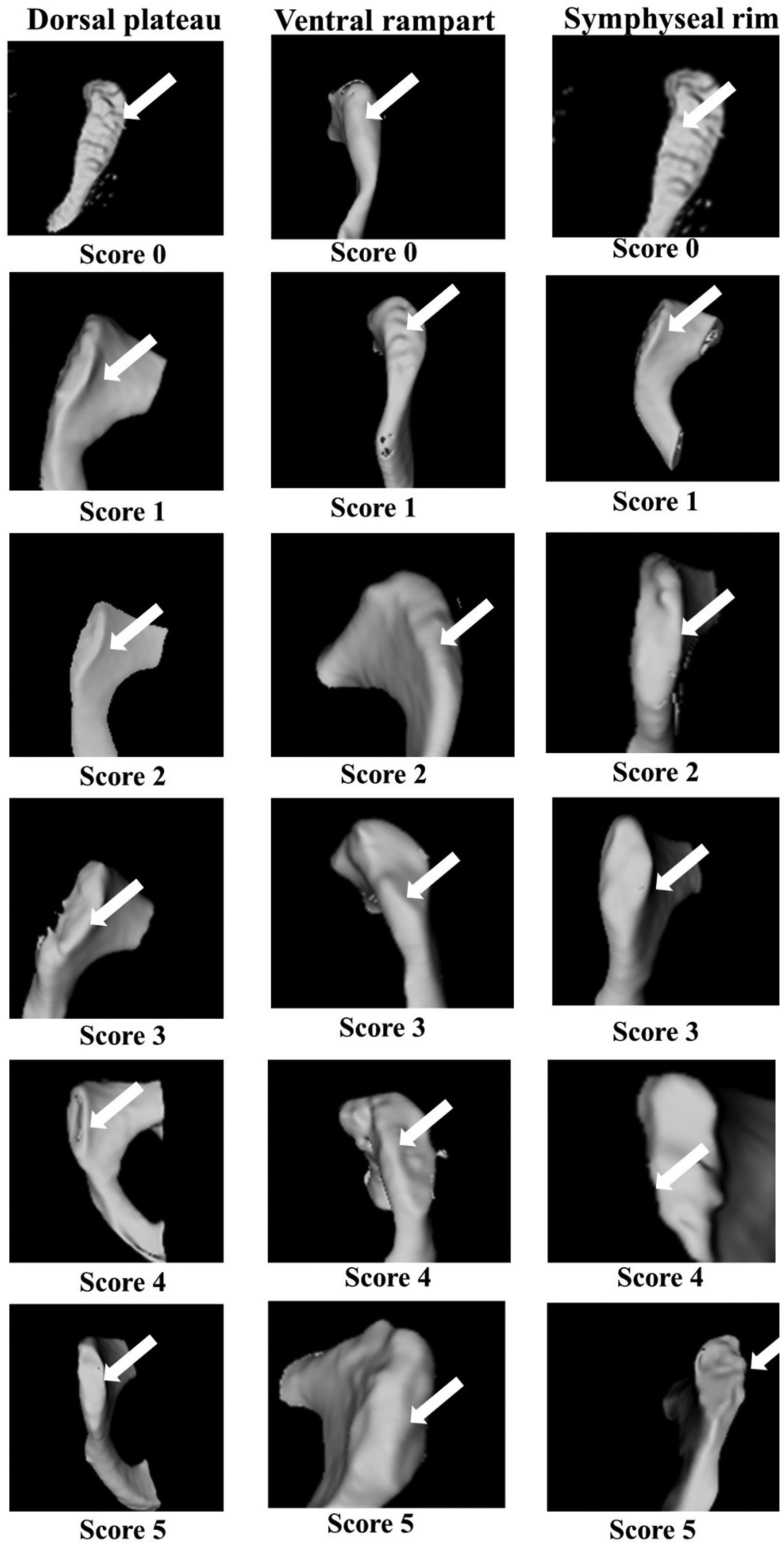
Morphological features defined by McKern-Stewart. 18
Relevant papers were retrieved (see subsections on search strategy and eligibility criteria), their overall quality was evaluated, and the risk of bias was established. The mean ages reported in these studies were extracted and subjected to meta-analysis to evaluate the applicability of the method for age estimation.
Materials and methods
The review question “To determine the applicability of the McKern-Stewart method for age estimation from pubic symphyseal changes” was constituted. Methodology for the present systematic review was developed in accordance with Preferred Reporting Items for Systematic Reviews and Meta-Analysis (PRISMA) guidelines.47,48 The review was subsequently registered with the PROSPERO database (International Prospective Register of Systematic Reviews) under the ID number CRD42020159557.
Search strategy
The search strategy was formulated with the objective of deriving scientific papers employing the McKern Stewart method to estimate and/or verify the age-at-death of skeletal remains. Search equations generated using a combination of MeSH terms and relevant free terms in amalgamation with specific Boolean operators, were keyed into four different databases- PubMed, CENTRAL, Google Scholar and ScienceDirect by two independent researchers (VW and RS). The final search equations generated for extracting relevant studies were:
“age estimation” AND “pubic symphysis” “forensic age estimation” AND “pubic symphysis” “age estimation” AND “McKern Stewart” AND “pubic symphysis” “forensic age estimation” AND “McKern-Stewart” AND “pubic symphysis” “accuracy” AND “reliability” AND “McKern-Stewart”
Eligibility criteria
Observational studies pertaining to the McKern-Stewart method, conducted in or before 2019 were included for review and meta-analysis. Editorials, literature reviews, case reports and research articles employing methods other than the McKern-Stewart method for pubic symphyseal age estimation were excluded.
No language restrictions were defined at the time of data search and extraction. Duplicate articles across the different databases were excluded. Each of the two researchers (VW and RS) conducted an initial assessment of all extracted titles. Articles wherein the title and abstract did not present sufficient information were retrieved to assess its inclusion/exclusion in the review. From these retrieved articles, original studies which did not employ the McKern-Stewart method for pubic symphyseal age estimation were excluded. Reference lists of relevant original studies were re-evaluated so as to identify any original research overlooked during the initial search. All the articles which addressed the review objective and satisfied the pre-decided inclusion and exclusion criteria were subjected to qualitative and quantitative analysis. Unanimity between the two reviewers was ensured at each stage of this systematic extraction of research articles.
Data extraction
A spreadsheet was created to standardize the process of data extraction. Information pertaining to the title of the paper, research objectives described within the abstract, study population, sample size, URL, description and details of the articles extracted, were noted in a systematic manner. Data in terms of age ranges, mean age and standard deviation pertaining to the total cumulative score ranging from 0–15, as well as, individual scores for each component (0–5) defined by McKern-Stewart was extracted wherever possible. Relevant articles with restricted access, or, where pertinent data was missing were marked. Authors of such original articles were contacted, and, at the end of a nine-month waiting period available data was subjected to qualitative and quantitative assessment.
Assessment of risk of bias of studies
The methodological quality of the available studies was established independently by two researchers (VW and RS) using the QUADAS-2 tool. 49 QUADAS-2 uses four parameters to evaluate the risk of bias of any study: patient selection, index test, reference standard, and flow and timing. The first three parameters additionally indicate the applicability of these selected studies in answering the review question. Risk of bias was established by answering specific signalling questions in the form of “Yes” or “No” or “Unclear,” provided within the tool. The risk of bias analysis was evaluated using ROBVIS. 50
Meta-analysis
Meta-analysis of the extracted data was carried out using IBM Statistical Package for Social Sciences (SPSS v 23.0). Differences between mean ages corresponding to each cumulative score (0–15) reported across different studies were analysed using the Kruskal-Wallis test For studies which reported mean ages for scores of each component (0–5), an additional Kruskal-Wallis test was performed. Boxplots corresponding to the same representing the median, minimum, maximum values and interquartile range were plotted.
Results
An initial search across the four databases yielded a total of 1711 articles. These were subsequently reduced to 1040 articles following the removal of duplicates. 1010 articles did not fall within the limits of the inclusion criteria pertaining specifically to the type of study, and were thus excluded. An exhaustive screening of the remaining 30 papers based on the information contained within the title, and, abstract further reduced the search results to 10 original articles. From these remaining 10 observational studies, three additional research articles had to be excluded due to restricted access to their complete versions. Thus, a total of seven original articles fulfilling the inclusion criteria, and, addressing the review question were obtained. These seven observational studies were then subjected to a rigorous qualitative evaluation. Figure 2 illustrates a flow chart with various steps of the Systematic Review (in accordance with PRISMA guidelines). 47
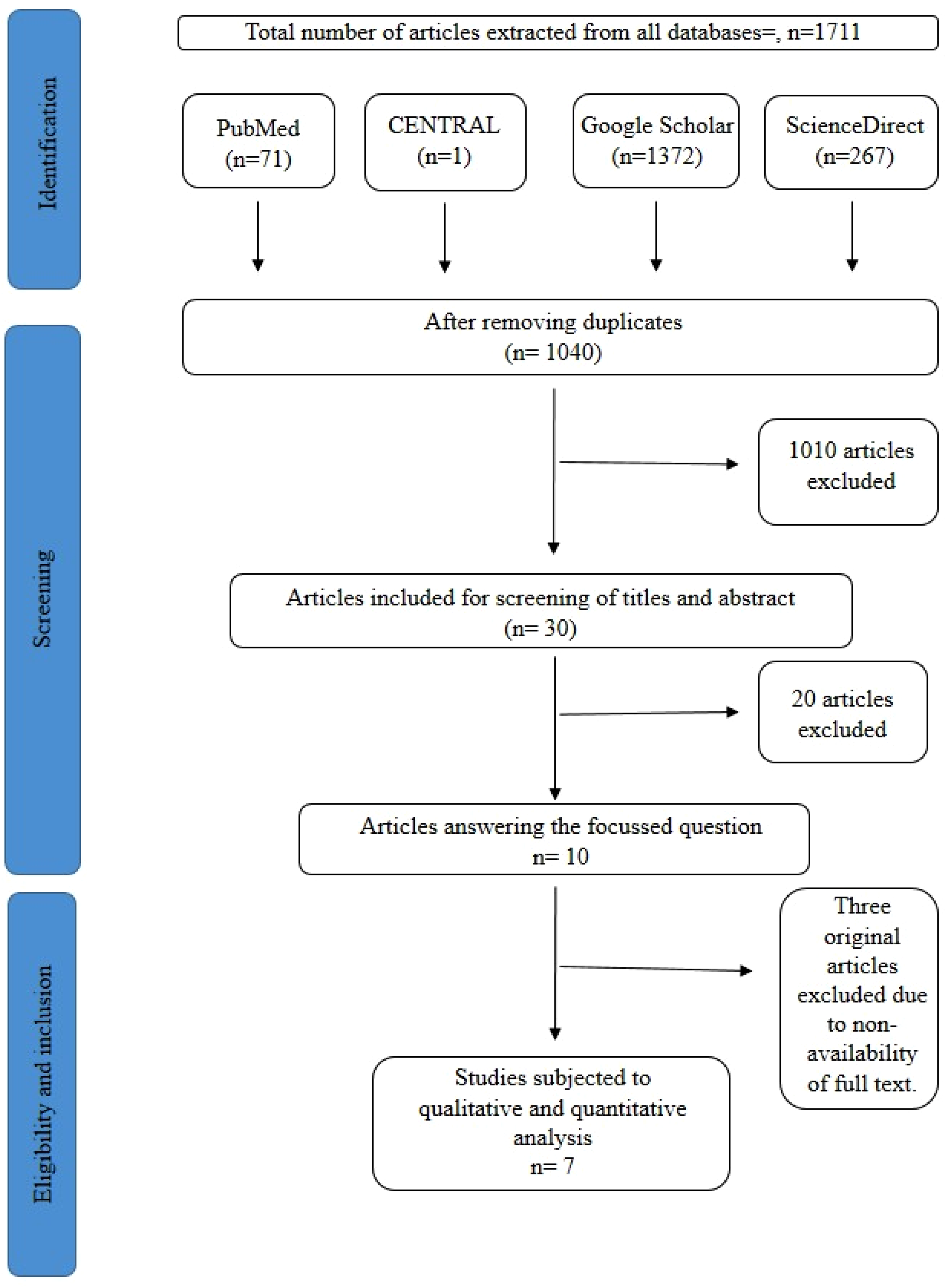
Flow chart with various steps of the systematic review along the guidelines proposed by the preferred reporting items for systematic reviews (PRISMA).
In order to determine the applicability of the McKern-Stewart method, data extracted from the seven studies was meta-analysed alongside the data contained within McKern-Stewart's original study. Thus, meta-analysis was conducted on a total of eight studies.
Characteristics of included studies
All studies included herein, barring the primary study by McKern and Stewart are prospective cross-sectional studies carried out within the Indian subcontinent. The age groups included within these studies ranged between 12–75 years. McKern and Stewart 18 analysed 349 skeletal remains of the male sex pertaining to a varied age range. Their obtained findings were represented in terms of age range, mean age, mode and standard deviation, for, cumulative scores (0–15). Sinha and Gupta 30 applied the McKern-Stewart method on male skeletal remains and demonstrated their observations in the form of mean age, standard deviation and associated age ranges. Sharma et al. 31 analysed remains of both sexes using the McKern-Stewart method and commented on the obtained mean ages and standard deviation. Kumar et al., 32 established the mean age for each cumulative score (0–15). Kumar 33 reported the mean age, standard deviation and corresponding age range of 32 remains belonging to the male sex. Prasad et al. 35 described the mean age and standard deviation for scores 0–15 based on a thorough examination of 80 male skeletal remains. Singh et al., 34 and Selvamurugan et al. 36 reported mean ages for scores 0–15 after a thorough analysis of 53 and 100 skeletal remains, respectively. Thus, a total of 8 studies had reported mean ages for scores 0–15, including McKern-Stewart's original research.
McKern-Stewart in their original study had additionally reported the mean age for scores 0–5 for each of the three components. 18 Similar work was undertaken later by Kumar, 33 Singh et al., 34 and Selvamurugan et al. 36 Thus, a total of 4 studies had reported mean ages corresponding to scores 0–5 for each of the three components.
Meta-analysis had to be restricted to mean age data obtained from each study, as, other associated descriptive statistics of range and standard deviation were not always reported.
Risk of bias of studies
All seven studies included for qualitative analysis had reported their patient selection procedure, and inappropriate exclusion of samples was avoided in all these studies. Sharma et al. 31 and Singh et al. 34 reported blinding at the time of analysis. Proof of age, wherever employed, included police and municipal records, often followed by confirmation from relatives of the deceased. All seven studies being prospective cross-sectional studies, information pertaining to sex, wherever applicable, was known to the investigators. Across all seven papers, there was adequate information regarding flow and timing. None of the studies reported a high risk of bias, with most of them indicating some concerns regarding risk of bias wherein relevant information had not been provided in order to comment on certain aspects. The assessment of risk of bias is shown in Table 1.
Risk of bias assessed using QUADAS-2.

Quality of evidence assessed using GRADEpro
In order to confer the undertaken review additional authenticity, the quality of evidence was analysed and graded using GRADEpro. 51 The present systematic review includes a total of 795 participants spread across seven cross-sectional studies. No serious indicators for high risk of bias, or, indirectness was observed. Lack of significant differences between mean ages for each score, across studies, indicates high precision and consistency. There is no direct evidence of publication bias or other associated biases, as, age estimation was carried out by an objective analysis of skeletal remains. An overall moderate to high evidence in favour of use of the McKern-Stewart method for age estimation in skeletal remains was obtained. (Table 2).
Quality of evidence evaluation of studies using GRADEpro.

*Quality assessment parameters.
Meta-analysis
Kruskal-Wallis test for distribution of mean ages corresponding to cumulative scores 0–15 across different studies
The Kruskal-Wallis test yielded no significant difference between mean ages across studies for cumulative scores 0–15 (p > 0.05) as shown in Table 3. Additionally, no sexual dimorphism was observed for distribution of mean ages corresponding to scores 0–15 (p > 0.05).
Differences in mean ages for cumulative scores (0–15) and scores for each component (0–5) across different studies.

*Significance level of p=0.05.
Kruskal-Wallis test for distribution of mean age for each component defined by McKern-Stewart
The distribution of mean ages for scores 0–5, for each of the three components (dorsal plateau, ventral rampart, and, symphyseal rim), across studies, showed no significant difference (p > 0.05) as shown in Table 3.
Boxplots for distribution of mean age corresponding to scores 0–15
Boxplots for scores 0–15 representing the median, minimum and maximum values, along with the interquartile range associated with each cumulative score has been shown in Figure 3. The obtained minimum-maximum age range pertaining to a total score of 0 was 17.29–20.99 years. Morphological changes associated with score 1–2 were observed between 17.00–25.00 years. Score 3 yielded an age range of 17.00–25.25 years. For the cumulative score 4–5, the corresponding age range was 20.33–25.50 years. With a score of 6–7, an age range of 22.42–30.00 years was observed. Boxplots for the scores 8–9, and, 10 indicated ranges of 24.14–31.87 years, and 24.50–35.50 years, respectively. The age group showing the desired morphological characteristics associated with the score 11–13, across all the studies, was 29.18–38.83 years. For score 14, the obtained age range was 35.84–48.88 years. For a cumulative score of 15, the associated age range was observed to be 49.00–59.40 years, with 41.00 years representing the outlier for this specific group.
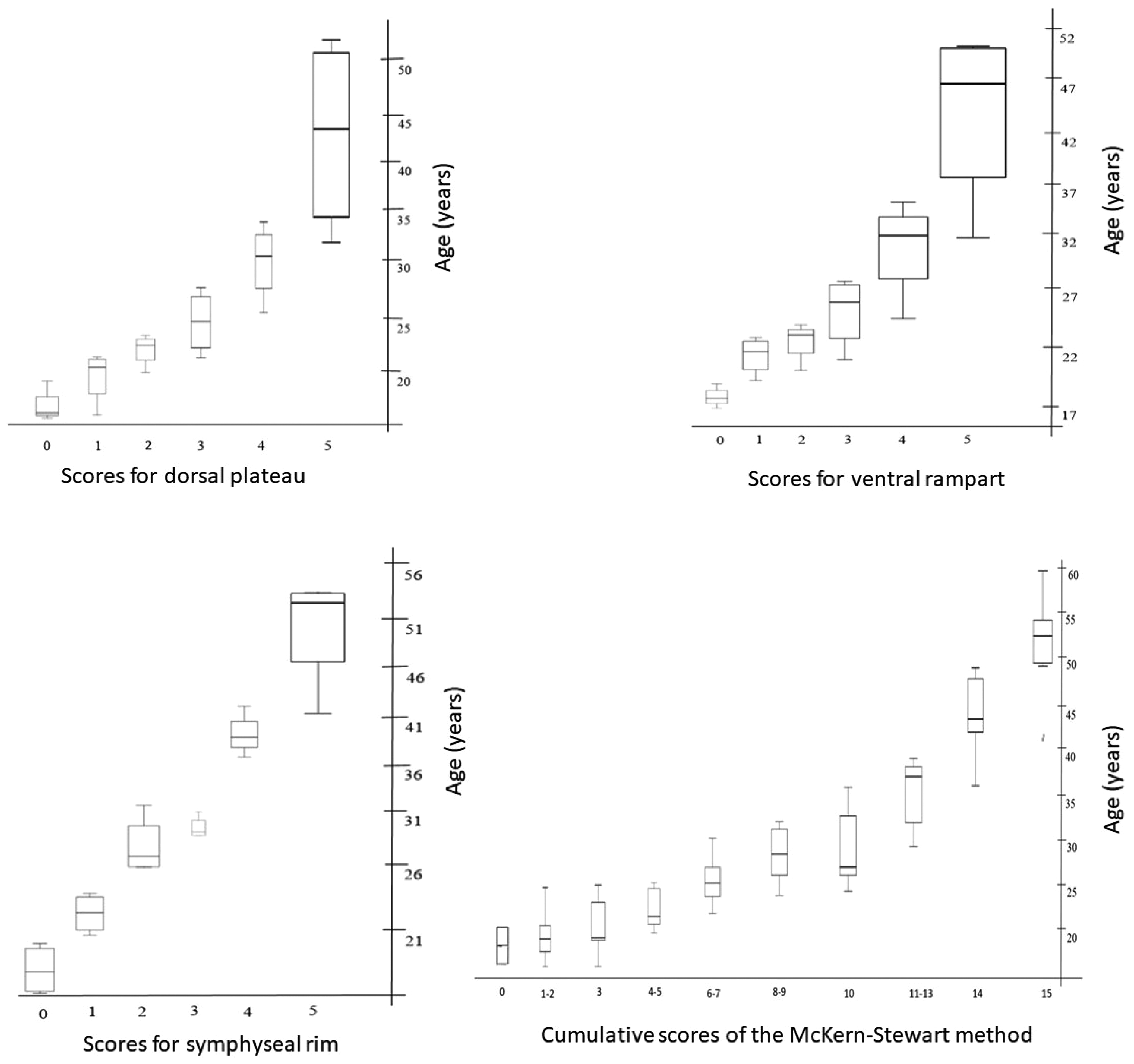
Box and whisker plots showing distribution of mean age corresponding to the scores defined by McKern-Stewart 18 across all studies.
Boxplots corresponding to scores 0–5 for dorsal plateau
The minimum-maximum age range for characteristics pertaining to score 0, across studies, was observed to be 15.67–19.30 years. For score 1, the obtained age range was 16.00–21.70 years. With score 2, an age range of 20.00–23.75 years was observed. For a score of 3, the defined morphological characteristics were observed within an age range of 21.50–28.90 years. A score of 4 yielded a corresponding age range of 26.10–34.42 years. Lastly, with the score 5, 32.90–51.87 years, was the obtained range. (Figure 3)
Boxplots corresponding to scores 0–5 for ventral rampart
A score of 0 yielded an age range of 16.87–18.30 years. For score 1, 18.90–23.00 years represented the associated age range. With score 2, the minimum-maximum age range for the defined set of morphological characteristics was observed to be 19.80–24.00 years. The obtained range for score 3 was 20.10–28.12 years. Score 4 yielded a range of 23.30–35.25 years and 31.90–49.84 years was the range corresponding to score 5. (Figure 3)
Boxplots corresponding to scores 0–5 for symphyseal rim
Score 0 yielded a minimum-maximum range of 16.20–19.20 years. With score 1, the obtained age range was 20.95–23.80 years. 25.00–31.00 years was the corresponding range associated with score 2. For score 3, 28.08–30.30 years was the obtained range. With score 4, onset and progression of the defined set of morphological characteristics was observed between 35.90–42.00 years. Lastly, for score 5 of symphyseal rim, the obtained age range was 41.10–53.80 years (Figure 3).
Discussion
The McKern-Stewart method 18 renders pubic symphyseal age estimation a more objective and simpler approach. They methodically grouped morphological changes ensuing within the pubic symphysis originally described by Todd 21 into three distinct components. The onset and progression of morphological changes transpiring within each component was further broken down and assigned scores ranging from 0–5. The cumulative score computed using the three components were employed to assign an age range to skeletal remains presenting for examination. Subsequently, numerous researchers embarked on age estimation studies using the McKern-Stewart method.30–44
All the studies included in the present systematic review reported a low to moderate risk of bias, a plausible explanation for which is the lack of information concerning index and reference tests. An overall moderate to high evidential quality was indicated. Studies by Sinha and Gupta 30 indicated lower age ranges for cumulative scores 1–5, and higher age ranges for scores 6–13, when compared to McKern-Stewart's original study. An upper limit of 69.175 years was obtained corresponding to a score of 15. Component wise analysis by Kumar 33 similarly yielded higher mean age values for scores 0–5 than those defined by McKern and Stewart.
The distribution of mean ages for cumulative scores (0–15) across these studies, including McKern-Stewart's original work shows no significant difference, suggesting that there is a certain uniformity in the prevalence of age related pubic symphyseal changes. Component-wise analysis also points towards a certain concordance in the distribution of mean ages corresponding to scores (0–5) for each component, across different studies. Boxplots for mean ages pertaining to scores 0–15 for all included studies indicates that it is possible to assign an age range to remains based on the observed changes. Boxplots for scores 0–5 for each of the three components suggest a similar trend. Even though an overlap is observed for age ranges corresponding to scores 0–15, a minimum age can successfully be assigned to the remains in question. This is of particular forensic significance in medico-legal cases including missing person identification.
Kumar et al. 32 reported a lack of sexual dimorphism in the distribution of mean ages for cumulative scores (0–15). This contradicts the findings of Sharma et al., 31 Berg 23 and Gilbert-McKern, 24 who reported a significant sex difference in the expression of morphological changes within the pubic symphysis. However, a significantly lower number of female remains were analysed within this study, necessitating the need to further scrutinize and address concerns regarding the applicability of the McKern-Stewart method for female skeletal remains.
Since the meta-analysed studies scrutinized the method on Indian populations, it can be assumed that the distribution of mean ages for this biogeographical region falls within the limits of McKern and Stewart's findings. Thus, the McKern-Stewart method can successfully be employed for age estimation on Indian populations. However, the sample size of observational studies included for meta-analysis comprised of a comparatively narrower age range. Additionally, specific sexual dimorphism targeted research, estimating the overall accuracy of the McKern-Stewart method are currently wanting. Further evidence pointing towards the applicability of the method for age estimation requires carrying out population specific studies around the globe. Age-related hormonal changes, modifications in bone texture, and, other lifestyle factors are known to influence the observed morphological changes alongside biogeography and demography, warranting the need to carry out studies targeting different populations. 52
The component-based analysis, in consortium with the observed uniformity in the distribution of mean ages for cumulative scores (0–15) across studies points towards the reliability of morphological features described by McKern-Stewart. However, with increasing cumulative score, broadening of age ranges associated with these scores is observed. A similar broadening of age ranges is observed with increasing scores for each individual component described by McKern-Stewart. A possible explanation for this is that McKern and Stewart's study sample for establishing their method of pubic symphyseal age estimation inadvertently comprised of younger individuals. This resulted in narrower yet overlapping age ranges for scores 0–13, and extremely broad ranges for scores 14, and 15, dampening the role of the method in forensic age estimation. From a forensic point of view, accuracy and precision is, and continues to remain, a longstanding challenge faced by forensic anthropologists. Accurate age estimates, along with other parameters of sex and stature are vital to the process of human identification. Broader overlapping age cohorts associated with the McKern-Stewart method pose a drawback to ascertaining the identity of skeletal remains presenting for medico-legal investigations.
One way to overcome this drawback and achieve greater accuracy is through a multifactorial approach to age estimation, by employing additional age markers within the pelvis. The acetabulum and auricular surface of the ilium, present as favourable, durable evidences which allow for accurate age estimation in older individuals.9,15–17 Such a multifactorial approach has the potential to greatly improve the reliability associated with age estimation. 53 Regression-based models offer another efficient alternative over the current application of the McKern-Stewart method. 42 Such a regression-based approach can improve applicability of the method by reducing the associated error rate resulting from the use of broader age cohorts. Additionally, regression models generated using individual components can aid in age estimation without mandating the need to visualize and score even the highly degraded features. Population specific studies employing the use of such regression models need to be undertaken as they can help confer additional validity to the McKern-Stewart method in favour of its applicability in forensic investigations. An alternative approach is to estimate the varying accuracy of each morphological feature towards the overall process of age estimation. Taking into consideration such a differential contribution can help generate more accurate age estimation models using the McKern-Stewart method. The use of such models, in consortium with a CT-based age estimation approach can further improve the implementation process by doing away with the time and resource intensive maceration process.
Conclusion
The findings of the present systematic review suggest uniformity in the distribution of mean ages corresponding to a defined set of morphological characteristics defined by McKern and Stewart, across multiple studies. No significant differences between mean ages were obtained for each cumulative (0–15), as well as, individual score (0–5) in these studies. Thus, it can be concluded that the McKern-Stewart method can be applied successfully to an Indian population for the purpose of age estimation. Broader age cohorts for higher scores, as well as, overlapping values for age ranges in relation to the cumulative scores, however, can be considered a limitation for its applicability in forensic case work. It can be overcome through the development of population specific regression models, and by incorporating a multifactorial approach to the process of age estimation.
Footnotes
Acknowledgements
This research article is a part of the ongoing doctoral research being conducted by the principal author (Varsha Warrier) at the Department of Forensic Medicine and Toxicology, All India Institute of Medical Sciences, Jodhpur, India. The principal author is grateful to the University Grants Commission, New Delhi for awarding research fellowship (UGC-JRF) for pursuing PhD. The authors are also thankful to the individuals who participated in this study, and to the authorities of the institution for allowing us to conduct this research.
Declaration of conflicting interests
The author(s) declared no potential conflicts of interest with respect to the research, authorship, and/or publication of this article.
Funding
The review was methodologically registered under the PROSPERO database (International Prospective Register of Systematic Reviews) with the ID number CRD42020159557. No support in the form of grants.
