
Abstract
Noma or cancrum oris is an orofacial gangrene causing progressive mutilating destruction of the infected tissues. It mainly affects malnourished children with poor oral hygiene and concurrent debilitating systemic illnesses. It is a polymicrobial infection and borrelia vincentii and fusobacterium are the most important pathogens known. We present a case of a boy aged 2.5 years with noma where klebsiella was grown and was the initial cause of failure of empiric therapy.
Introduction
Noma rapidly causes destruction of soft tissues, as well as muscle and bone, and may be fatal. Its aetiology is not fully understood, but is believed to be consequent to opportunistic infection due to an imbalance in normal oral microbiotic flora, seen predominantly in immunosuppressed or debilitated patients. 1 Several organisms have been implicated, though bacteroides and spirochetes are the primary causative agents. Klebsiella is not normally found, although it is implicated in cases of necrotising fasciitis.
Case report
A boy aged 2.5 years presented with swelling and ulceration of his lower lip associated with drooling and purulent discharge. He had had a febrile illness ten days before, after which a blackish discoloration on the mucosal aspect of the lower lip was noticed. This rapidly increased to involve the surrounding area over the next three days.
On examination, the boy had a swollen lower lip with an irregular punched-out ulcer involving the surrounding perioral area and extending to the gingiva but sparing the commissures (Figure 1a). There was concomitant anaemia (Hb = 70 g/L) and an elevated erythrocyte sedimentation rate, while HIV serology was negative.
(a) Lower lip swelling with an irregular punched out ulcer with central eschar. (b) Progression of the lesion after four days of empirical antibiotic therapy.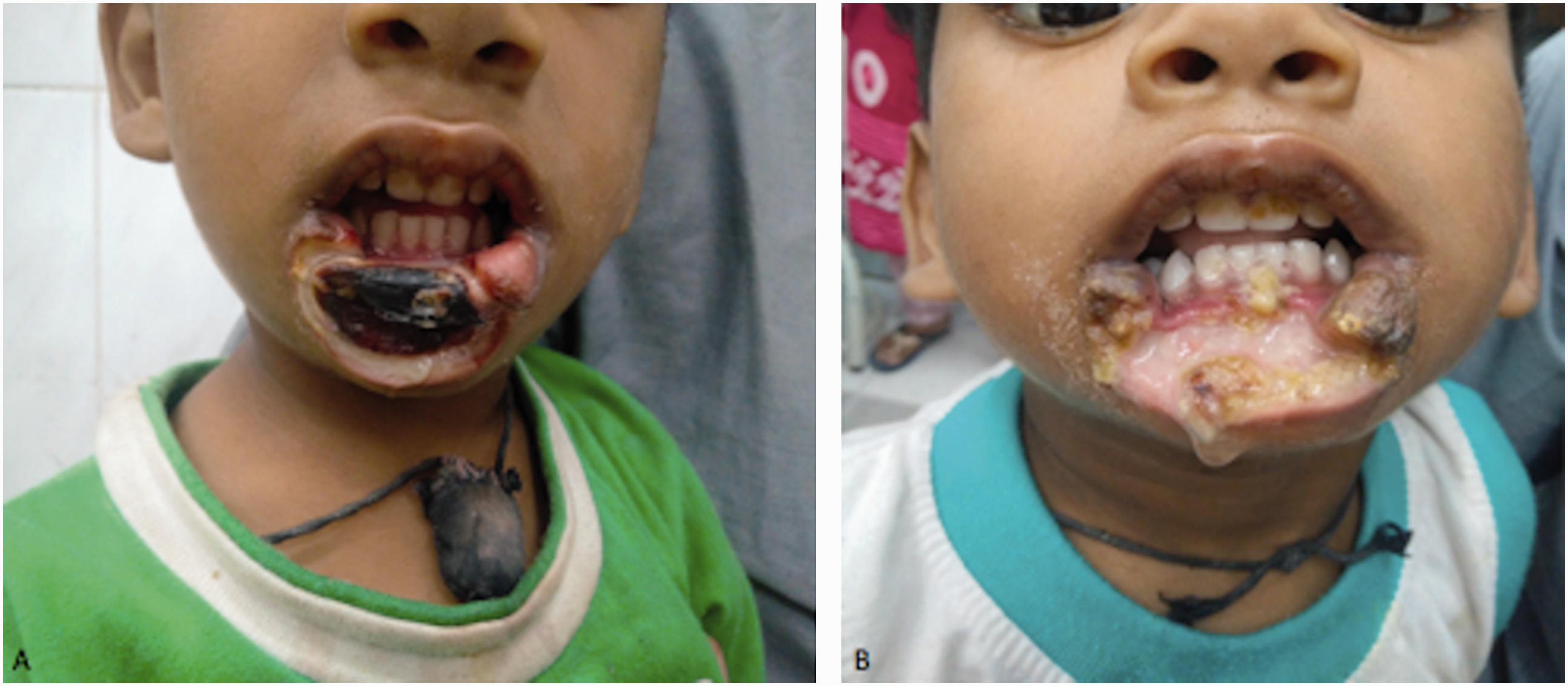
The clinical picture and course was suggestive of noma and, pending culture, oral metronidazole 80 mg tds and amoxicillin-clavulanic acid 200 mg b.i.d. was started. Even after four days, no improvement was seen (Figure 1b). Culture revealed Klebsiella pnuemoniae while anaerobic cultures were sterile. Based on the sensitivity report, the previous therapy was abandoned and ceftriaxone 425 mg b.i.d. was infused intravenously. This, together with hydrogen peroxide mouthwashes four times daily, led to a dramatic improvement within one week (Figure 2). The tissues healed, leaving a defect which requires reconstructive surgery.
(a) Improvement after one week of ceftriaxone. (b) At four-week follow-up.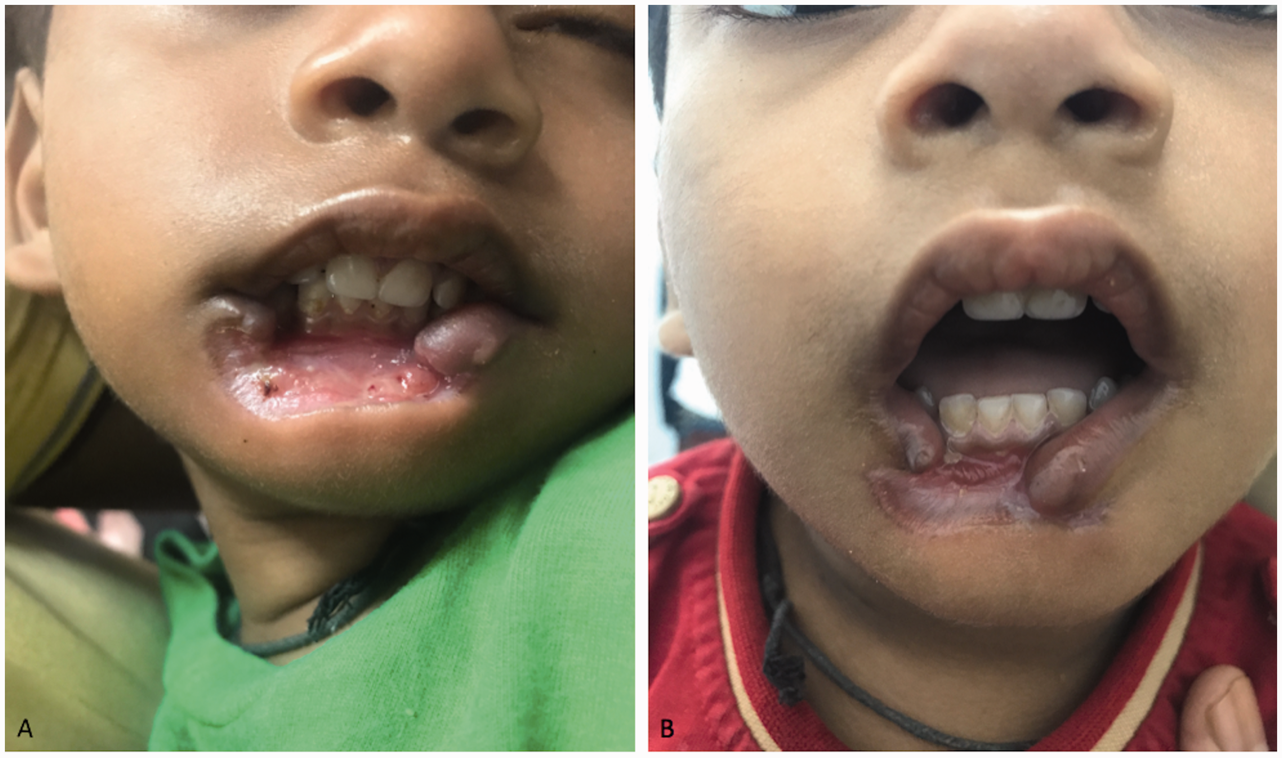
Discussion
Noma is derived from the Greek word ‘nomien’ which means to devour. It typically affects children during the weaning period, which often corresponds to a period of growth retardation in deprived children. Noma is considered to be the ‘face of poverty’ with a peak incidence in a belt extending from Mauritania to central Sudan, an arid zone with low a developmental index. Cases are exceedingly rare in adults except where some degree of immunosuppression exists.
Lowered host resistance and disruption of the gingival mucosal barrier allows entry of microorganisms responsible for the disease process. It was earlier believed that noma starts as a necrotising ulcerative gingivitis, later extending to involve the labiogingival fold and then the mucosal surface of the cheek and lip but this view is now disputed. According to recent reports, many viruses might begin the process, which typically may occur in the mouth of malnourished children after measles infection.
It is important to differentiate noma from ecthyma gangrenosum and necrotising fasciitis.
The former is classically associated with Pseudomonas aeruginosa sepsis and affects critically ill and immunocompromised patients. Unlike noma, the disease has a strong predilection for the so-called ‘apocrine’ areas, producing multiple lesions usually but rare involvement of the face. A characteristic tender erythematous halo is seen around the central eschar preceded by a bullous or pustular lesion.
The latter is a polymicrobial infection of the fascial planes and subcutaneous tissues, causing extensive tissue destruction. It most commonly involves the lower extremities, trunk and perineum, and very rarely involves the face, with only 35 cases reported worldwide.2,3 The disease is mainly seen in adults with an underlying predisposition, such as diabetes or alcoholism, and very rarely occurs in children. The hallmark symptom is intense pain and tenderness in the involved areas, out of proportion to the physical findings. The patient is toxic and there is a woody induration of the subcutaneous tissue with the presence of crepitus. All these features help to distinguish this entity from noma.
There is lack of a clear consensus on the first line antibiotic therapy for noma and most authors recommend ampicillin-cloxacillin and metronidazole for a minimum of 14 days. 4 While spirochaetes and fusobacterium are commonly implicated, their presence may just reflect the stage of the disease and the antibiotic profile used. Furthermore, it is plausible that antibiotics used change the bacterial population during the progression of the disease.
Klebsiella pneumonia was cultured from necrotic tissue, which is typically associated with fasciitis rather than necrotising gingivitis. However, the failure of first-line antibiotics and the isolation of Klebsiella, an uncommonly isolated organism, nonetheless suggests it being the primary aetiology. At any rate, the value of modifying treatment related to a second microbiological culture is highlighted.
Footnotes
Declaration of conflicting interests
The author(s) declared no potential conflicts of interest with respect to the research, authorship, and/or publication of this article.
Funding
The author(s) received no financial support for the research, authorship, and/or publication of this article.