






Abstract
Objective
The aim of our study is to determine if a fresh cadaver model (FCM) for the instruction of ultrasound (US)–guided fine-needle aspiration (FNA) of thyroid nodules is a practical method for instruction.
Study Design
Pre- and postinstruction assessment of medical students’ ability to perform US-guided FNA of artificially created thyroid nodules placed adjacent to the thyroid gland of a fresh cadaver.
Setting
University-based fresh cadaver laboratory.
Subjects and Methods
Study participants included a total of 17 first- and second-year medical students with minimal US training. Technical skills were assessed using a 10-item checklist. In addition, a cognitive assessment regarding the indications, contraindications, and complications of the procedure was completed. A postinstruction assessment was provided for participants 5 weeks after their initial assessment. Differences between pre- and postinstruction assessment scores of technical skills were analyzed using McNemar’s test. The mean cognitive knowledge gain was analyzed using a paired 2-sample t test.
Results
Eight of 10 items on the skills checklist were statistically significant between pre- and postinstruction skills assessment (P < .05). There was a statistically significant change in cognitive knowledge gain regarding the contraindications of the procedure (P = .001), but not for indications or complications (P = .104 and P = .111, respectively).
Conclusion
US-guided FNA continues to be an important diagnostic procedure in the workup of thyroid nodules, making it an essential skill to integrate into surgical skills lab. Our FCM for the instruction of US-guided FNA is the first of its kind, and this pilot study shows this is a viable method for instruction.
Completion of ultrasound (US)–guided fine-needle aspiration (FNA) of the thyroid is an integral component in the workup of thyroid nodules and has been the procedure of choice for the past 4 decades. 1 The prevalence of thyroid nodules varies based on the type of screening method used. 2 However, previous autopsy studies have found the prevalence to be up to 50% among those with no history of thyroid disease and up to 67% with the use of high-resolution US.3,4 FNA of the thyroid is the most sensitive and specific tool used in the workup of thyroid nodules; sensitivity is reported to be 65% to 98%, while the specificity is 72% to 100%. 5 While there is still some discussion regarding what type of specialists should perform US-guided FNA, several fields in medicine, including endocrine surgeons, otolaryngologists, pathologists, endocrinologists, and radiologists perform the procedure. 6 Due to the high prevalence of thyroid nodules and the breadth of specialties that perform the procedure, medical schools and residency programs will greatly benefit from the incorporation of training models that accurately simulate the experience of performing US-guided FNA.
Fresh cadavers have been used in the instruction of several procedures, including chest tubes, central lines, intubation, and, more recently, breast aspiration and biopsy of palpable lesions.7,8 The use of a cadaver has been found to be an effective method for the instruction of various surgical skills and these experiences are highly rated among participants. 9 Models that attempt to accurately represent human anatomy have been created for the instruction of thyroid FNA; however, a model that uses a fresh cadaver has yet to be described in the literature. 10 Our study aims to explore the use of a fresh cadaver model (FCM) for the instruction of US-guided FNA of thyroid nodules.
Methods
Throughout medical school, all students at our institution rotate through a fresh cadaver laboratory for training in outpatient and intensive care unit procedures. This study was integrated into the preexisting fresh cadaver laboratory curriculum. For the purpose of this research, all participants were first- and second-year medical students with minimal US experience. The fresh cadavers used for this project were obtained through our institution’s Willed Body Program. This study was approved by the University of Arizona Institutional Review Board.
The equipment used to complete this model includes the following: a fresh cadaver, US with linear probe, 10-cc syringe, 25-gauge needle, No. 15 blade scalpel, silk ties, latex-free gloves, water with food color added, and a marking pen. The artificial lesions used to simulate thyroid nodules are created using the tips of latex-free gloves, which are injected with 3 to 5 cc of colored water to simulate a cystic nodule. The tip of the glove is closed by twisting the base and using a silk tie to tightly secure the glove tip ( Figure 1 ). The silk tie is then left long so the artificial nodule can easily be withdrawn from the neck after the student has successfully punctured and aspirated fluid from the nodule.
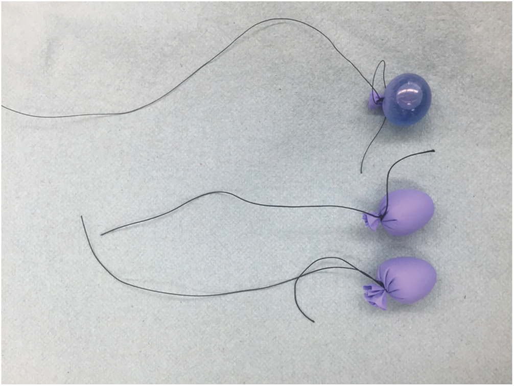
Artificial thyroid nodules created from the tips of latex-free gloves, which are then filled with colored water so the students can accurately determine if they are aspirating from the artificial nodule.
After the nodules are created, a careful dissection of the neck is performed. A horizontal incision extending from the sternocleidomastoid (SCM) to the contralateral SCM is created through the epidermis and dermis using a No. 15 blade scalpel. The incision is carried on through the subcutaneous fat to the level of the platysma muscle. Subplatysmal flaps are created and the strap muscles are identified, including the sternohyoid and sternothyroid muscles. The strap muscles are preserved as they will aid in stabilization of the artificially created thyroid nodules. After the strap muscles and the midline raphe are identified, the strap muscles are separated and parted from the capsule of the thyroid. Upon identification of the thyroid gland, lateral dissection is completed to where it meets with the carotid sheath fascia. The thyroid gland is then revealed and the nodules are placed into the adjacent perithyroidal tissue ( Figure 2 ).

Dissection of the neck is completed to reveal the thyroid and adjacent structures, so artificial nodules can be secured. GV, great vessels; SCM, sternocleidomastoid muscle; SPSF, subplatysmal skin flap; TC, thyroid cartilage.
During the preinstruction assessment period, students arrive and are presented with the materials needed to complete US-guided FNA of the thyroid using the described FCM; no instruction is given during this period. Among the materials provided to the students, they have the option of choosing between 3 US probes and 3 needle gauges to assess their understanding of the materials needed to complete an US-guided FNA ( Figure 3 ). The student is instructed to palpate the neck and select the appropriate US probe to identify the artificial thyroid nodule under US ( Figure 4 ). Upon identification of the nodule on US, the student is instructed to select the correct needle gauge and perform an US-guided FNA. While the participant is completing the exercise, an author is scoring his or her preinstruction skills assessment ( Table 1 ). After completion of the skills assessment, the senior author then goes through the procedure step by step, from selecting the proper tools to ensuring that the participant can complete all 10 items on the checklist independently prior to leaving the laboratory. Participants are instructed to return for the postinstruction skills assessment approximately 5 weeks later. This time frame was selected to ensure there was proper transference of skill, but was also based on the scheduling needs related to the existing curriculum. There is no opportunity to practice the procedure in between the pre- and postinstruction skills assessment. The 10-item checklist used for participant skills assessment is a modified checklist that was amended as appropriate for the anatomical location of the thyroid.11-13
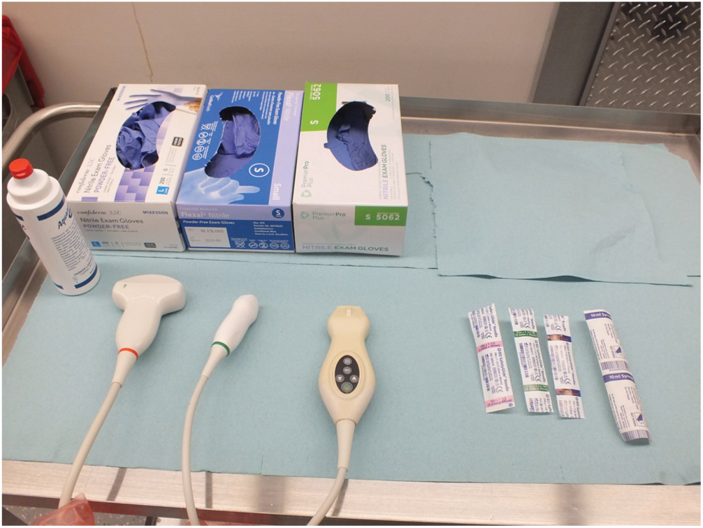
Students were provided 3 ultrasound (US) probes (convex, micro convex, and linear) and 3 needle gauges (18, 21, and 25 gauge) to choose from upon entering the laboratory to perform an US-guided fine-needle aspiration.
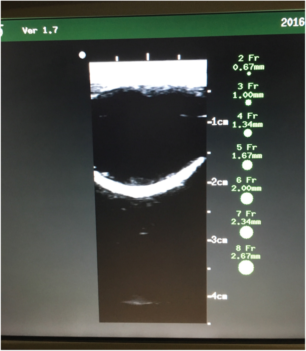
Appearance of the cystic, water-filled nodule under ultrasound.
Pre- and Postinstruction Skills Assessment.

Our study also included a pre- and postinstruction cognitive assessment, which evaluated the participants’ ability to retain information related to 3 major indications, contraindications, and complications of the procedure ( Table 2 ).14-16 The cognitive assessment was given at the beginning of the preinstruction laboratory, and all participants were then provided a short lecture after the fresh cadaver session regarding the indications, contraindications, and complications of US-guided FNA of the thyroid. A follow-up cognitive assessment was completed upon return to the laboratory 5 weeks later. Statistical analysis of the pre- and postinstruction skills assessment data was completed using a McNemar’s test to assess differences in the binomial paired data. A paired 2-sample t test was used to compare the mean pre- and postassessment scores for both the technical skills and cognitive assessments. Statistical significance was set at P < .05.
Pre- and Postinstruction Cognitive Assessment.
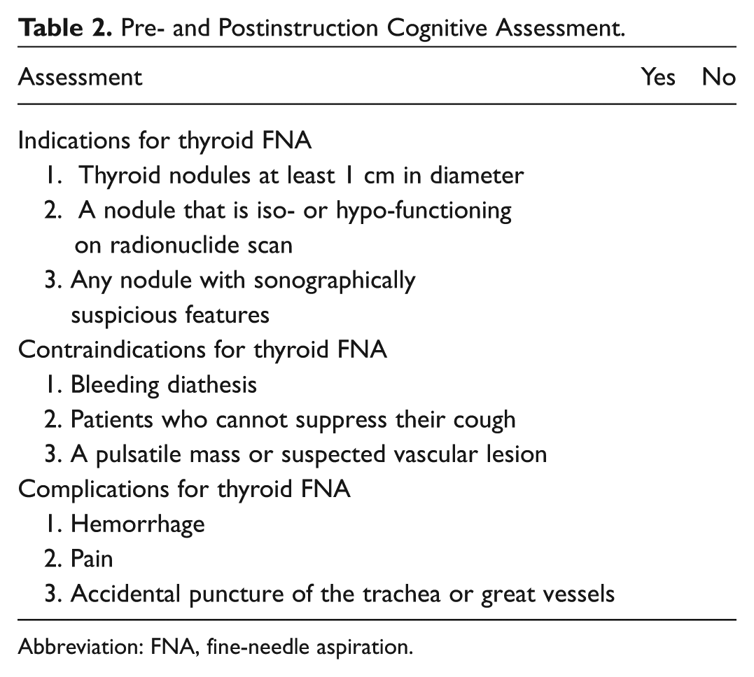
Abbreviation: FNA, fine-needle aspiration.
Results
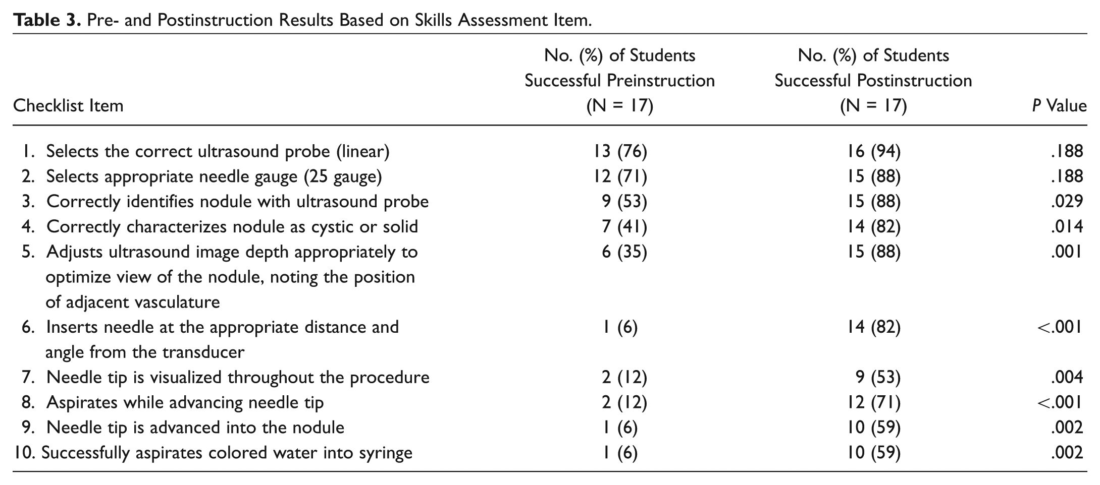
During January 2016, a total of 17 first- and second-year medical students participated in this study. Students had little exposure to US, and no students had previous experience related to thyroid or neck US. When comparing the 10 pre- and postinstruction skills assessment items, 8 skills were found to be statistically significant (P < .05) ( Table 3 ). There were no differences between the pre- and postinstruction skills assessment with regard to the ability of students to select the correct US probe or needle gauge (P = .188 for both items 1 and 2 on checklist). The preinstruction group mean (standard deviation [SD]) was 3.12 (1.54) compared with the postinstruction group mean (SD) of 7.53 (2.45) (P < .001). There was a statistically significant change in cognitive knowledge gain regarding the mean number of known contraindications of the procedure (P = .001), but not for indications or complications (P = .104 and P = .111, respectively) ( Table 4 ).
Pre- and Postinstruction Results Based on Skills Assessment Item.
Pre- and Postinstruction Results of Cognitive Assessment.

Discussion
Thyroid nodules are considered common pathologies encountered by those who manage diseases of the head and neck. US-guided FNA of the thyroid is considered a safe procedure, but there are potential risks, including bleeding with the ability to cause acute tracheal compression. 17 The use of a fresh cadaver laboratory may serve to diminish any deficiency in US-guided FNA skills, as the cadaver model has been shown to enhance the confidence levels of students learning procedural skills. 18 Furthermore, skills learned using a FCM are directly transferable to patient care. Martin et al 19 educated junior residents on critical invasive skills using a competency-based cadaver laboratory and found that these residents were capable of completing the learned skills rapidly and with minimal complications. Ultimately, FCMs allow the learner to gain some mastery of this important skill prior to performance on a living individual, which is beneficial to patient safety.
A previous technique used for the instruction of US-guided FNA of thyroid nodules includes the use of phantom models to simulate the procedure. Richardson et al 10 created 2 gelatin models used for the instruction of FNA; one was a flat, rectangular model used for initial practice, while the second was a cylindrical model that simulated human anatomy using a pig laryngotracheal complex. The main drawback of gelatin models is the lack of accurate simulation of anatomical structure and tissue consistency, making it difficult to create a training experience that teaches users how to avoid iatrogenic injury of vessels or surrounding structures. 10 The major benefit of the FCM is the ability to give trainees the most realistic experience possible, working with human tissue and anatomy, while avoiding the safety and ethical concerns of training on living patients. Gelatin models do have the advantage of being low cost compared with FCMs. According to Richardson et al, 10 the 2 gelatin models cost less than $40 to create and lasted up to 2 weeks. With each use, however, the models decrease in quality since track marks form, making US visualization more difficult and decreasing the integrity of the medium. Tabas et al 20 found that the cost of a fresh cadaver ranges from $800 to $3000 depending on location. While gelatin models degraded with more uses, the FCM maintained appropriate tissue consistency, and therefore, the quality of instruction was not compromised with continued use. This justifies the overall cost of a cadaver, knowing that it can teach many individuals a vast amount of procedures.
Beyond the use of phantom models, other types of models used for instruction of US-guided FNA of the thyroid include a virtual reality simulator, which is created using a 3-dimensional model of the thyroid gland and the neck. 21 This innovative model allows the trainee to experience real-time feedback via a phantom probe, which provides a haptic response when the simulation needle enters various tissues. 21 Simulation has also been used for the instruction of US-guided needle puncture among interventional radiology trainees. Vidal et al 22 used data input from computed tomography (CT) scans of patients to create specific models in which trainees could use a virtual US scanner to guide the needle to the correct location. Models like these allow the novice to repeatedly practice the procedure before completing it on a living individual, which leads to increased confidence and procedural organization. While there is a long-term cost benefit to the utilization of a virtual reality simulator, the fresh cadaver adds a component of tactile realism, preserved tissue planes, and realistic tissue handling that is not experienced by other training methods. 23 The use of a fresh cadaver also provides a 3-dimensional experience with a haptic response, but is less limiting than a virtual reality simulator in that the learner can perform any head and neck procedure from the very start when an incision is made to completion.23,24 Ultimately, trainees greatly benefit from the integration of several training methods, which encourages the use of a fresh cadaver in conjunction with simulator training to reinforce important surgical concepts.
While simulation has an important role in surgical training, the benefits for more senior-level learners are less clear. 24 Significant benefits have been demonstrated for beginning learners, especially when simulation is aimed toward perfecting laparoscopic and open procedures.25,26 This also draws to question the importance of how low-fidelity vs high-fidelity models affect surgical training. Among novice learners, low-fidelity models have proven to be as effective as high-fidelity models. Friedman et al 27 found that a low-fidelity model using corrugated tubing was just as effective as a high-fidelity full-scale simulator in teaching junior residents cricothyrotomy. However, simulation training for more senior-level learners is an area requiring more research to determine how these high-fidelity models, like fresh cadavers, play a role in their surgical training. 24 While low-fidelity models have an important role in educating novice learners, the experience one gains from working with a fresh cadaver is unprecedented, since one cadaver serves to educate an individual in various life-saving skills, including intubation and cricothyrotomy, to more clinic-based procedures like US-guided FNA.
Our model shows great promise in improving strategies for instruction of US-guided FNA of the thyroid, but there are several limitations to our study. The study included a small population of 17 first- and second-year medical students. Although this group was used for availability, our results show that those further in training (ie, residents) are also able to use this model. As mentioned, this procedure is performed by numerous specialists, and introducing the model to students enables medical schools to ensure graduates are not only cognitively prepared but technically proficient as well. While assessing medical students made it possible to evaluate our model on US-naive learners, future studies with a larger sample size and participants from various levels of training would improve the evaluation of this model. Our model was only evaluating each student’s ability to complete US-guided FNA of a cystic nodule; thus, implementing alternative artificial lesions that simulate solid and malignant lesions would increase the simulation experience. Cost and transportation of the cadaver are also possible barriers to the use of this model. Our institution shares the cost with the Department of Surgery, which decreases the financial burden and increases the ability to use each fresh cadaver to its fullest potential. Although there could be perceived limitations on how this model could be broadly applied to medical students, there are approximately 100 Willed Body programs in the United States (most are university or state based), which makes access to cadavers less of a restraint. 28
Due to the sharp decline in technical procedures performed by medical students during their clinical years, creating innovative ways to instruct students on common surgical skills is of the utmost importance. 7 Based on our results, this FCM is a viable method for the instruction of US-guided FNA of thyroid nodules and is the first educational thyroid FNA model to use a fresh cadaver. While this model led to significant improvement in technical skill, there was not a meaningful increase in cognitive knowledge gain, which encourages integration of this model with appropriate didactic training. Future directions for this work include incorporating this model into appropriate resident education to determine if the results are reproducible among otolaryngology residents and how the skills learned from this model are translated to the clinical setting.
Author Contributions
Disclosures
Footnotes
Acknowledgements
Thank you to the individuals who have selflessly donated their body to science and helped advance medical education among our medical students. Special acknowledgment to Jared Alvarado with the University of Arizona College of Medicine Willed Body Program for his support in making this project possible.
No sponsorships or competing interests have been disclosed for this article.
This article was presented at the 2016 AAO-HNSF Annual Meeting and OTO EXPO; September 18-21, 2016; San Diego, California.