
Abstract
Objective:
Patients with congenital and acquired heart diseases or arteriopathy require small-diameter vascular grafts for arterial reconstruction. Autologous veins are the most suitable graft, but when absent, an alternative is necessary. This work addresses the issue.
Background:
Tissue-engineering efforts to create such grafts by modifications of acellular natural scaffolds are considered a promising area.
Methods:
Homologous saphenous veins harvested from cadavers and organ donors were processed by decellularization with detergent and enzymatic digestion, followed by crosslinking by dye-mediated photooxidation. They were validated for acellularity, mechanical strength, and crosslink stability. In-vitro and in-vivo cytotoxicity and hemocompatibility studies were conducted. Collagen conformity was studied by Fourier transform infrared spectroscopy, and heat stability by differential scanning calorimetry. A limited large animal study was performed.
Results:
The processing method delivered biocompatible, hemocompatible, effectively crosslinked grafts, with high heat stability of 126 ℃, an enthalpy value of 183.5 J·g−1, and collagen conformity close to that of the native vein. The mechanical strength was 250% better than the native vein. The presence of extracellular matrix proteins allowed the acellular vein to become a triple-layered vascular structure in the sheep venous system.
Conclusion:
Crosslinking after decellularization by the dye-mediated photooxidation method could be reproduced in any human vein to obtain a small-diameter vascular grafts.
Keywords
Introduction
The small-diameter vascular grafts (SDVG) currently used in clinical practice have limitations due to restricted availability and the secondary morbidity of autologous vein harvested, lifelong anticoagulation (synthetic), neointimal proliferation, and aneurysm formation. The various forms used are arterial and venous autografts, allografts, xenografts, alloplastic prostheses, and tissue-engineered products. Synthetic vascular grafts <6 mm in diameter tend to be thrombogenic and hence are suspect in terms of usage. However, they are frequently required in various situations such as peripheral vascular diseases, neurosurgical cases (<2–3 mm grafts), arteriovenous fistula conduits, shunt surgeries, and most significantly, in coronary artery bypass surgery. 1 Occasionally, aortovisceral bypasses (especially kidney or great vessels of the neck) require these conduits. Therefore, the design and construction of a suitable SDVG is an active area of research.
In 9 randomized trials, the autologous saphenous vein scored best for primary patency rate among graft materials. Subsequently, polyethylene terephthalate was developed by DuPont as Dacron. 2 Crimp was introduced to produce flexibility and distensibility, and a velour technique for tissue incorporation. To combat the excessive porosity, preclotting gelatin and collagen impregnation were considered. 3 Heparin bonding of grafts has been attempted with encouraging results, and polytetrafluoroethylene (PTFE), the biostable inert negatively charged material launched by DuPont, has had some success as a vascular graft. 4 In recent times, reoperations after coronary artery bypass grafting have become common, and it is often hard to find a suitable autologous conduit; SDVG < 5 mm are the appropriate sizes. Latent needs for such grafts in cerebral vessel aneurysm and hand injuries have surfaced as technology and imaging systems have advanced. Available biodegradable and bioabsorbable scaffolds such as poly(ɛ-caprolactone) (PCL) and polyhydroxybutyrate, which are under trial, although advantageous in terms of reactivity, fall short hemodynamically. 5
Certain basic properties are required of a vascular graft, such as low thrombogenicity, high biocompatibility, compliance, ability to withstand the continuous stress of arterial blood flow and pressure, resistance to neointimal hyperplasia, and a cell adhesion property to expedite endothelialization. Once endothelialization is achieved, the grafts acquire superior hemocompatible properties. Moreover, vascular grafts should be readily available in a variety of sizes and lengths, resistant to infection, and easy to suture.
Saphenous vein homografts have been tried as SDVG in emergencies, along with immunosuppression, with poor results despite the presence of an endothelial cell lining. 6 The poor outcome is probably due to intimal denudation, ABO incompatibility, and the preservative agents used. Because antigenicity is the main problem with allogenic grafts, decellularization of small-diameter allogenic and xenogenic veins has been considered, without much success. We proposed that the decellularization and dye-mediated photooxidation method of crosslinking a saphenous venous graft (SVG) would make it completely hemocompatible, biocompatible, and result in integration into the site of implantation, so that the graft can behave like a native blood vessel. This might be achieved by a novel approach to create SDVG from fresh cadaver saphenous veins, which are available off the shelf.
Materials and methods
SVG were co-harvested during the harvest of hearts for transplantation or obtained from cadavers within 48 h of death, according to the homograft retrieval protocol for heart and blood vessels followed in our institution. Saphenous veins were harvested with an average length of 120 mm (range, 90 to 140 mm) and diameters of 3–5 mm. Appropriate clearance from the institutional research and ethics committee was obtained prior to the harvest, and a proper consent form for harvesting was designed and approved by the committee.
Tissue processing involved complete decellularization followed by crosslinking and anti-thrombosis and anti-calcium treatment. Complete decellularization was achieved with a detergent (deoxycholic acid; Himedia, Mumbai, India) and enzymatic digestion (deoxyribonuclease and ribonuclease; Sigma-Aldrich, MO, USA) by incubating for 50 h at 37 ℃. 7 The SVG were crosslinked using the dye-mediated photooxidation method (DP). In DP, the acellular venous graft was treated with 0.01% methylene blue in nonionic water containing heparin sulphate (100 U·mL−1) and O2 was delivered to the container at 2 L·min−1 to churn the fluid with 0.125 mL of clinical grade H2O2. The treated graft was kept at 20 ℃ under a medium wavelength of ultraviolet light for 2 h, and the pH of the solution was maintained at 8.5.
The native and crosslinked SVG were fixed in 10% neutral-buffered formalin before embedding in paraffin wax. The tissues were sectioned using a microtome (RM2235, Leica Microsystems, Wetzlar, Germany) at 5 µm thickness onto polylysine-coated glass slides. The samples were stained with hematoxylin and eosin and elastic van Gieson stain, according to the standard protocol. The mechanical tensile strength of native and crosslinked SVG (n = 5) were determined using a uniaxial tensile testing machine (model #3354; Instron Corporation, MA, USA). The specimens were cut into dumb-bell shapes using an ISO 527-2:1993(E) specimen-type 5B die, and stress-loaded to rupture at a constant speed of 10 mm·min−1, using a 100 N load cell.
Blood from a human volunteer was collected in citrate phosphate dextrose, and centrifuged at 2500 r/min for 5 min to prepare platelet-rich plasma (PRP). Platelet-poor plasma was prepared by centrifuging blood at 4000 r/min for 15 min. It was used to adjust the platelet count in PRP to 2.0–2.5 × 10⁸·mL−1. Crosslinked and native SVG were washed 4 times with normal saline through the lumen of the specimen. The perfusion circuit and materials were treated with phosphate-buffered saline before perfusion with PRP; 4 mL of PRP was added for each perfusion, using a peristaltic pump at a flow rate of 40 mL·min−1 (shear strength, 10 dynes·cm−2). Two perfusion tubes with no material were exposed to PRP as a reference. The platelet count was assessed in the initial and 30-min samples, using a hematology analyzer (Sysmex-K 4500, IL, USA). The equipment calibration was verified with a traceable standard reference. The amplitude of platelet aggregation in response to adenosine diphosphate and collagen was determined in the initially exposed samples. Partial thromboplastin time in each sample was measured using a reagent kit (Diagnostica Stago, Paris, France) and a Start 4 coagulation analyzer. Fibrinogen was measured in each sample, using a reagent kit (Diagnostica Stago, Paris, France) and a Start 4 coagulation analyzer. A hemocompatibility study was performed in accordance with ISO 10993-4 standards. The native and DP-treated saphenous veins were taken as test material, and 2 empty polystyrene culture dishes were exposed to blood as a reference.
The in-vitro cytotoxicity of crosslinked SVG was determined in accordance with ISO 10993-5 standards. Small pieces (approximately 1 cm in length) of DP-crosslinked SVG and control (native) SVG were seeded in triplicate with approximately 5 × 10⁵ BALB/c3T3 cells (NCCS, Pune, India) and the cultures were incubated with a complete medium (for direct contact method) and manganese tricarbonyl transfer reagent for a toxicity assay (Sigma, MO, USA), according to the manufacturer’s protocol, for 24 h and 7 days at 37 ℃ in a humidified CO2 atmosphere. The cells with culture medium but no test material acted as negative controls. The cell culture incubated with polyurethane in complete medium was considered a positive control. After 24 h and 7 days of incubation, the manganese tricarbonyl transfer medium was withdrawn from the all culture test samples and controls. An equal quantity of solubilizing reagent (Sigma, MO, USA) was added and mixed by gentle shaking to enhance dissolution. The absorbance of the medium was analyzed spectrophotometrically at 570 nm.
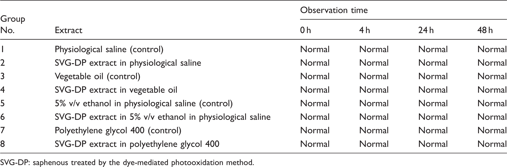
The cells incubated along with the test materials and controls for 24 h were evaluated with an inverted microscope for morphological changes such as cell lysis, reduction of cell growth, vacuolization, clarity of nucleus, intracytoplasmic granulation, and fibroblastic morphology. The reactivity of test samples was qualitatively graded according to ISO standards. After morphological analysis, the cells were trypsinized, and a viability test was performed using a trypan blue dye exclusion method, and graded. Systemic toxicity of the crosslinked SVG was tested using 40 Swiss albino mice. Extracts of the SVG were prepared using physiological saline (extract 1), vegetable oil (extract 2), a 5% v/v solution of ethanol in saline solution (extract 3), and polyethylene glycol 400 (extract 4). Eight groups of mice, each comprising 5 males, were treated with the control and test extracts. The route of administration was intravenous for extracts 1 and 3 and intraperitoneal for extracts 2 and 4. The animals were observed for mortality and morbidity over a period of 3 days after dosing. All animals were observed immediately after the injection (0 h) and at 4, 24, 48, and 72 h for any toxic clinical signs. Native and DP crosslinked SVG were examined using a scanning electron microscope to characterize their surfaces. The sections were also subjected to laser scanning confocal microscopy to study the surface morphology of crosslinked SVG. Both native and crosslinked SVG were characterized structurally by Fourier transform infrared (FTIR) spectroscopy to check for any changes in the functional groups in the extracellular matrix proteins after decellularization and crosslinking. The FTIR spectrum was obtained using a Nicolet 5700 spectrometer. The native and processed SVG were studied for absorption in the range of 3500–500 cm−1. The denaturation temperature of native and crosslinked SVG was measured with a differential scanning calorimeter (DSC-2920; TA Instruments, Inc., USA). The test was conducted on specimen in an aluminium pan, with an empty aluminium pan as a reference. The heating rate was 10 ℃ per minute, and the temperature was increased from 20 ℃ to 200 ℃ under a nitrogen atmosphere. The degradation temperature was taken as the peak height of the heating endothermic curve.
For immunohistochemistry, the native and crosslinked SVG were fixed in 10% neutral-buffered formalin before embedding in paraffin wax. Tissues were sectioned using a microtome (RM2235; Leica Microsystems, Germany) at 5-µm thickness onto polylysine-coated glass slides. These sections were incubated with primary antibodies: monoclonal anticollagen type I clone COL-1 and monoclonal anticollagen type IV clone COL-94 (Sigma, MO, USA), mouse monoclonal antibody NCL-laminin and mouse monoclonal antibody NCL-fibronectin (Novacastra, Newcastle-upon-Tyne, UK), which were diluted 1:100. Their binding was exposed by subsequent incubation with anti-mouse immunoglobulin (whole molecule) peroxidase conjugate (Sigma-Aldrich, MO, USA). The staining procedure was performed according to the manufacturer’s instructions. For a burst test, the vessels were pressurized from inside by pumping normal saline at a constant pressure rise rate of 50 mm Hg·s−1 until the vessels burst. Only those in which the samples tore at the center (not close to the clamped or tied ends) were accepted as valid tests. For protein adsorption studies, 4-cm 2 samples of an ePTFE graft, the DP-processed venous lumen, and native saphenous venous lumen were exposed to human serum albumin, and the amount of albumin adsorbed was determined quantitatively. Native saphenous vein was taken as the control.
One large-animal experiment was performed in a male sheep (aged 18 months, weight 24.5 kg). This animal received DP-treated SVG as an interposition graft, 130-mm long, in its internal jugular vein on the left side. The animal was premedicated with intravenous ketamine 10 mg·kg−1 and xylazine 0.1 mg·kg−1, and given a prophylactic dose of antibiotic (intravenous cefazolin 10 mg·kg−1). Locally, 1% lidocaine was infiltrated over the skin to be incised, and also instilled into the deeper plane. The contralateral native jugular vein was preserved. A small portion of the patent graft was explanted after 24 weeks, and the conduit continuity was retained by end-to-end anastomosis.
Statistical analysis was performed using SPSS version 16 for Windows software (SPSS, Inc., Chicago, IL, USA). All results are presented as mean ± standard error of the mean with the 95% confidence interval. Parameters were compared using analysis of variance, with subgroup analyses using the Bonferroni correction for multiple testing, or paired t tests, as appropriate. A p value <0.05 was considered statistically significant.
Results
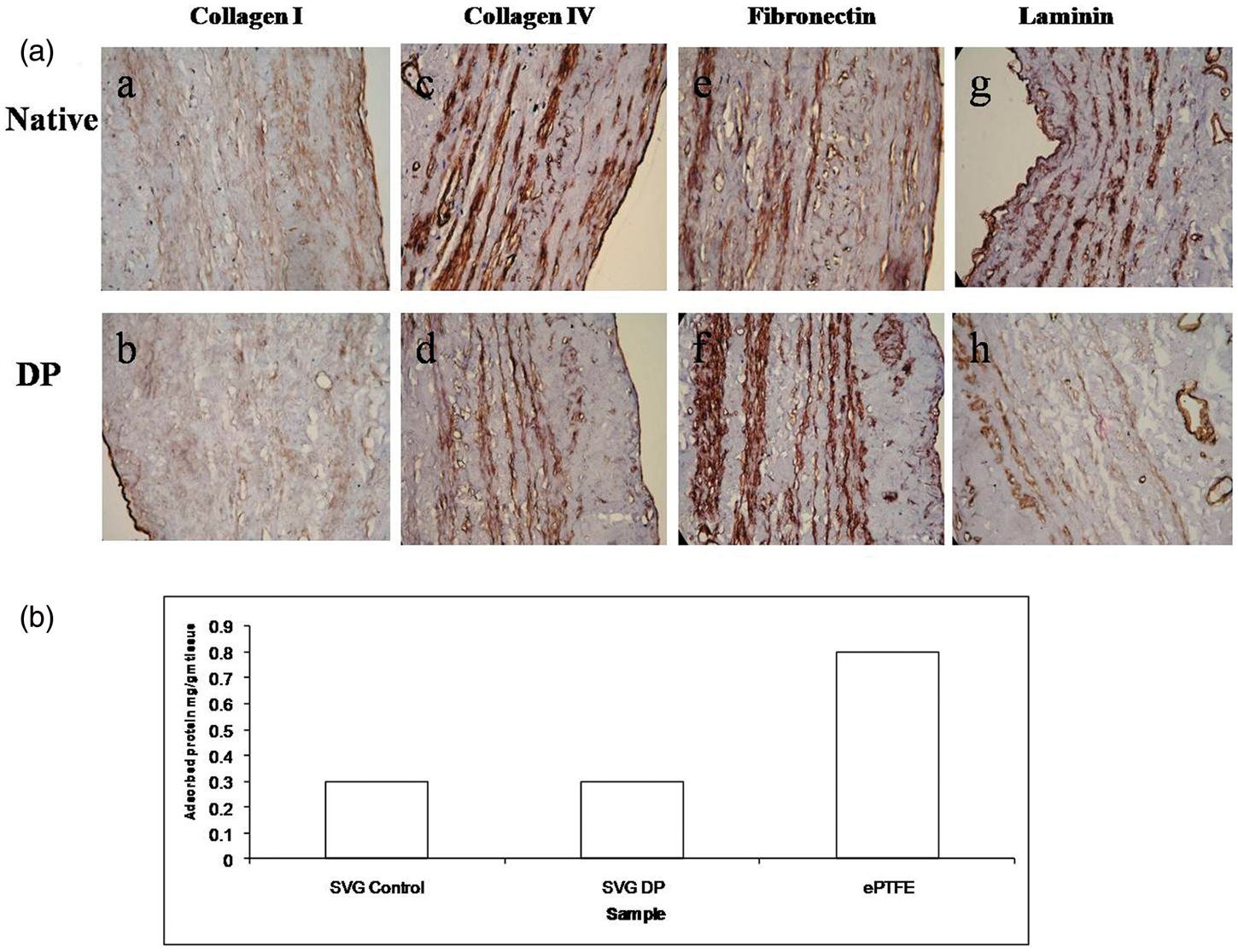
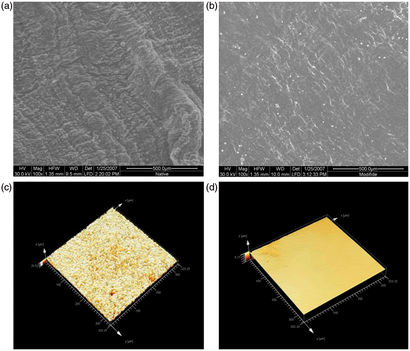
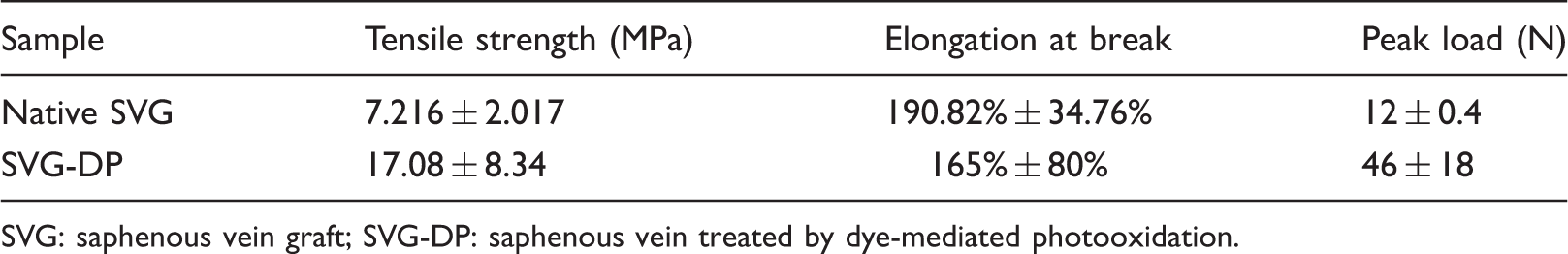
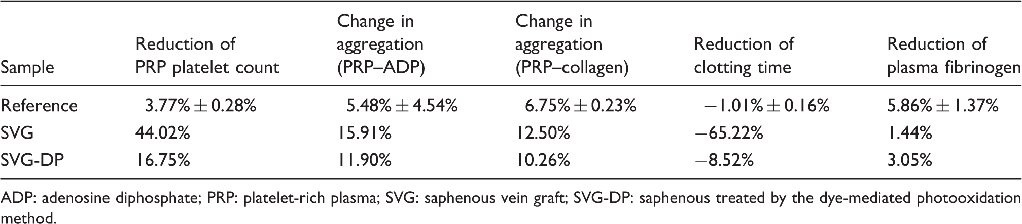
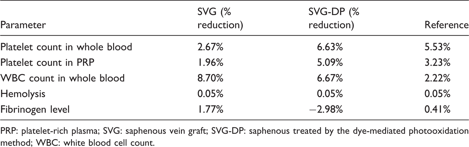
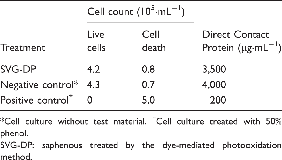
Treatment of SVG with deoxycholic acid along with deoxyribonuclease and ribonuclease enzyme digestion achieved complete decellularization of the veins. The temperature was kept constant at 37 ℃ to achieve complete decellularization with no endothelial lining and no nucleus in the endothelium and medial layer. The basic architecture of the vein was preserved while rendering it acellular, which was confirmed by hematoxylin and eosin staining (Figure 1(a)). Elastic van Gieson stain showed highly organized and uniformly distributed elastin fibres in SVG crosslinked by DP (Figure 1(b)). Immunohistochemical sections of DP-treated SVG and native SVG were compared for the presence of essential extracellular matrix proteins such as collagen type I, collagen type IV, fibronectin, and laminin (Figure 2(a)). The architecture of the tissue was found to be well preserved even after processing, without washing away the matrix elements. The processing efficiency with respect to retention of extracellular matrix protein (laminin, fibronectin, collagen I and IV) appeared to be present in the DP-processed veins. Protein (human albumin) adsorption by native SVG and DP-treated SVG was quite similar; compared to a frequently used synthetic vascular conduit (ePTFE; WL Gore, USA) of similar diameter, they had 8-times less adsorption (Figure 2(b)). Confocal microscopic images of DP-treated SVG (Figures 3(c) and (d)) showed that its surface was smooth, and light penetration was 1 µm, whereas the light penetration of the native saphenous vein was 20.6 µm; smoothness was reconfirmed by scanning electron microscopy (Figure 3(a) and (b)). Collagen type I conformation in the DP-treated SVG did not differ much from collagen type I in the native vein, which was substantiated by the FTIR spectrum. FTIR spectra of DP-treated SVG showed type I collagen by the peaks at 1635, 1452 and 1239 cm−1, which correspond to amide I, amide II, and amide III bonds, respectively, and the infrared ratio of 0.92 substantiated the integrity of the collagen molecular structure (data not shown). Differential calorimeter scans were used to study heat stability for further substantiation of crosslinking efficiency. DP-treated SVG demonstrated a smooth curve of heat stability >120 ℃, with an enthalpy value of 181.8 J·g−1 (data not shown). This enthalpy value indicates crystallinity of the material, which explains the better mechanical strength. The burst pressure of native SVG was found to be 1680 ± 720 mm·Hg, and the DP-treated SVG showed significantly increased burst strength of 4475 ± 1330 mm·Hg. The tensile strength of DP-treated SVG was significantly better than native SVG (Table 1). Hemocompatibility test results appeared to be more favorable for DP-treated SVG, which were even superior to those of the control (Table 2). The results for platelet consumption, hemolysis, and percentage reduction of fibrinogen in DP-processed vein were comparable to the reference values and those of native saphenous vein (Table 3). In-vitro cytotoxicity studies showed that the cells exposed to DP-treated SVG were normal; no change in the morphology was observed. There were no significant qualitative changes in the cells compared to native tissue (Table 4). There was a significant increase in cell proliferation after 7 days compared to 24 h of cell seeding (p < 0.05; Figure 4(A)). No significant changes in cell proliferation or cytotoxicity observed in native and DP-treated tissue after 7 days. The body weight of the control and test animals did not show any change 72 h after the treatment in systemic toxicity studies in mice. No abnormal clinical signs were observed in any of the animals in the control and treated groups during the observation period (Table 5). No mortality was observed in either group.
(a) Hematoxylin and eosin stained sections showing the absence of cells. (b) Elastic van Gieson stained sections of saphenous vein grafts treated with dye-mediated photooxidation, showing the presence of elastin with intact architecture. (a) Immunohistochemical sections of native and dye-mediated photooxidation processed saphenous vein grafts stained for collagen type I (a, b), collagen type IV (c, d), fibronectin (e, f), and laminin (g, h), showing their presence after processing. (b) Protein adsorption studies showing no significant difference in the amount of adsorbed protein between saphenous vein grafts (SVG) treated with dye-mediated photooxidation (DP) and native saphenous vein. Higher protein adsorption was seen in commercially available expanded polytetrafluoroethylene (ePTFE). Scanning electron microscopy showing the homogeneity of (a) native saphenous vein and (b) dye-mediated photooxidation (DP)-processed saphenous vein grafts. Laser light penetration thickness as a parameter of crosslinking and also the surface appearance of the tissue with respect to processing under laser confocal microscopy. Dye-mediated photooxidation processed saphenous vein grafts had less penetration of light and smoother surfaces: (c) native saphenous vein (5.74 µm), (d) dye-mediated photooxidation processed saphenous vein grafts (1 µm). (A) Manganese tricarbonyl transfer tests indicate survival of BALB/c3T3 in dye-mediated photooxidation-treated and native saphenous vein grafts 24 h and 7 days after cell seeding. (B) Hematoxylin and eosin stained sections showing complete decellularization (a, b) as well as autologous cell deposition in explanted tissue from the sheep, having the appearance of a blood vessel with 3 distinct zones in the vascular microscopic structure; (c) and (d) represent immunohistochemical sections of explanted tissue showing the presence of smooth muscle actin and von Willebrand factor for endothelialization. Biomechanical properties of saphenous veins. SVG: saphenous vein graft; SVG-DP: saphenous vein treated by dye-mediated photooxidation. Hemocompatibility of crosslinked saphenous veins. ADP: adenosine diphosphate; PRP: platelet-rich plasma; SVG: saphenous vein graft; SVG-DP: saphenous treated by the dye-mediated photooxidation method. Thrombogenicity by static mode after improvisation of the dye-mediated photooxidation method (n = 3). PRP: platelet-rich plasma; SVG: saphenous vein graft; SVG-DP: saphenous treated by the dye-mediated photooxidation method; WBC: white blood cell count. Mean viable cell counts and protein values after in-vitro cytotoxicity studies. Cell culture without test material. †Cell culture treated with 50% phenol. SVG-DP: saphenous treated by the dye-mediated photooxidation method. Systemic toxicity studies in mice. SVG-DP: saphenous treated by the dye-mediated photooxidation method.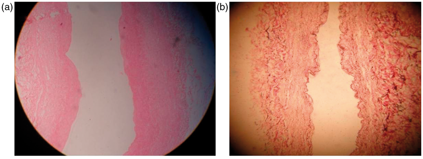
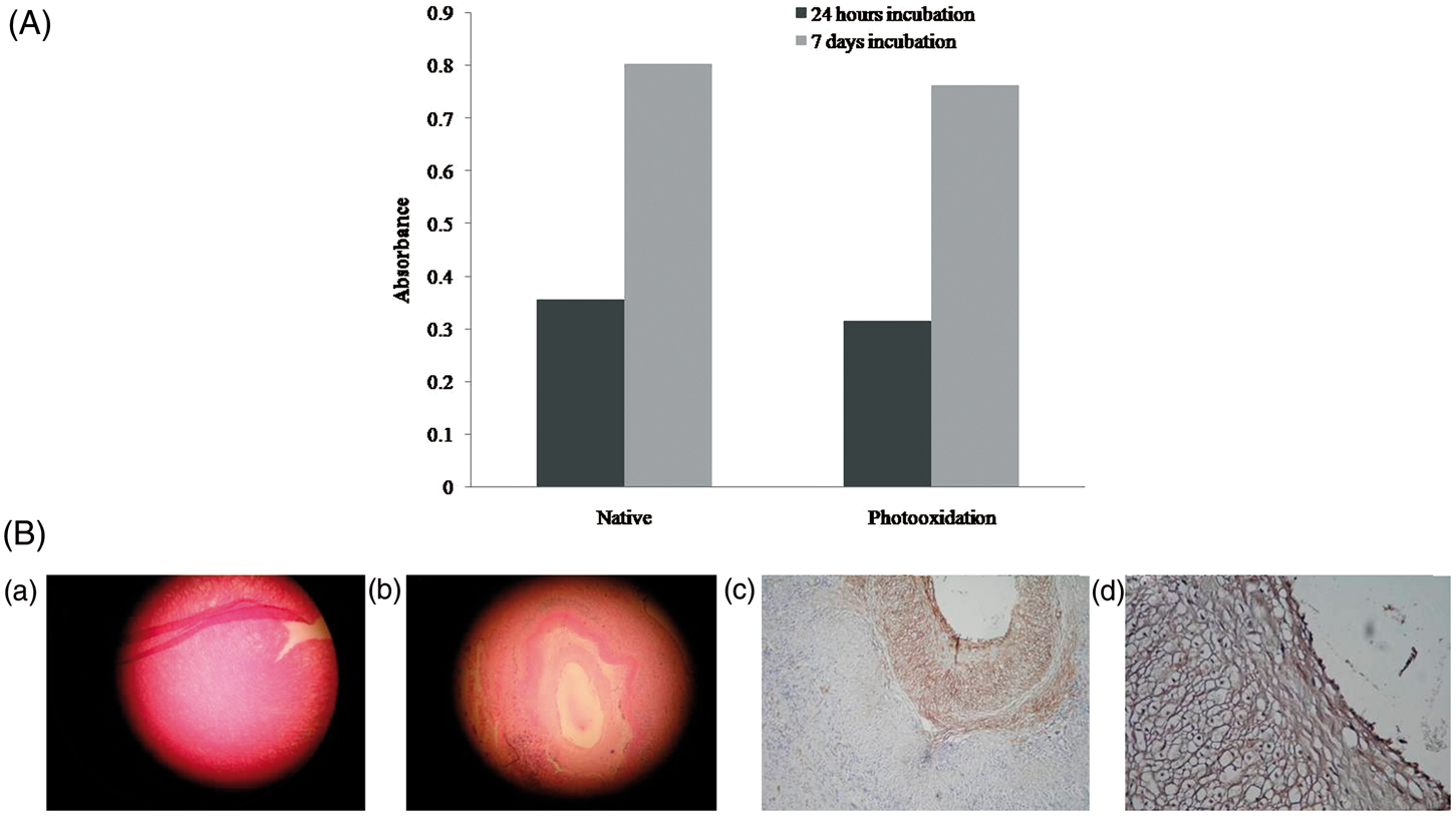
In the large-animal experiment, the sheep had a normal recovery post-surgery, and was under observation for 24 weeks. There were no obvious symptoms or signs of inflammation at the site of surgery. After 6 months, the DP-treated SVG was seen to be patent, and a small portion of the center was explanted. The luminal diameter remained unchanged, and no thrombosis was noticed on visual observation; re-cellularization was observed in microscopic sections (Figure 4(b)). The acellular scaffold appeared to be trizonal with endothelial cell growth and a smooth muscle layer, which was shown by immunohistochemistry (Figure 4(b)).
Discussion
SDVG is a necessity in various fields of vascular surgery. Although catheter intervention has largely taken on the therapeutic role in vascular obstruction, in certain situations such as redo CABG, the necessity of these grafts is undeniable. Every year, more than 700,000 CABG operations require SDVG. When using a small synthetic vessel, post-implant complications such as thrombosis and stenosis are the main drawbacks. The most important factor implicated in graft failure is the lack of endothelial cells lining the lumen of the graft. 7 Polymers such as PTFE and Dacron are usually used as large-diameter vascular grafts, and have worked efficiently. Although these grafts have poor elasticity and tissue compliance and require anticoagulation while in situ, they are still preferred in global clinical practice. However, when these polymers are used as grafts for small-diameter (<6 mm) vessels, they occlude rapidly after implantation.1,9 SDVG, mainly from autologous saphenous veins as well as internal mammary artery or radial artery, are used for vascular reconstructive surgery. 10 They are extensively used in CABG, arteriovenous fistulas prior to renal dialysis, and in peripheral vascular diseases as interposition grafts for replacement of diseased arteries. 11 This requires an additional surgical procedure under the same anesthetic condition. Not only the extra surgery but also the stringent indications for usage of autologous arteries very often limit their applications.
To design and fabricate an SDVG out of a natural biomaterial and make it available off the shelf was the goal of this study. The graft developed in this study was extracted from human vein, and antigenicity was removed by making it completely acellular, but at the same time, scaffold architecture remained intact and compact, which was evident by hematoxylin and eosin as well as elastic van Gieson staining studies. Elastic van Gieson stain showed the presence of elastin. Elasticity is a desired property of arterial grafts for durability and continuous blood flow stress in systole and diastole. Natural tissue with elastin to some extent is important, and may be the viable tissue engineering tool for vascular grafts. The mechanical testing studies were conducted on acellular crosslinked and native saphenous veins, where the tensile strength was considerably higher in the DP-processed veins, with fair stress relaxation. Noticing the favorable increase in strength and thermal stability in the processed veins, hemocompatibility comparisons with the native veins were conducted. The excellent hemocompatibility results following ISO 10993-4 protocol proved that the efficacy of DP-treated SVG was as good as the native veins. The DP method of crosslinking was chosen so that the inherent property of in-vivo cell deposition and survival for favorable remodelling of the graft without thrombogenicity was retained. Because aldehyde is cytotoxic and the probable cause of calcification, DP was chosen as an effective crosslinking procedure, although aldehyde crosslinking is the method commonly followed in tissue-based products available in the clinical field. The FTIR studies revealed amide and imide peaks similar to the original tissue, without denaturation of basic collagen structure.
Having identified these favorable characteristics with the DP method, DP-treated veins were subjected to heat stability studies, which revealed a melting temperature of 126 ℃ and an enthalpy value of 183.5 J·g−1 with a smooth curve. The enthalpy value indicates the crystallinity of the material, indirectly reflecting improved mechanical strength, unique thermal behavior, and increased fatigue strength. 12 The high temperature with a homogeneous curve signifies a high level of crosslinking, which provide a lower thrombogenic property as well as stability to the tissue. Our major objective was to address the issues of thrombogenicity and mechanical sturdiness with preservation of the elastic component of the grafts under development from homologous veins, so that off-the-shelf availability of the physiological grafts becomes feasible. We preserved the basic extracellular matrix of human vein without cells, and strengthened it by crosslinking and heparin bonding. Heparin bonding simultaneously improved hemocompatibility. During the crosslinking procedure, a base solution containing heparin sodium was introduced. Heparin crosslinking is largely a surface phenomenon by an electrostatic method. Crosslinking with heparin prevents early tissue deterioration by forming an artificial matrix of heparin-protein complex. Heparin has a high affinity for these kinds of membranes, with formation of firm ionic associations with the collagen structure. Studies have already shown that heparin can make collagen thromboresistant. 13 In our study, dye-mediated photooxidation in the base solution of heparin produced oxidized heparin, and immobilized the heparin, thereby reducing the collagen-induced platelet procoagulant activity. 14
There are many reported complications associated with the use of synthetic and biodegradable SDVG, such as graft occlusion, aneurysm at the site of anastomosis, distal embolization, and erosion of adjacent structures. 7 In our study, we compared the DP-processed graft with ePTFE in terms of protein adsorption studies in vitro, where native saphenous vein was taken as a control. Protein adsorption of DP grafts was almost similar to that of control (native) saphenous veins, whereas the ePTFE showed 8-times more adsorption of albumin, which signifies a higher probability of thrombosis. Synthetic conduits, due to their high protein adsorption, are known to cause a thrombogenic response in vivo.15,16 The collagenase digestion study was performed to compare the DP-treated graft with native saphenous vein. Bacterial collagenase was used for the study, and the native vein seemed to be more digestible. The hydroxyproline assay of the DP graft showed minimal collagen extraction by collagenase. This again proves the stability of the graft (data not shown).
Many more biodegradable polymers and bioresorbable polymers are being studied as material for SDVG. Polyglycolic acid and polylactic acid are worth mentioning in this regard. Polyglycolic acid, being highly hydrophilic, loses its mechanical strength in 42 h; 1 whereas polylactic acid is more stable, semi-crystalline, and hydrophobic, and used in scaffold making. Polyglycolic acid vascular grafts were shown to result in dilatation and aneurysm formation. The homologous tissue appeared to be quite dependable in this respect, and manipulating it by making the tissue acellular did not take away the basic property and collagen structure, which was evidenced by FTIR studies. 17
Dye-mediated photooxidation was used for crosslinking; the dye is biocompatible and used therapeutically in vasoplegic conditions or for lymphatic delineation. Even the burst test result was effectively high (almost two-and-half times that of normal saphenous vein) indicating the efficient crosslinking, which can probably withstand the continuous pulsatile stress of blood flow, although this needs to be proved. In the initial experimentation, thrombogenicity studies showed that the DP-treated graft was not completely hemocompatible (data not shown). Certain changes were made in the DP processing pertaining to pH, temperature, and the time for photooxidation during crosslinking. The DP process was optimal at a more alkaline pH. The static method of hemocompatibility reinforced the claim of non-thrombogenicity of the material (Table 2). To alleviate the chances of calcification, ethyl alcohol was chosen one of the components of the preservative solution, to prevent valve cusp calcification.18,19 The mechanism of ethanol inhibition may result from its ability to alter multifactorial components in the tissue, such as eliminating phospholipids and cholesterol. The enhanced stability was evident by increased resistance to collagenase digestion.
Studies on cell-seeded bioabsorbable grafts require a highly specialized laboratory with cyclic guanosine monophosphate facilities, and yet the reproducibility is questionable in certain laboratories. In our study, we considered in-vivo cell seeding, considering the body as the best bioreactor, and DP-processed veins were examined for cell adhesion extracellular matrix proteins such as collagen I, collagen IV, laminin, and fibronectin by immunohistochemistry. 20 These proteins were abundant although less so than in the native saphenous vein. The sheep jugular vein was chosen to observe thrombogenicity in vivo. Usually, due to diet, sheep possess hypercoagulability, and the sluggish flow in the venous system might increase coagulability. Considering this possibility, one such experiment was conducted, and surprisingly, without any postoperative anticoagulation, the processed vein remained patent and had become a triple-layered vascular structure 24 weeks after implantation, with a smooth muscle layer with complete endothelialization. This phenomenon can be related to the presence of cell adhesion molecules. Excessive matrix formation was not noted in DP veins after this animal experiment. Orthotopic implantation into the sheep arterial system will be a future step, and a series of experiments have been planned.
The technique of processing fresh cadaver saphenous vein resulted in a hemocompatible, non-calcifying, biocompatible, biological vascular graft that is conducive to in-vivo cell adhesion and possesses high burst strength and elasticity. It carries a great promise to fill the lacunae in the field of SDVG. Therefore, we propose that any vein from a fresh cadaver without infection can be transformed into an SDVG. Any number and size of grafts with respect to length and diameter may be a possibility, and would fill the deficiency in this area with off-the-shelf availability.
Footnotes
Funding
This study had been conducted with the partial funding of the Department of Biotechnology, Govt. of India, under SBIRI Scheme.
Acknowledgments
The authors thank Mr Janardhan Reddy for preparing histological sections, Sheerin Begam Nasser for assistance with decellularization, Dillip Kumar Bishi for his scientific inputs, and A Pandian for technical help.
Conflicts of interest statement
None declared.