






Abstract
Background
Recent studies have shown that tissue damage in neuromyelitis optica (NMO) is not limited to the spinal cord and optic nerve but can also appear in the brain. Previous magnetic resonance imaging (MRI) studies have reported controversial findings regarding the presence of white matter atrophy in NMO patients.
Purpose
To investigate regional white matter changes in NMO using voxel-based morphometry (VBM).
Material and Methods
Conventional MRI and T1-weighted three-dimensional MRI were performed on 20 patients with NMO and 20 age- and sex-matched normal controls (NCs). The data were analyzed by statistical parametric mapping 5 (SPM5) to generate white matter concentration maps, and regional white matter concentrations were compared between the two groups. Relationships between the white matter concentration in regions with significant group differences and the Expanded Disability Status Scale (EDSS) and disease duration were further explored.
Results
Compared to NCs, NMO patients had decreased white matter volumes in the right precentral gyrus, right postcentral gyrus, left middle and medial frontal gyrus, right superior frontal gyrus, bilateral inferior and superior parietal lobule, right angular gyrus, right middle occipital gyrus, and left precuneus. No significant correlation was found between white matter regions with volume reduction and the EDSS and disease duration in NMO.
Conclusion
We found white matter atrophy in several brain regions in the frontal, parietal, and occipital lobes in NMO, suggesting that subtle white matter damage relevant to the motor, vision, and cognition systems exists in NMO patients. The pattern of white matter atrophy in NMO is different from that in multiple sclerosis (MS).
Introduction
Neuromyelitis optica (NMO) is a severe form of demyelinating disease characterized by the selective involvement of the spinal cord and optic nerves (1,2). Previous studies have shown that occult brain damage might exist in normal-appearing brain tissue in NMO patients (3–7). Among them, grey matter abnormalities in NMO have been reported by Rocca et al., who used diffusion tensor imaging (DTI) and magnetization transfer imaging (MTI) (5). However, they found no significant difference between NMO patients and controls in DTI or MTI metrics in normal-appearing white matter (NAWM). Recently, several studies have reported the absence of metabolic changes in NMO patients using magnetic resonance (MR) spectroscopy; these studies also reported that diffuse brain damage was absent in NMO (8–10). However, our group identified brain damage in both NAWM and normal-appearing grey matter (NAGM) in NMO patients (6). In particular, widespread and subtle white matter abnormalities in NMO patients have been reported by studies utilizing tract-based spatial statistics and structural network analysis (11,12). To the best of our knowledge, it is unclear whether white matter atrophy is present in NMO.
Voxel-based morphometry (VBM) involves spatially normalizing high-resolution images from all the subjects in a study into the same stereotactic space (13). It can overcome problems of intra- and inter-observer bias and sensitivity as well as the need for the a priori definition of structures of interest. VBM studies have only recently been conducted for demyelinating diseases of the central nervous system (14–17). Using VBM, our group found mild grey matter atrophy in several regions of the frontal and temporal lobes and the insula in NMO patients compared to normal controls and found extensive grey matter atrophy in multiple sclerosis (MS) patients (18). However, few studies (19) have reported white matter volume changes in NMO, and therefore, the current study aimed to determine whether regional white matter brain volume changes are present in NMO patients and to explore the correlations between regions with significant group differences and clinical variables such as the Expanded Disability Status Scale (EDSS) and disease duration.
Material and Methods
Patients
We studied 20 patients with NMO (2 men, 18 women; mean age, 37.2 years; standard deviation [SD], 12.1 years) and 20 age- and sex-matched healthy subjects (mean age, 36.9 years; SD, 11.2 years). All subjects were right-handed. The institutional review board of Xuanwu Hospital approved the study, and written informed consent was obtained from each participant. All patients fulfilled the revised diagnostic criteria for NMO (1). All patients had episodes of optic neuritis and myelitis. None of the participating patients had lesions on brain T2-weighted (T2W) images, and MR imaging (MRI) showed a longitudinally extensive spinal cord T2 lesion spanning at least three vertebral segments. The median disease duration was 3.1 years (range, 0.4–8.0 years), and the median EDSS was 3.0 (range, 1.0–6.0). None of the participating patients with NMO had been treated with related medications (e.g. corticosteroids and immunosuppressants) within 3 months of the MR images being obtained.
MRI acquisitions
Imaging was performed on a 1.5T MR scanner (Sonata; Siemens Medical Systems, Erlangen, Germany) in the Department of Radiology, Xuanwu Hospital, Capital Medical University. A standard head coil was used with foam padding to restrict head motion. The routine axial slices were positioned to run parallel to a line that joins the most inferoanterior and inferoposterior parts of the corpus callosum, with: an identical field of view (FOV), 240 × 210 mm; number of sections, 30; section thickness, 4 mm; and intersection gap, 0.4 mm. These were used to determine whether lesions in the brain existed: (i) axial T2W turbo spin-echo (repetition time [TR], 5500 ms; echo time [TE], 94 ms; number of signals acquired, 3; echo train length, 11; matrix size, 256 × 224; (ii) axial fluid-attenuated inversion recovery (FLAIR) sequences (TR/TE, 8500/150 ms; inversion time [TI], 2200 ms; number of signals acquired, 1; echo train length, 8; matrix size, 256 × 224); and (iii) sagittal three-dimensional (3D) volumetric T1-weighted (T1W) magnetization-prepared rapid acquisition gradient echo (MP-RAGE) (TR/TE, 1970/3.9 ms; TI, 1100 ms; flip angle, 15°; FOV, 219 × 250 mm; matrix size, 256 × 256; slice thickness, 1.7 mm; voxel dimensions, 0.5 × 0.5 × 1.7 mm) images were obtained.
Data analysis
All structural MRI postprocessing was performed by a single experienced observer, who was unaware of the patient’s identity. Regional volumetric measurements were performed on the 3D T1W MP-RAGE images, using an optimized VBM approach and the statistical parametric mapping (SPM5) software provided at www.fil.ion.ucl.ac.uk/spm. The 3D MR data-sets of all patients and controls were preprocessed using the following main steps: (i) segmentation: all images were registered to the Montreal Neurological Institute (MNI) template and segmented into grey matter, white matter and cerebral spinal fluid (CSF); (ii) normalization: segmented images were normalized to the MNI template, and the parameters were then applied to normalize individual T1 images separately; (iii) re-segmentation: the fully normalized images were segmented once again into grey matter, white matter and CSF; (iv) modulation: the normalized white matter was modulated by the Jacobian determinants based on the voxel; (v) smoothing: the normalized and modulated white matter images were smoothed using a 12 mm3 full-width half-maximum (FWHM) Gaussian kernel, as previously reported (15).
Statistical analysis
A two-sample t-test was used to compare the global brain volume and white matter volumetric measurements between the NMO patients and normal controls (NCs). We used the AlphaSim programme at P < 0.05 at a voxel level across the whole brain. In addition, only clusters with 30 or more contiguous voxels were considered. Finally, the MNI coordinates corresponding to areas with significant differences were converted to Talairach coordinates. Significant clusters were anatomically localized using the atlas of Talairach and Tournoux (20). A Pearson correlational analysis was used to assess correlations between regional white matter loss and clinical variables (disease duration and EDSS).
Results
No difference in the global brain volume was observed between the NMO and NC groups (t = 1.354, P = 0.186). We found no white matter regions with significantly increased volume in NMO patients relative to the controls. Significant clusters of reduced white matter volume were observed in NMO patients relative to the controls (Table 1, Fig. 1). The areas of white matter atrophy included the right precentral gyrus, right postcentral gyrus, left middle and medial frontal gyrus, right superior frontal gyrus, bilateral inferior and superior parietal lobule, right angular gyrus, right middle occipital gyrus, and left precuneus.
Statistical parametric mapping (SPM) regions with a decreased white matter concentration, overlain on a high-resolution T1W image, comparing NMO patients with NCs (AlphaSim corrected P < 0.05). SPM regions with significant white matter loss: right precentral and postcentral gyrus, left middle and medial frontal gyrus, right superior frontal gyrus, bilateral inferior and superior parietal lobule, right angular gyrus, right middle occipital gyrus, and left precuneus. Brain areas with significant white matter differences between normal controls and NMO patients.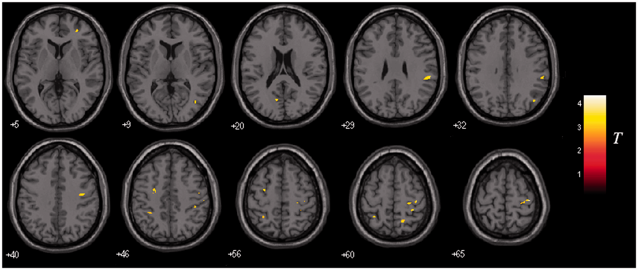
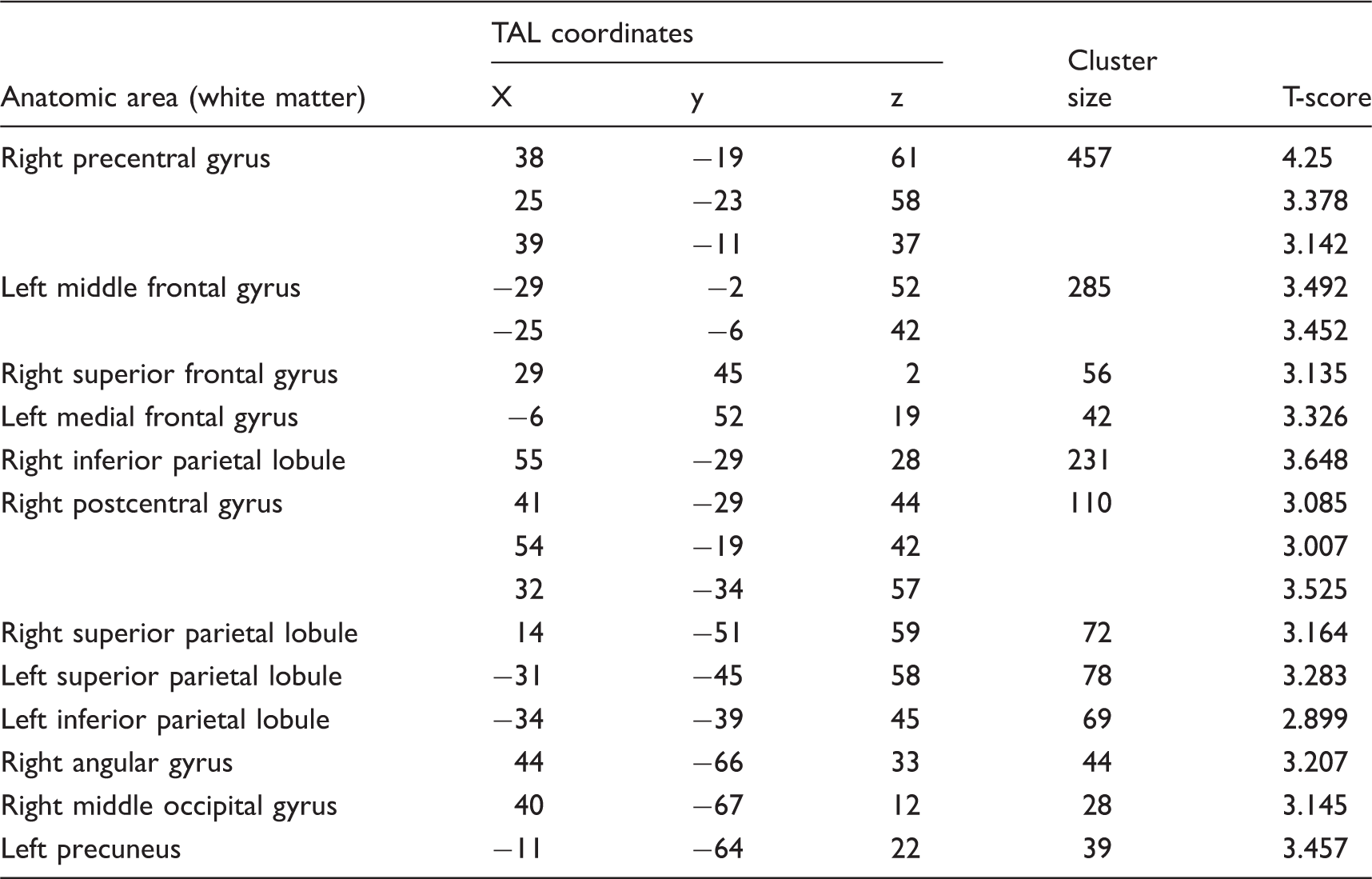
No significant correlation was found between regional white matter atrophy and the EDSS or disease duration in NMO patients.
Discussion
To our knowledge, this is one of the first reports of white matter atrophy in NMO patients using VBM. In this study, we found subtle regional white matter atrophy in the frontal, parietal, and occipital lobes. No significant correlation was found between regional white matter atrophy and the EDSS or disease duration in NMO.
Compared with NCs, NMO patients had decreased white matter volume in several regions of the frontal, parietal, and occipital lobes. The most significant regions of atrophy were the right precentral and postcentral gyrus, bilateral inferior parietal lobule, and left middle frontal gyrus. The precentral gyrus and the postcentral gyrus represent the primary motor and somatosensory centres, respectively. White matter volume changes in the precentral gyrus and the postcentral gyrus were most likely induced by motor and sensory abnormalities due to myelitis. The inferior parietal lobule (IPL) consists of several areas that are involved in the analysis of specific aspects of visual information, alone or in combination with the somatosensory area (21). The occipital lobe is the visual processing center of the brain containing most of the anatomical region of the visual cortex (22). Visual problems in NMO patients might be responsible for the volume reduction in the IPL and the occipital lobe.
A recent article using VBM described white matter atrophy in NMO patients and its correlation with cognitive impairment (19). Similar to that study, we observed white matter atrophy in the precentral gyrus, postcentral gyrus, superior frontal gyrus, and middle and medial frontal gyrus. Unlike the previous study, we did not observe white matter atrophy in the optic chiasm, pons, cerebellum, or corpus callosum. The discordant results may be due to the different clinical characteristics of the NMO patients, including a longer disease duration and higher EDSS score in Blanc’s study (mean duration, 11.9 years; mean, EDSS 3.42) than in the present study (mean duration, 3.1 years; mean EDSS, 3.0) or different patient inclusion criteria.
Recent functional MRI studies performed by our group have found that neural activity in the resting state is altered in patients with NMO. In these studies, we have reported decreased amplitude of low frequency fluctuation (ALFF) in the precuneus, increased ALFF in the middle frontal gyrus (23), and decreased regional homogeneity (ReHo) in the medial frontal gyrus (24). In the present study, we found white matter atrophy in the above regions. The precuneus and IPL have also been shown to compose the default-mode network (DMN), which is highly active in the resting state (25,26). A volume reduction in these regions might be the structural basis of the abnormal functional changes, thus contributing to the cognitive impairment.
A previous study from our group reported mild grey matter atrophy in several regions of the frontal, temporal lobes, and insula in NMO patients compared to normal controls by means of VBM, whereas MS patients showed extensive and severe grey matter atrophy (18). In this study, subtle atrophy was limited to the specified regions of NAWM in NMO patients. Pathologic and radiologic studies have already revealed diffuse white matter damage in MS (27,28). Therefore, the patterns of white matter damage are distinct in each disease.
We found no significant correlation between the regions of white matter atrophy and the EDSS or disease duration in NMO patients. Several reasons may explain this finding: (i) the small sample size of the group; (ii) motor and sensory symptoms are also related to lesions in the spinal cord, but we did not evaluate lesions in the spinal cord; (iii) the EDSS is only a general score that focuses primarily on motor and visual function. Further studies are required using improved clinical scores such as the MS functional composite and detailed cognitive scores.
Our study had several limitations. First, cognitive neuropsychological tests were not performed in this study, which prevents the correlation of cognitive test scores with the VBM results. Second, because serum NMO-IgG was not tested, correlations between the serum NMO-IgG level and the regional volume decrease could not be considered in this study. Third, to evaluate brain damage in NMO patients, combined structural and functional techniques should be used in future studies.
In conclusion, regional white matter atrophy was identified in NMO patients in the precentral and postcentral gyrus, inferior parietal lobule, and middle frontal gyrus. These data suggest that the white matter damage relevant to the motor, vision, and cognition systems exists in NMO patients. The pattern of the subtle white matter atrophy in NMO was different from the diffuse white matter atrophy previously observed in MS.
Footnotes
Funding
This work was supported by the National Natural Science Foundation of China (No. 81101038, 30930029), Beijing Natural Science Foundation (No. 7133244), and Beijing Nova Program (No. xx2013045).