






Abstract
Background:
Shoulder stiffness is a common complication after arthroscopic rotator cuff repair. However, there is no consensus on the treatment of stiffness after repair. Although one treatment option is an intra-articular steroid injection, it may negatively affect repair integrity, and there is a paucity of literature regarding the timing of intra-articular injections for stiffness after repair and its effect on repair integrity.
Purpose:
To compare repair integrity and clinical outcomes after an intra-articular steroid injection administered at 6 weeks and 12 weeks postoperatively for shoulder stiffness after arthroscopic rotator cuff repair.
Study Design:
Cohort study; Level of evidence, 3.
Methods:
Patients who developed stiffness after arthroscopic rotator cuff repair were given a series of 3 intra-articular steroid injections every 4 weeks from 6 (6-week group) and 12 weeks (12-week group) postoperatively. The control group had rotator cuff tears but neither stiffness nor injections. Shoulder range of motion (ROM), the Korean Shoulder Scoring System (KSS) score, and the University of California, Los Angeles (UCLA) shoulder score were assessed preoperatively with a minimum of 2-year follow-up. Repair integrity was assessed using magnetic resonance imaging at 6 months postoperatively.
Results:
Seventy-four of 209 patients (35.4%) who underwent arthroscopic rotator cuff repair developed stiffness. There were no significant differences in retear rates among the 6-week (5.7%, 2/35 patients), 12-week (10.3%, 4/39 patients), and control groups (14.1%, 19/135 patients) (P = .374). Both the 6- and 12-week groups showed significant improvement in ROM (both P < .001), KSS scores (both P < .001), and UCLA scores (both P < .001) at the final follow-up. The 6-week group showed significantly better ROM (P < .001), KSS scores (P < .001), and UCLA scores (P < .001) than the 12-week group at 3 months postoperatively. However, both the 6- and 12-week groups showed significantly lower KSS (81.3 ± 12.0 [P = .004] and 83.4 ± 8.6 [P = .035], respectively) and UCLA (29.3 ± 4.6 [P = .006] and 30.0 ± 3.3 [P = .042], respectively) scores than the control group (90.4 ± 13.6 and 32.3 ± 4.7, respectively) at the last follow-up.
Conclusion:
An intra-articular steroid injection administered at 6 weeks postoperatively for shoulder stiffness after arthroscopic rotator cuff repair may be effective for reducing patients’ pain and improving shoulder ROM at 3 months postoperatively without compromising repair integrity.
Shoulder stiffness is one of the most common complications of rotator cuff repair, and some surgeons consider it as a shoulder-tightening process independent of the repair technique and efforts at physical therapy.11,14,33 The prevalence of shoulder stiffness after arthroscopic rotator cuff repair ranges from 1.5% to 32.7%.3,5,8,22,34,38 Surgeons may consider limited range of motion (ROM) inevitable after rotator cuff repair. 48 Although adhesive capsulitis has a favorable natural course in which the limitation of ROM improves as time goes by,35,39 stiffness after repair is often recalcitrant to treatment, resulting in dissatisfaction with pain management and gradual loss of shoulder ROM.11,14,25,33 Despite successful rotator cuff repairing, the occurrence of stiffness after repairing may make patients experience dissatisfaction, and both the surgeon and the patient may go through a distressing situation, particularly at the early postoperative period.
Traditionally, oral medication, intra-articular corticosteroid injections, physical therapy, and surgical treatment, such as arthroscopic capsular release, have been ways to treat stiffness after repair.12-14,19,25 However, oral medication has inferior effects compared with other nonoperative interventions and no effect on the course of shoulder stiffness, and stiffness after repair is often resistant to physical therapy.13,47 Arthroscopic capsular release improves ROM and is generally advocated in patients who respond poorly to nonoperative management. 32 However, the patient and the surgeon have to face the stressful situation of undergoing a second operative procedure. Capsular release for postoperative shoulder stiffness improves ROM but tends to remain painful, with lower patient satisfaction and functional activity compared with idiopathic and postfracture shoulder stiffness.2,24
There are few reports on the timing of intra-articular steroid injections, and its effect on repair integrity, for stiffness after repair to acquire pain relief and ROM without risking repair integrity. Our goal was to intervene as soon as possible to relieve patients’ pain and improve shoulder ROM for early stiffness after repair, with the main concern for the effect on repair integrity.
The purpose of this study was to compare repair integrity and clinical outcomes in patients who received an intra-articular corticosteroid injection at 2 distinct time points (6 weeks postoperatively vs 12 weeks postoperatively) for shoulder stiffness after arthroscopic rotator cuff repair. We hypothesized that stiffness after repair treated with an intra-articular corticosteroid injection at 6 weeks postoperatively would not have negative effects on repair integrity and would reveal better clinical outcomes compared with injection at 12 weeks.
Methods
Inclusion and Exclusion Criteria
The inclusion criteria were patients with full-thickness tears of the supraspinatus with or without full-thickness tears of other rotator cuff tendons who underwent arthroscopic rotator cuff repair from August 2013 to October 2014. The exclusion criteria were partial-thickness tears of the supraspinatus, regardless of full-thickness tears of other rotator cuff tendons; moderate to severe glenohumeral arthritis requiring total shoulder arthroplasty; revision rotator cuff surgery; any previous shoulder surgery; outside magnetic resonance imaging (MRI) not eligible for measurements in the current study; additional long head of the biceps procedure (tenotomy or tenodesis) or distal clavicle resection; and patients who did not meet the follow-up period of a minimum of 2 years. Also, patients with rotator cuff tears concomitant with preoperative stiffness were also excluded. Institutional review board approval was obtained before the initiation of the present study, and data were reviewed retrospectively.
Patients
Arthroscopic single-row repair was performed for tears ≤1-cm, and arthroscopic transosseous-equivalent suture bridge repair was performed for tears >1 cm. Shoulder stiffness after arthroscopic rotator cuff repair was diagnosed at 6 weeks postoperatively in all patients when they had either <140° of passive forward flexion or <40° of passive external rotation. 11 However, patients with diagnosed stiffness after repair during the first half of this study (August 2013 to March 2014) were given a series of 3 intra-articular steroid injections every 4 weeks from 12 weeks postoperatively (at 12, 16, and 20 weeks postoperatively; 12-week group), and patients with stiffness after repair during the second half of this study (April 2014 to October 2014) were given a series of 3 intra-articular steroid injections every 4 weeks from 6 weeks postoperatively (at 6, 10, and 14 weeks postoperatively; 6-week group). No patients were given additional treatments for stiffness outside of routine medicine and rehabilitation before intra-articular steroid injection therapy. The control group consisted of patients who underwent arthroscopic rotator cuff repair and did not show stiffness.
We evaluated symptom duration before surgery, smoking, prevalence of diabetes and hypothyroidism, mediolateral and anteroposterior tear sizes, fatty infiltration according to the Fuchs modification 15 of the Goutallier classification, 18 and tendon involvement (type 1: supraspinatus only, type 2: supraspinatus and subscapularis, type 3: supraspinatus and infraspinatus, and type 4: all 3 tendons). 26 Tendon involvement was classified only with full-thickness tears, and partial-thickness tears did not affect the classification. Mediolateral tear size was measured on T2 coronal MRI by using a curvilinear line from the edge of the tendon to the superior aspect of the greater tuberosity. Soft tissue stumps on the lateral edge were disregarded. Anteroposterior tear size was measured on T2 sagittal MRI in a curvilinear line from the posterior to anterior edge of the tendon. The greatest length of round and tapered edges was calculated in both measurements. 23
Evaluation of Shoulder Stiffness
We defined shoulder stiffness as <140° of passive forward flexion or <40° of passive external rotation. Patients meeting any 1 of these 2 criteria were considered to have shoulder stiffness. 11 Shoulder ROM was assessed with a goniometer for active and passive ROM in forward flexion and external rotation at the side. Forward flexion was measured in degrees between the arm and the thorax with the elbow held straight, and external rotation with 0° of shoulder abduction was measured with the elbow in 90° of flexion between the thorax and the forearm.
Outcome Assessment
The Korean Shoulder Scoring System (KSS) score and University of California, Los Angeles (UCLA) shoulder score were assessed preoperatively; at 3, 6, and 12 months postoperatively; and during the last follow-up (a minimum of 2 years).
Injection Protocol
The intra-articular injection was administered using an ultrasound-guided posterior approach with the patient in the lateral decubitus position and the probe positioned at the myotendinous junction of the infraspinatus muscle inferior to the scapular spine. 9 The needle was inserted in-plane parallel to the long axis of the transducer and advanced in the joint between the humeral head and the posterior glenoid labrum, and the injection was given with 1 mL triamcinolone (20 mg), 4 mL of 0.5% bupivacaine, and 5 mL of normal saline.
Rehabilitation Protocol
All patients underwent an identical rehabilitation program starting the day after the operative procedure with active finger, wrist, and elbow ROM exercises; pendulum exercises; and passive shoulder ROM. An abduction brace was applied for 6 weeks, and active ROM, stretching, and rotator cuff isometric exercises were allowed after 6 weeks. All the patients included in this study underwent the same postoperative rehabilitation program, regardless of the tear size. Rotator cuff strengthening exercises were allowed after 3 months postoperatively in all patients. Return to sports activity and manual labor were allowed after 6 months postoperatively in all patients.
Integrity After Repair
The integrity of the repaired rotator cuff tendon was examined using MRI at postoperative 6 months. We defined a retear as Sugaya types IV (presence of a minor discontinuity) or V (presence of a major discontinuity). 42
Statistical Analysis
Demographics, MRI measurements, and retear rates between the 6-week and 12-week groups were compared using the independent t test, chi-square test, or Fisher exact test. Preoperative and last follow-up KSS and UCLA scores were compared with a paired t test. Changes in ROM and KSS and UCLA scores over time were compared between the 6-week group and 12-week group by repeated-measures analysis of variance, and post hoc testing was performed by the independent t test with adjusted levels of significance by the Bonferroni method to compare the 6-week and 12-week groups at every time point. For comparison among the 6-week group, the 12-week group, and the control group, analysis of variance was used for the parametric variables, and the Kruskal-Wallis H test was used for the nonparametric variables. SPSS Statistics (version 20.0; IBM) was used for all statistical analyses, and P < .05 was considered statistically significant.
Results
Of the 358 patients who underwent arthroscopic rotator cuff repair between August 2013 and October 2014, 149 patients were excluded as per the exclusion criteria. They included 12 cases of a partial-thickness tear of the supraspinatus, regardless of full-thickness tears of other rotator cuff tendons, 5 cases of moderate to severe glenohumeral arthritis requiring total shoulder arthroplasty, 11 cases of revision rotator cuff surgery, 15 cases of any previous shoulder surgery, 45 cases of outside MRI not eligible for measurements, 40 cases of additional long head of the biceps procedure (tenotomy or tenodesis) or distal clavicle resection, and 21 patients who did not meet the follow-up period of a minimum of 2 years. Of the 209 remaining patients, 74 patients developed stiffness after repair; overall rate of stiffness after repair was 35.4%.
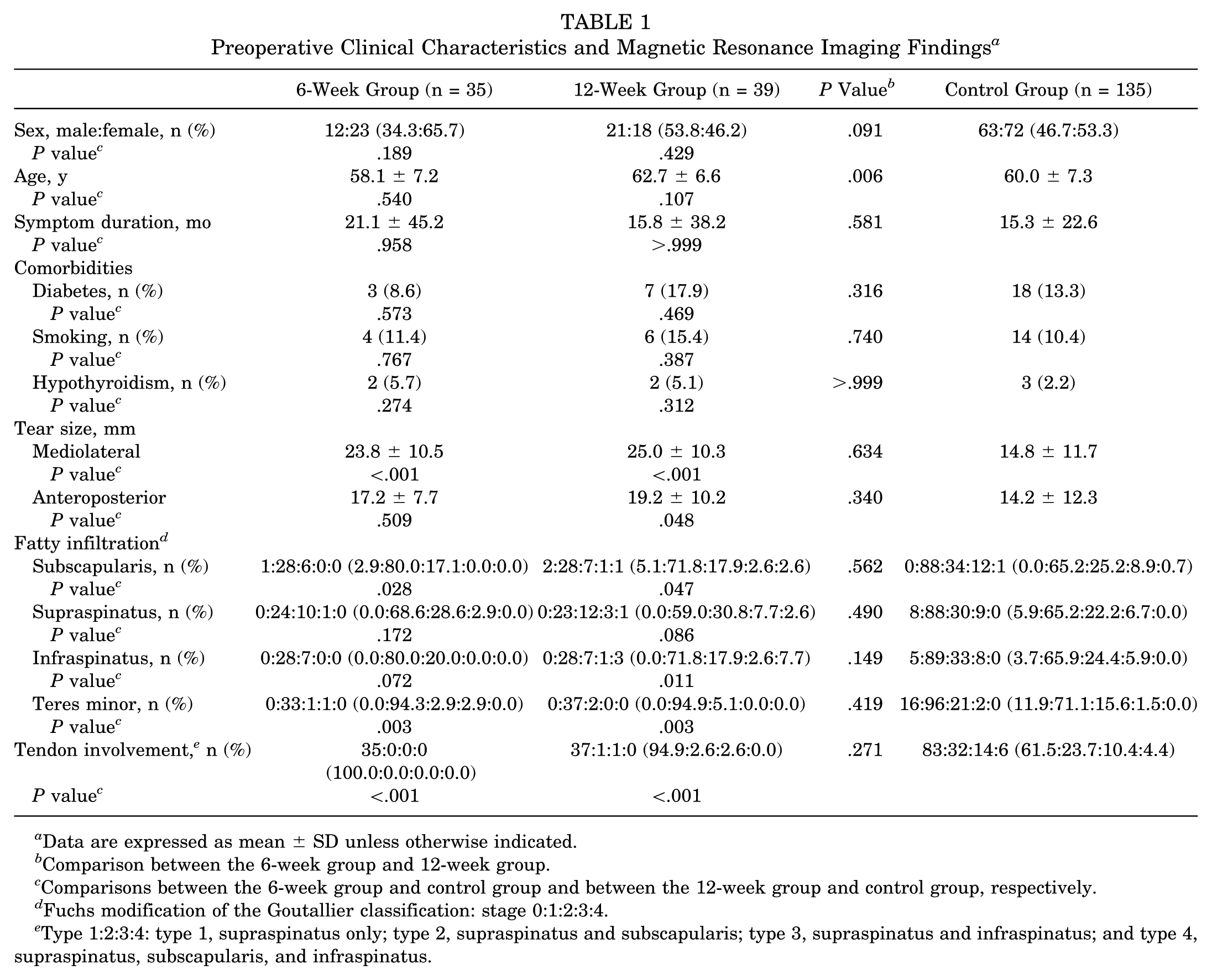
The 6-week group consisted of 35 patients (12 men and 23 women), and the 12-week group consisted of 39 patients (21 men and 18 women). The 12-week group (mean age, 62.7 ± 6.6 years) was significantly older than the 6-week group (mean age, 58.1 ± 7.2 years) (P = .006). However, there were no significant differences between the 6-week and 12-week groups in sex; preoperative symptom duration; smoking; prevalence of diabetes and hypothyroidism; mediolateral and anteroposterior tear sizes; fatty infiltration of the subscapularis, supraspinatus, infraspinatus, and teres minor; and tendon involvement (all P > .05) (Table 1). The control group consisted of 135 patients (63 men and 72 women). There were no significant differences in age, sex, symptom duration before surgery, comorbidities, and fatty infiltration of the supraspinatus between the 6-week group and control group and between the 12-week group and control group. However, there were significant differences in mediolateral tear size (23.8 ± 10.5 mm vs 14.8 ± 11.7 mm [P < .001] and 25.0 ± 10.3 mm vs 14.8 ± 11.7 mm [P < .001], respectively), fatty infiltration of the subscapularis (stage 0:1:2:3:4, 1:28:6:0:0 vs 0:88:34:12:1 [P = .028] and 2:28:7:1:1 vs 0:88:34:12:1 [P = .047], respectively) and teres minor (stage 0:1:2:3:4, 0:33:1:1:0 vs 16:96:21:2:0 [P = .003] and 0:37:2:0:0 vs 16:96:21:2:0 [P = .003], respectively), and tendon involvement (type 1:2:3:4, 35:0:0:0 vs 83:32:14:6 [P < .001] and 37:1:1:0 vs 83:32:14:6 [P < .001], respectively) between the 6-week group and control group and between the 12-week group and control group. The 12-week group also showed significant differences in anteroposterior tear size (19.2 ± 10.2 mm vs 14.2 ± 12.3 mm [P = .048], respectively) and fatty infiltration of the infraspinatus (stage 0:1:2:3:4, 0:28:7:1:3 vs 5:89:33:8:0 [P = .011], respectively) compared with the control group (Table 1).
Preoperative Clinical Characteristics and Magnetic Resonance Imaging Findings a
Data are expressed as mean ± SD unless otherwise indicated.
Comparison between the 6-week group and 12-week group.
Comparisons between the 6-week group and control group and between the 12-week group and control group, respectively.
Fuchs modification of the Goutallier classification: stage 0:1:2:3:4.
Type 1:2:3:4: type 1, supraspinatus only; type 2, supraspinatus and subscapularis; type 3, supraspinatus and infraspinatus; and type 4, supraspinatus, subscapularis, and infraspinatus.
Clinical Outcomes
Both the 6-week and the 12-week groups showed significant improvement in the final assessment compared with the preoperative assessment of KSS and UCLA scores. For the 6-week group, the KSS score improved from 61.2 ± 11.7 to 81.3 ± 12.0 (P < .001), and the UCLA score improved from 22.0 ± 4.2 to 29.3 ± 4.6 (P < .001). For the 12-week group, the KSS score improved from 58.5 ± 17.0 to 83.4 ± 8.6 (P < .001), and the UCLA score improved from 20.6 ± 5.2 to 30.0 ± 3.3 (P < .001). For the control group, the KSS score improved from 66.3 ± 15.0 to 90.4 ± 13.6 (P < .001), and the UCLA score improved from 22.0 ± 4.8 to 32.3 ± 4.7 (P < .001) (Table 2).
KSS and UCLA Scores a
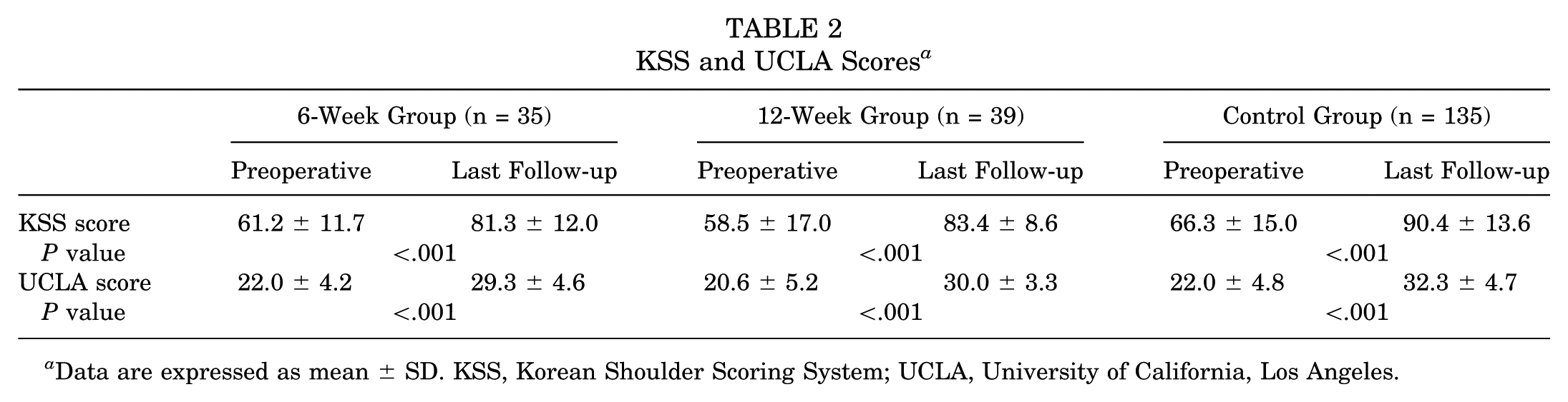
Data are expressed as mean ± SD. KSS, Korean Shoulder Scoring System; UCLA, University of California, Los Angeles.
The 6-week group showed significant differences compared with the 12-week group in active forward flexion (132.0° ± 29.4° vs 104.1° ± 25.3° [P < .001], respectively) and external rotation (29.0° ± 12.1° vs 21.2° ± 9.4° [P = .003], respectively) at 3 months postoperatively. Compared with the control group, the 6-week group and 12-week group showed no significant differences in active forward flexion from 3 months postoperatively (138.8° ± 26.0° vs 132.0° ± 29.4° [P = .543], respectively) and from 6 months postoperatively (147.2° ± 34.5° vs 136.3° ± 25.2° [P = .272], respectively) and in passive forward flexion from 6 months postoperatively (160.3° ± 12.8° vs 153.1° ± 17.5° [P = .297], respectively) and from 1 year postoperatively (162.7° ± 8.1° vs 157.2° ± 14.4° [P = .129], respectively). However, both the 6-week group (34.0° ± 9.3° [P = .157]) and the 12-week group (34.6° ± 10.5° [P = .234]) showed no significant differences in active external rotation only at 6 months postoperatively compared with the control group (39.3° ± 13.3°). Furthermore, passive external rotation in both the 6-week group and the 12-week group was significantly lower than that in the control group from 6 weeks postoperatively to the last follow-up (Figure 1).
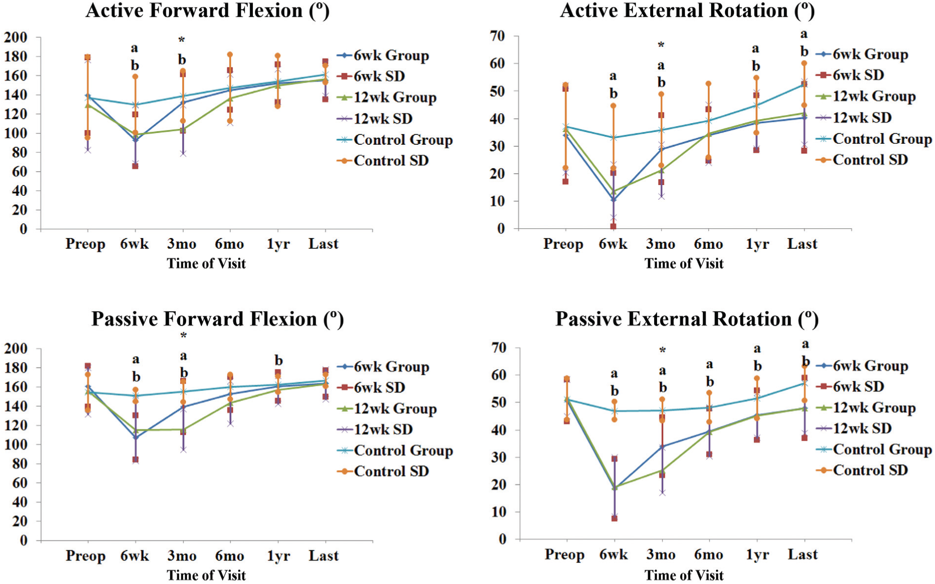
Preoperative and postoperative range of motion showing a significant improvement of the 6-week group compared with the 12-week group at 3 months postoperatively. *P < .05 between the 6-week group and 12-week group, a P < .05 between the 6-week group and control group, and b P < .05 between the 12-week group and control group.
Additionally, the 6-week group showed significant differences compared with the 12-week group in the KSS (71.5 ± 11.9 vs 58.2 ± 10.9 [P < .001], respectively) and UCLA (27.0 ± 4.0 vs 22.2 ± 4.4 [P < .001], respectively) scores at 3 months postoperatively. These significant differences in ROM and KSS and UCLA scores at 3 months postoperatively became insignificant after 6 months postoperatively. For the KSS score, compared with the control group, the 6-week group showed no significant differences only from 3 months postoperatively (69.3 ± 12.2 vs 71.5 ± 11.9 [P = .990], respectively) to 6 months postoperatively (80.2 ± 10.2 vs 75.3 ± 10.9 [P = .060], respectively). However, the 12-week group showed significant differences from 3 months postoperatively to the last follow-up compared with the control group. For the UCLA score, the 6-week group and 12-week group showed no significant differences from 3 months postoperatively (27.1 ± 4.0 vs 25.7 ± 4.6 [P = .268], respectively) to 1 year postoperatively (29.0 ± 4.4 vs 31.1 ± 4.6 [P = .094], respectively) and at 1 year postoperatively (29.5 ± 3.2 vs 31.1 ± 4.6 [P = .265], respectively) compared with the control group (Figures 2 and 3).
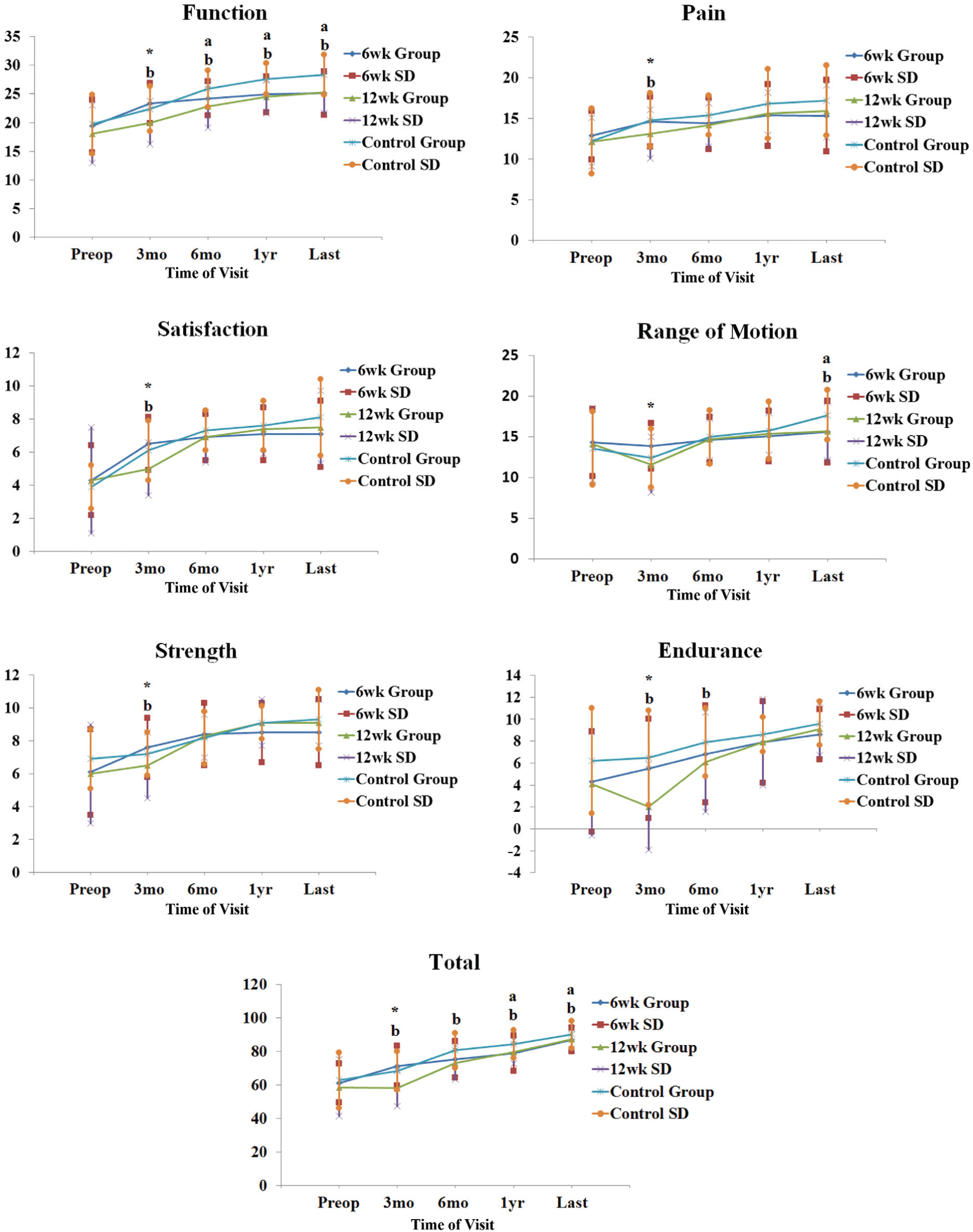
Preoperative and postoperative Korean Shoulder Scoring System scores showing a significant improvement of the 6-week group compared with the 12-week group at 3 months postoperatively. *P < .05 between the 6-week group and 12-week group, a P < .05 between the 6-week group and control group, and b P < .05 between the 12-week group and control group.
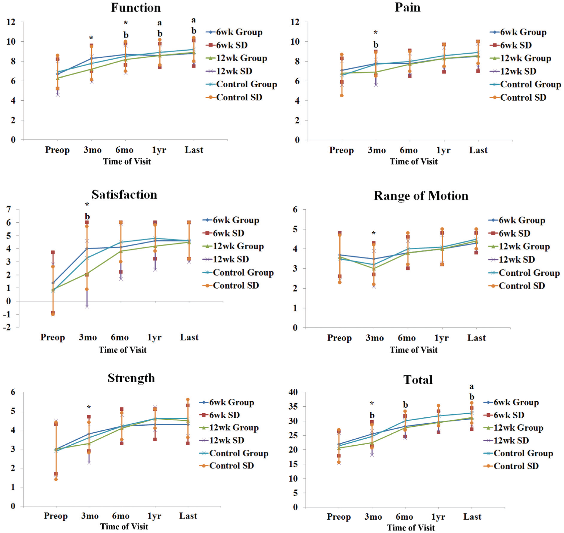
Preoperative and postoperative University of California, Los Angeles, shoulder scores showing a significant improvement of the 6-week group compared with the 12-week group at 3 months postoperatively. *P < .05 between the 6-week group and 12-week group, a P < .05 between the 6-week group and control group, and b P < .05 between the 12-week group and control group.
Repair Integrity
The overall retear rate was 12.0% (25/209 patients), and there was no significant difference in retear rates among the 6-week group (2/35 patients, 5.7%), the 12-week group (4/39 patients, 10.3%), and the control group (19/135 patients, 14.1%) (P = .374). Moreover, the retear rates between the 6-week group and the 12-week group showed no significant difference (P = .677).
Number of Involved Tendons
In patients with only supraspinatus tendon tears, there was no significant difference in retear rates between the 6-week group (2/35 patients) and 12-week group (4/37 patients) (P = .675) and no significant differences compared with the control group. However, there was no patient in the 6-week group and only 2 patients in the 12-week group with ≥2 tendon tears, so the comparison of retear rates between those 2 groups was not possible (Table 3).
Retear Rates According to the Number of Involved Tendons a
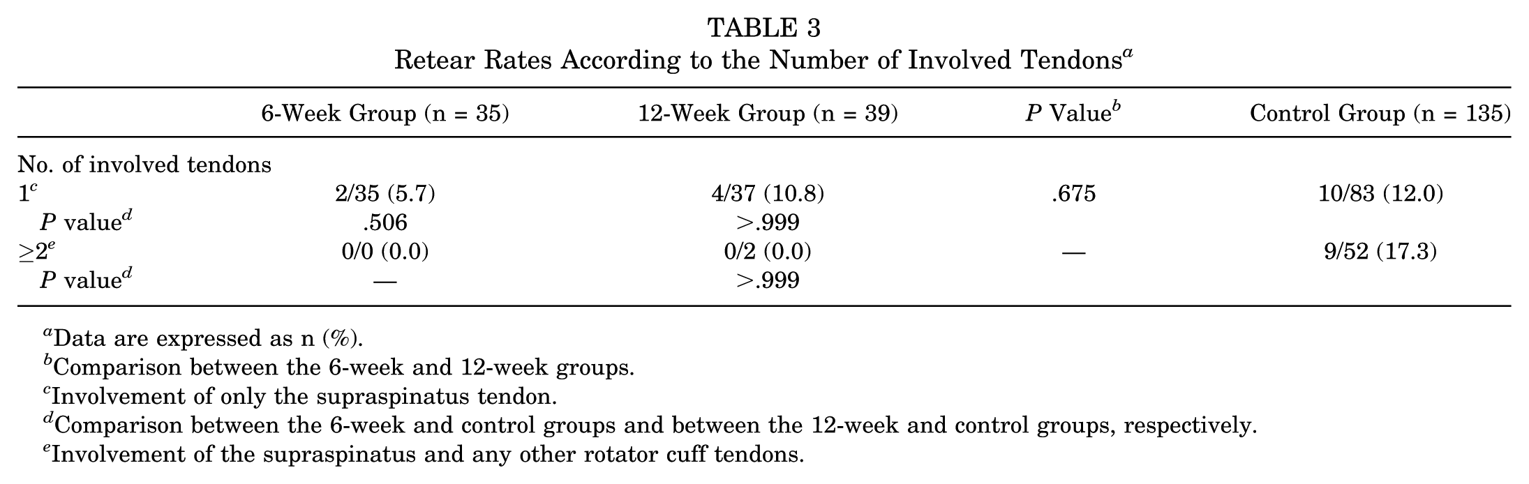
Data are expressed as n (%).
Comparison between the 6-week and 12-week groups.
Involvement of only the supraspinatus tendon.
Comparison between the 6-week and control groups and between the 12-week and control groups, respectively.
Involvement of the supraspinatus and any other rotator cuff tendons.
Age
Both in patients <65 years old (3.4% vs 4.3% [P > .999], respectively) and in patients ≥65 years old (16.7% vs 18.8% [P > .999], respectively), there were no significant differences in retear rates between the 6-week group and the 12-week group. Compared with the control group (10.9% in patients <65 years old and 23.5% in patients ≥65 years old), the 6-week group and 12-week group also showed no significant differences in retear rates both in patients <65 years old (P = .298 and P = .463, respectively) and ≥65 years old (P > .999 and P > .999, respectively) (Table 4). Also, a multiple logistic regression analysis was performed to control for age, which may be a confounding variable because it differs between the 6-week and 12-week groups. Age (odds ratio, 1.131 [95% CI, 0.993-1.288]; P = .063) and the 12-week group itself (odds ratio, 1.227 [95% CI, 0.194-7.949]; P = .828) were not independent predictors of a retear.
Retear Rates According to Age and Mediolateral and Anteroposterior Tear Sizes a
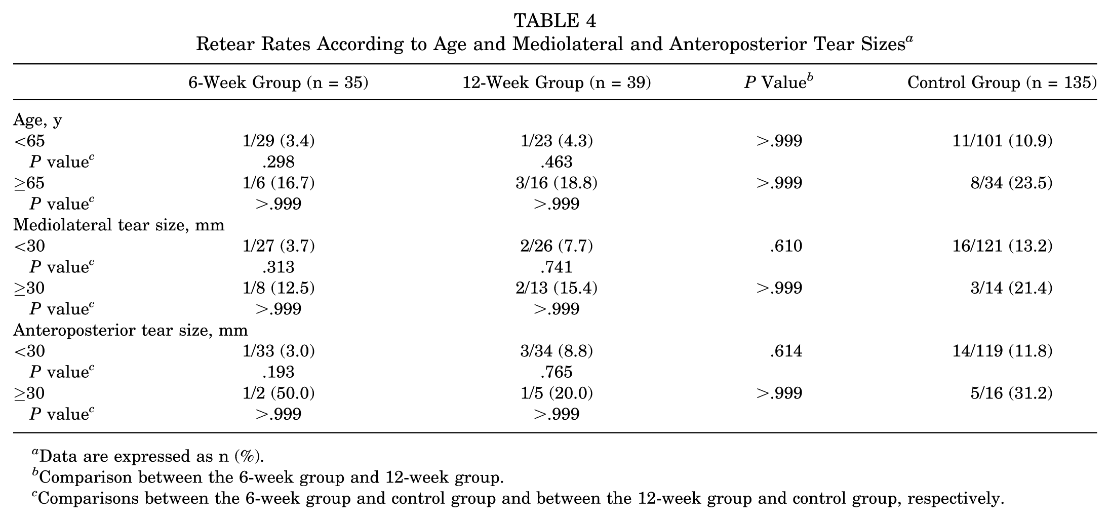
Data are expressed as n (%).
Comparison between the 6-week group and 12-week group.
Comparisons between the 6-week group and control group and between the 12-week group and control group, respectively.
Tear Size
Both in patients with <30-mm (3.7% vs 7.7% [P = .610], respectively) and in those with ≥30-mm (12.5% vs 15.4% [P > .999], respectively) mediolateral tear sizes, there were no significant differences in retear rates between the 6-week group and the 12-week group. Compared with the control group (13.2% in patients with <30-mm mediolateral tear size and 21.4% in patients with ≥30-mm mediolateral tear size), the 6-week group and 12-week group also showed no significant differences in retear rates both in patients with <30-mm (P = .313 and P = .741, respectively) and in those with ≥30-mm mediolateral tear sizes (P > .999 and P > .999, respectively) (Table 4). Also, both in patients with <30-mm (3.0% vs 8.8% [P = .614], respectively) and in those with ≥30-mm (50.0% vs 20.0% [P > .999], respectively) anteroposterior tear sizes, there were no significant differences in retear rates between the 6-week group and the 12-week group. Compared with the control group (11.8% in patients with <30-mm anteroposterior tear size and 31.2% in patients with ≥30-mm anteroposterior tear size), the 6-week group and 12-week group also showed no significant differences in retear rates both in patients with <30-mm (P = .193 and P = .765, respectively) and in patients with ≥30-mm mediolateral tear sizes (P > .999 and P > .999, respectively) (Table 4).
Discussion
The retear rates after intra-articular steroid injections for shoulder stiffness after arthroscopic repair of full-thickness rotator cuff tears between the 6-week and 12-week groups showed no significant difference. Significant improvement was demonstrated at the last follow-up in both the 6-week and the 12-week groups in ROM (forward flexion and external rotation) and KSS and UCLA scores after intra-articular steroid injections for stiffness after repair. The 6-week group showed significantly better ROM and KSS and UCLA scores at 3 months postoperatively than the 12-week group, although these differences became insignificant after 6 months postoperatively. Compared with the control group, the 6-week group and 12-week group gained similar ROM in active and passive forward flexion from 3 months and 6 months postoperatively and from 6 months and 1 year postoperatively, respectively. However, both groups did not gain similar ROM in both active and passive external rotation compared with the control group, except for the insignificant differences in active external rotation only at 6 months postoperatively. We believe that this is because the extent of increase in ROM of external rotation that both the 6-week group and 12-week group gained fell short of the extent that the control group gained, although both groups showed continuous improvement in ROM of external rotation. Compared with the control group, the 6-week group and 12-week group showed similar results in the pain, satisfaction, strength, and endurance domains of the KSS and in the pain, satisfaction, ROM, and strength domains of the UCLA score. In particular, both groups showed results similar to the control group in the pain and satisfaction domains of both the KSS and UCLA score right after the injection. However, despite the insignificance compared with the control group in the majority of the domains, the 6-week group and 12-week group showed significantly lower KSS and UCLA scores at the last follow-up. We believe that this is because the insignificant differences in every domain added up to the significant differences.
Conventional treatment for stiffness after arthroscopic rotator cuff repair includes oral medication, corticosteroid injections, physical therapy, and surgical capsular release. The first-line treatment is usually nonoperative treatment that includes oral steroids and nonsteroidal anti-inflammatory drugs. 14 Oral steroids have short-term benefits that are not maintained beyond 6 weeks. 4 Physical therapy is a commonly used treatment for shoulder stiffness. However, there is still no consensus on the effect, intensity, and timing of physical therapy for stiffness after repair.20,24,37,47 Furthermore, shoulder stiffness occurring after rotator cuff repair is often resistant to medication and physical therapy probably because of extra-articular and capsular adhesion. 47 Nonoperative treatment for stiffness after repair may be a long agonizing wait for enhanced results, and surgical interventions may be needed. Arthroscopic capsular release is reported to be a safe and reliable method for idiopathic, surgical, or posttraumatic stiffness. 24 Traditionally, arthroscopic capsular release was considered appropriate for the treatment of shoulder stiffness when a 6-month regimen of nonoperative treatment has failed. 20 However, waiting for 6 months with nonoperative treatment may be a long and painful period for patients who have to face the stress of a second operative procedure. Even after arthroscopic capsular release, the shoulder may remain painful.
An intra-articular corticosteroid injection is a common treatment for primary frozen shoulder, leading to satisfactory results with improved ROM and pain reduction. 29 The use of intra-articular steroid injections for adhesive capsulitis resulted in an increase in forward flexion before starting physical therapy at postoperative 7 days. 41 However, to our knowledge, there is no literature on the timing of corticosteroid injections for shoulder stiffness after arthroscopic rotator cuff repair and its relation to repair integrity. Our main concern was the possibility of retears by disrupting tendon healing with intra-articular steroid injections at an early postoperative period of 6 weeks.27,45 However, an intra-articular steroid injection at 6 weeks postoperatively was thought to be a safe method without compromising repair integrity.
Subacromial prednisolone injections in a rat model have shown that biomechanical changes normalized after the inflammatory phase (2-3 weeks).6,28,30,44 In a clinical study, subacromial triamcinolone injections after the inflammatory phase at the first postoperative month did not result in tendon deterioration of the rotator cuff during the recovery phase. 40 This prior study reported that the mean visual analog scale for pain score significantly decreased from 7.7 ± 1.2 to 2.3 ± 1.4 at the end of the first month after the injection and to 1.2 ± 1.8 at 3 months after the injection. However, there was no significant difference (P = .06) in the retear rate between patients with (6.8%) and without (18.4%) an injection. To treat shoulder stiffness after repair, subacromial injections are also an option. Although the prevalence of shoulder stiffness among patients with postoperative pain after rotator cuff repair was not determined, and this study did not analyze the cause of postoperative pain after rotator cuff repair, many patients with postoperative pain are likely to have shoulder stiffness, and a subacromial injection can be considered as an option to treat stiffness after repair. Our goal was to provide an early intervention without compromising repair integrity with an interval of 4 weeks.
From many patient factors in our study, all of them showed no significant differences except for age in which the 12-week group had a significantly higher mean age than the 6-week group. Tendon degeneration may be an inevitable progression due to aging that eventually leads to fatty infiltration and atrophy. 21 However, some literature mentions the insignificance of age to repair integrity. No significant difference was shown in retear rates between patients >70 years old (51.1%, 24/47 patients) and those <70 years old (39.8%, 76/191 patients) after arthroscopic rotator cuff repair. 36 There was an insignificantly higher retear rate of 21.2% (14/66 patients) in patients >65 years old compared with 13.6% (11/81 patients) in patients <65 years old after arthroscopic rotator cuff repair. 10 Tear size affects the results, and age itself can be considered less contributive to repair integrity, with retears in 34 of 81 patients (42.0%) aged ≥65 years after arthroscopic rotator cuff repair at 6 months’ follow-up. 7 In the current study, even though age showed statistical significance between the 6-week and 12-week groups, age itself was not an independent predictor of a retear. However, we believe that the result derived from this logit model cannot be generalized.
The definition of stiffness is still controversial. Orthopaedic surgeons are aware that most patients develop a certain degree of stiffness after rotator cuff repair, whether it is mild or severe. 48 There is no consensus on the amount of motion deficit needed to establish the criteria for shoulder stiffness after arthroscopic rotator cuff repair.1,33,38,43 Stiffness is defined by adding the passive ROM deficits of abduction, forward flexion, external rotation, and internal rotation, with 0°-20° considered as mild, 25°-70° as moderate, and >70° as severe. 43 This definition simply adds ROM deficits without considering various possible clinical outcomes resulting from each ROM deficit itself. 33 The definition of stiffness is crucial, and the outcomes may differ because of inaccurate diagnoses and the establishment of inappropriate treatment strategies. However, the definition of stiffness is difficult to establish because the criteria may be subjective.
Hand-behind-the-back ROM may not accurately assess active internal rotation of the shoulder. 17 Moreover, hand-behind-the-back movement is reported to be affected by elbow and wrist motion, it includes shoulder extension and abduction, and 62% of shoulder internal rotation occurs when the thumb reaches the sacrum. 46 Therefore, internal rotation was excluded from the diagnostic criteria of stiffness after repair and the evaluation during follow-up in the current study.
One of the strengths of this study is that, to our knowledge, the current study is the first study on repair integrity after intra-articular steroid injections for shoulder stiffness after arthroscopic rotator cuff repair at 2 distinct time periods. However, this study has several limitations. First, this is a retrospective study. Thus, it has an intrinsic limitation in terms of the methodology, which would have been better if it were a prospective randomized study. Second, the sample size is small, but all patients were selected during a certain period to reflect the actual situation. Third, follow-up of MRI for the assessment of repair integrity was obtained at 6 months postoperatively. We believe that 6 months may be adequate for the evaluation of repair integrity because retears are reported to occur within 6 months postoperatively and do not occur between 6 and 24 months postoperatively. 31 Moreover, a report on experimental rotator cuff repair in sheep showed that a repaired rotator cuff had 52% strength of an intact rotator cuff at 3 months postoperatively and 82% strength at 6 months postoperatively. 16 Accordingly, an examination of repair integrity using MRI at 6 months postoperatively would be helpful.
In conclusion, early intervention with an intra-articular corticosteroid injection at 6 weeks postoperatively for patients who develop shoulder stiffness after arthroscopic rotator cuff repair may relieve pain and enhance shoulder ROM without influencing retears of the repaired rotator cuff tendon. These results may help shoulder surgeons caring for patients with postoperative shoulder stiffness in regard to treatment planning and enhance patient satisfaction.
Footnotes
Acknowledgements
The authors thank Da Young Go for her valuable assistance with the collection and arrangement of patient data.
The authors declared that they have no conflicts of interest in the authorship and publication of this contribution.