
Abstract
Background:
Radial tears of the meniscus are a common knee injury, frequently resulting in osteoarthritis. To date, there are no established, effective treatments for radial tears. Adipose-derived stem cells (ASCs) may be an attractive cell source for meniscal regeneration because they can be quickly isolated in large number and are capable of undergoing induced fibrochondrogenic differentiation mediated by transforming growth factor β3 (TGF-β3). However, the use of ASCs for meniscal repair is largely unexplored.
Hypothesis:
ASC-seeded hydrogels with preloaded TGF-β3 will improve meniscal healing of radial tears, as modeled in an explant model.
Study Design:
Controlled laboratory study.
Methods:
With an institutional review board–exempted protocol, human ASCs were isolated from the infrapatellar fat pads of 3 donors, obtained after total knee replacement, and characterized. ASCs were encapsulated in photocrosslinkable methacrylated gelatin hydrogels to form 3-dimensional constructs, which were placed into tissue culture. The effect of TGF-β3—whether preloaded into the hydrogel or added as a soluble medium supplement—on matrix-sulfated proteoglycan deposition in the constructs was evaluated. A meniscal explant culture model was used to simulate meniscal repair. Cylindrical-shaped explants were excised from the inner avascular region of adult bovine menisci, and a radial tear was modeled by cutting perpendicular to the meniscal main fibers to the length of the radius. Six combinations of hydrogels—namely, acellular and ASC-seeded hydrogels supplemented with preloaded TGF-β3 (2 µg/mL) or soluble TGF-β3 (10 ng/mL) and without supplement—were injected into the radial tear and stabilized by photocrosslinking with visible light. At 4 and 8 weeks of culture, healing was assessed through histology, immunofluorescence staining, and mechanical testing.
Results:
ASCs isolated from the 3 donors exhibited colony-forming and multilineage differentiation potential. Hydrogels preloaded with TGF-β3 and those cultured in soluble TGF-β3 showed robust matrix-sulfated proteoglycan deposition. ASC-seeded hydrogels promoted superior healing as compared with acellular hydrogels, with preloaded or soluble TGF-β3 further improving histological scores and mechanical properties.
Conclusion:
These findings demonstrated that ASC-seeded hydrogels preloaded with TGF-β3 enhanced healing of radial meniscal tears in an in vitro meniscal repair model.
Clinical Relevance:
Injection delivery of ASCs in a TGF-β3-preloaded photocrosslinkable hydrogel represents a novel candidate strategy to repair meniscal radial tears and minimize further osteoarthritic joint degeneration.
Keywords
Menisci are essential for knee function and perform many important roles, including load transmission, shock absorption, stabilization, nutrient distribution, and proprioception. 11 The menisci are frequently damaged by sports injuries and age-related degeneration. 42 Unfortunately, the meniscus has a relatively low healing capability because of limited vascularization in the inner zone. As a result, meniscal tissue around the tear site is usually removed through meniscectomy, which greatly increases contact pressures on the tibial plateau and increases the risk of developing osteoarthritis.8,35 The extent of osteoarthritis relates to the volume of tissue resected. 9
There are several types of tear, such as horizontal, longitudinal, and radial tears. Radial tears most profoundly disrupt the ability of the meniscus to distribute the hoop stresses associated with weightbearing,12,15 and they constitute approximately 14% to 15% of all meniscal tears.15,17 Unfortunately, because all radial tears involve damage to the avascular zone, they are often the most challenging to treat. Historically, repairs of radial tears have not been recommended. Therefore, new strategies for regenerating meniscal tissue within a radial tear are needed to prevent accelerated osteoarthritic changes.
There have been many recent attempts to stimulate intrinsic healing of the menisci to improve the chances of healing the avascular zone. Strategies have included perforations to the vascular zone,51,52 synovial grafts,6,21 osseous perforations, 13 and the addition of fibrin clots,2,20 platelet-rich plasma, 19 or various growth factors.16,31 While some attempts have shown promise, they are generally limited by the hypocellularity of the avascular zone. However, it has been proposed that meniscal neotissue formation at the tear site may be promoted through the use of tissue-engineering approaches, which include the combination of cells, scaffolds, and growth factors, to restore or replace the damaged meniscus. 18
The use of adult mesenchymal stem cells (MSCs) as the cellular component for meniscus tissue engineering has been of particular research interest, as these cells are relatively abundant and capable of undergoing chondrogenic differentiation.39,49 The therapeutic potential of bone marrow–derived MSCs (BM-MSCs) has been established by many in vitro and in vivo studies reporting that they promoted healing of meniscal defects.49,50 Beyond bone marrow, MSCs can be obtained from many tissues. In particular, adipose-derived stem cells (ASCs) are an attractive cell source, as they can be more easily harvested than BM-MSCs, with larger yields. 54 For example, it is possible to harvest up to 40% of the entire infrapatellar fat pad (IPFP) volume, which in total measures about 25 cm3, without impairing joint function. 5 ASCs have been used extensively both in animal studies37,53 and in human clinical studies.14,24
According to many previous studies, growth factors are often necessary to promote robust neotissue formation. In particular, transforming growth factor β3 (TGF-β3) was shown to promote cartilage repair and accelerate cartilage differentiation, with numerous studies demonstrating chondrogenic differentiation of MSCs mediated by TGF-β3 supplementation.3,40 In particular, Lai et al 23 reported that ASCs seeded in 3-dimensional hydrogels underwent enhanced chondrogenic differentiation when cultured in medium supplemented with TGF-β3, as indicated by elevated collagen type II gene expression and glycosaminoglycan (GAG) production.
Last, hydrogels have been considered attractive biomaterial scaffolds for delivery of cells and growth factors. A hydrogel scaffold can fill an irregularly shaped defect and provide a stable microenvironment for cell growth and differentiation. 48 Furthermore, many hydrogels are injectable and could therefore be applied in the context of arthroscopic surgery, as most commonly performed clinically for meniscal tears. 38 An injectable hydrogel composed of methacrylated gelatin (mGL) was recently developed. 25 The mGL hydrogel was shown to support robust chondrogenic differentiation of encapsulated MSCs when cultured in medium supplemented with TGF-β3. 36 However, it not known whether mGL hydrogels preloaded with TGF-β3 can induce chondrogenic differentiation of encapsulated MSCs to the same degree as when TGF-β3 is added as a soluble supplement with each medium change.
In another recent study, a cell-seeded nanofibrous scaffold was shown to enhance healing of a radial meniscal tear, as simulated in an in vitro explant model. 41 However, cell attachment to the nanofibrous scaffolds required >24 hours of in vitro culture, and the scaffolds were wrapped around the tear site as sheaths, representing a challenging surgical approach. An alternative approach to consider is rapid suspension of cells in liquid hydrogels that can then be injected into the tear site with subsequent in situ curing. The objective of this study was to (1) assess the effect of TGF-β3 on ASC chondrogenesis in vitro when encapsulated within the mGL hydrogel or provided as medium supplement and (2) determine the effect of these ASC-seeded hydrogels on healing of radial meniscal tears, as simulated through an established in vitro model. We hypothesized that the inclusion of ASCs within the hydrogels would improve radial tear healing as compared with acellular hydrogels and that inclusion of TGF-β3 would promote chondrogenic differentiation and further enhance healing, whether encapsulated within the hydrogel or provided as a soluble medium supplement.
Methods
Cell Isolation
With institutional review board exemption, human ASCs (hASCs) were isolated from the IPFP procured after total knee arthroplasty from 3 female donors. Harvested IPFP tissue weighed an average of 6.8 g (range, 5-10 g) and was digested in 1 mg/mL of collagenase type I (Worthington Biochemical Corporation) with rotary agitation at 180 rpm for 3 hours at 37°C. The digested tissues were then centrifuged at 1500 rpm for 5 minutes to obtain the stromal vascular fraction pellet; the pellet was then resuspended and plated in T150 Flask as passage 0 hASCs. hASCs were cultured in growth medium (high-glucose Dulbecco’s Modified Eagle’s Medium [DMEM], 10% fetal bovine serum, 1% v/v antibiotic-antimycotic, 1-ng/mL fibroblast growth factor 2; Gibco). At 80% confluence, cells were detached with 0.25% trypsin in 1mM EDTA (Gibco) and passaged. All experiments were performed with passage 2 (p2) hASCs. To reduce donor-to-donor variation, cells from the 3 donors were pooled in equal number for all experiments.
Cell Characterization
p2 hASCs (1 × 10⁵) were seeded in a 6-well plate with 3 mL of growth medium per well. For differentiation assays (chondrogenesis, osteogenesis, and adipogenesis), after 2 days, when the cells reached ~70% confluency, growth medium was switched to
chondrogenic medium (DMEM, 1% v/v antibiotic-antimycotic, 10-µg/mL insulin-transferrin-selenium [ITS; Sigma-Aldrich], 0.1µM dexamethasone, 40-µg/mL proline [Sigma-Aldrich], 50-µg/mL ascorbic acid, and 10 ng/mL TGF-β3 [PeproTech])
osteogenic medium (DMEM, 1% v/v antibiotic-antimycotic, 10-µg/mL ITS, 0.1µM dexamethasone, 10mM β-glycerol phosphate, 50-µg/mL ascorbic acid)
adipogenic medium (DMEM, 10% v/v fetal bovine serum, 1% v/v antibiotic-antimycotic, 1-µg/mL ITS, 1µM dexamethasone, and 0.5mM 3-isobutyl-1-methylxanthine). 43
Cultures were then maintained for 21 days and stained with alcian blue for chondrogenesis, alizarin red for osteogenesis, or oil red O for adipogenesis. To characterize cell surface epitopes, hASCs at p2 were detached by trypsin-EDTA and incubated with propidium iodide and PE- or FITC-conjugated mouse (IgG1, k) anti-human antibodies for 30 minutes at 4°C. Antibodies included mouse anti-human CD31, CD34, CD44, CD45, CD73, CD90, and CD105 (BD Biosciences). Dead cells were excluded by positive propidium iodide staining. PE- or FITC-conjugated isotype-matched IgGs (BD Biosciences) were used as controls. After washing, the cells were sorted with the FACSAria II SORP flow cytometer (BD Biosciences) and data analyzed with DiVa (v 6) software. To assess colony-forming capacity, 100 p2 ASCs were plated in 100-cm2 dishes, cultured in growth medium for 14 days, and stained with 0.5% crystal violet in methanol for 5 to 10 minutes at room temperature.
Preparation of mGL Hydrogels
mGL was synthesized according to a procedure previously described with slight modification.25,29,47 Gelatin from bovine skin type B (15 g; Sigma-Aldrich) was dissolved completely in 500 mL of deionized H2O at 37°C, and then 15 mL of methacrylic anhydride was added dropwise. The mixture was placed in a 37°C shaker at 150 rpm for 24 hours. The mGL solution was dialyzed with 2000 NMWCO dialysis tubing (Sigma-Aldrich) for 4 days against H2O at room temperature to completely remove all low molecular weight by-products. After lyophilization, the mGL product was stored in a desiccator for future use. The methacrylation efficiency of the process was ~80% as assessed by 1H NMR.
The visible light (VL)–activated photoinitiator, lithium phenyl-2,4,6-trimethylbenzoylphosphinate (LAP), was synthesized as described by Fairbanks et al. 10 Briefly, dimethyl phenylphosphonite (Acros Organics) was reacted with 2,4,6-trimethylbenzoyl chloride (Sigma-Aldrich) via a Michaelis-Arbuzov reaction. At room temperature and under argon, 3.2 g (0.018 mol) of 2,4,6-trimethylbenzoyl chloride was added dropwise to an equimolar amount of continuously stirred dimethyl phenylphosphonite (3.0 g). The reaction mixture was stirred for 18 hours whereupon a 4-fold excess of lithium bromide (6.1 g) in 100 mL of 2-butanone (both from Sigma-Aldrich) was added to the reaction mixture from the previous step, which was then heated to 50°C. After 10 minutes, a solid precipitate formed. The mixture was cooled to ambient temperature, allowed to stand for 4 hours, and then filtered. This product is LAP. mGL was dissolved in Hank’s balanced salt solution (HBSS) at 10% (w/v), and after adjustment of pH to 7.4 with 1N NaOH, 0.15% w/v photoinitiator LAP and 1% v/v antibiotic-antimycotic were added. hASCs and TGF-β3 were added for subsequent experiments, as described later.
In Vitro TGF-β3 Release Studies
mGL solution (50 µL) with preloaded TGF-β3 (2 µg/mL) was distributed to silicone molds measuring 5 mm (diameter) × 2 mm (depth) and exposed to light (390-395 nm) for 30 seconds for photogelation. The hydrogels were cultured in 500 µL of HBSS (containing 0.1% bovine serum albumin [BSA]). At different points up to 21 days, HBSS was collected and completely replaced with fresh HBSS. The TGF-β3 content released from the mGL hydrogel was measured with an ELISA kit (Assay Biotech), according to the manufacturer’s instructions.
Effect of TGF-β3 on ASC Chondrogenesis
hASCs were suspended in the 10% mGL solution at a final density of 10 × 106 cells/mL. A 50-µL aliquot of the cell suspension was distributed to silicone molds measuring 5 mm (diameter) × 2 mm (depth) and exposed to light (390-395 nm) for 30 seconds for photogelation. The effect of TGF-β3 on ASC chondrogenesis was evaluated by comparing 3 culture conditions: (1) ASC-seeded hydrogels without TGF-β3, (2) ASC-seeded hydrogels with 100 ng of TGF-β3 (2 µg/mL) preloaded within hydrogel, and (3) ASC-seeded hydrogels with 10 ng/mL of TGF-β3 in the culture medium. With the exception of the third condition, hydrogels were cultured in differentiation medium consisting of DMEM with high glucose, 1% antibiotic-antimycotic, 0.1mM dexamethasone, 50 µg/mL of ascorbate 2-phosphate, 40 µg/mL of L-proline, and 10 µg/mL of ITS. Constructs of the third condition were cultured in differentiation medium additionally supplemented with 10 ng/mL of TGF-β3. Medium was changed twice a week in all conditions.
Constructs were analyzed on the basis of biochemical composition and histology at culture days 21 and 42. To determine sulfated GAG (sGAG) content, samples were papain digested and analyzed with the dimethylmethylene blue dye binding assay (Blyscan; Biocolor) according to the manufacturer’s instructions. Digested samples were also evaluated for total DNA content with the Picogreen assay (Invitrogen). For histological analysis, constructs were fixed in 4% paraformaldehyde, paraffin embedded, sectioned at 8-µm thickness, and stained with safranin O/fast green and alcian blue/fast red with standard protocols. Histologic images were captured with a microscope equipped with a digital camera (SZX 16; Olympus).
Fabrication of Nanofibrous Scaffold
Aligned electrospun poly-ϵ-caprolactone nanofibrous scaffold (NFS) was fabricated as described by Shimomura et al. 41 The NFS was used only to ensure early retention of the hydrogel in the tear site, as described later.
In Vitro Model of Radial Tear
The in vitro radial tear model was also adopted from Shimomura et al. 41 Adult bovine menisci were aseptically harvested from the hind-leg stifle of 2- to 3-year-old cows within 24 hours of slaughter (JW Treuth and Sons), and cylinder-shaped explants (5-mm diameter × 10-mm height) were then excised from the inner avascular zone with a sterile disposable biopsy punch (5 mm; Integra Miltex) (Figure 1, A and B). Three explants were harvested from 1 meniscus, with the long axis aligned along the direction of the meniscal circumferential main fibers (Figure 1A). A radial tear was mimicked by cutting perpendicular to the meniscal main fibers to the length of the radius (Figure 1C). Hydrogels (described later) were injected into the defect site (Figure 1D) and photopolymerized by VL (Figure 1E). The defect site was wrapped with the nanofibrous scaffold with the fiber direction paralleling that of the meniscal main fibers. The NFS was used as previously described to ensure retention of the hydrogel at the tear site but was removed before all analyses. 41 Six hydrogel/medium conditions were investigated: (1) acellular hydrogel, (2) ASC hydrogel, (3) acellular hydrogel with preloaded TGF-β3 (2 µg/mL), (4) ASC hydrogel with preloaded TGF-β3 (2 µg/mL), (5) acellular hydrogel with soluble TGF-β3 supplemented in culture medium, and (6) ASC hydrogel with soluble TGF-β3 supplemented in culture medium. Constructs of conditions 1 to 4 were cultured in differentiation medium, as described earlier, while constructs of conditions 5 and 6 were cultured in differentiation medium supplemented with 10 ng/mL of TGF-β3. Media were changed twice a week.
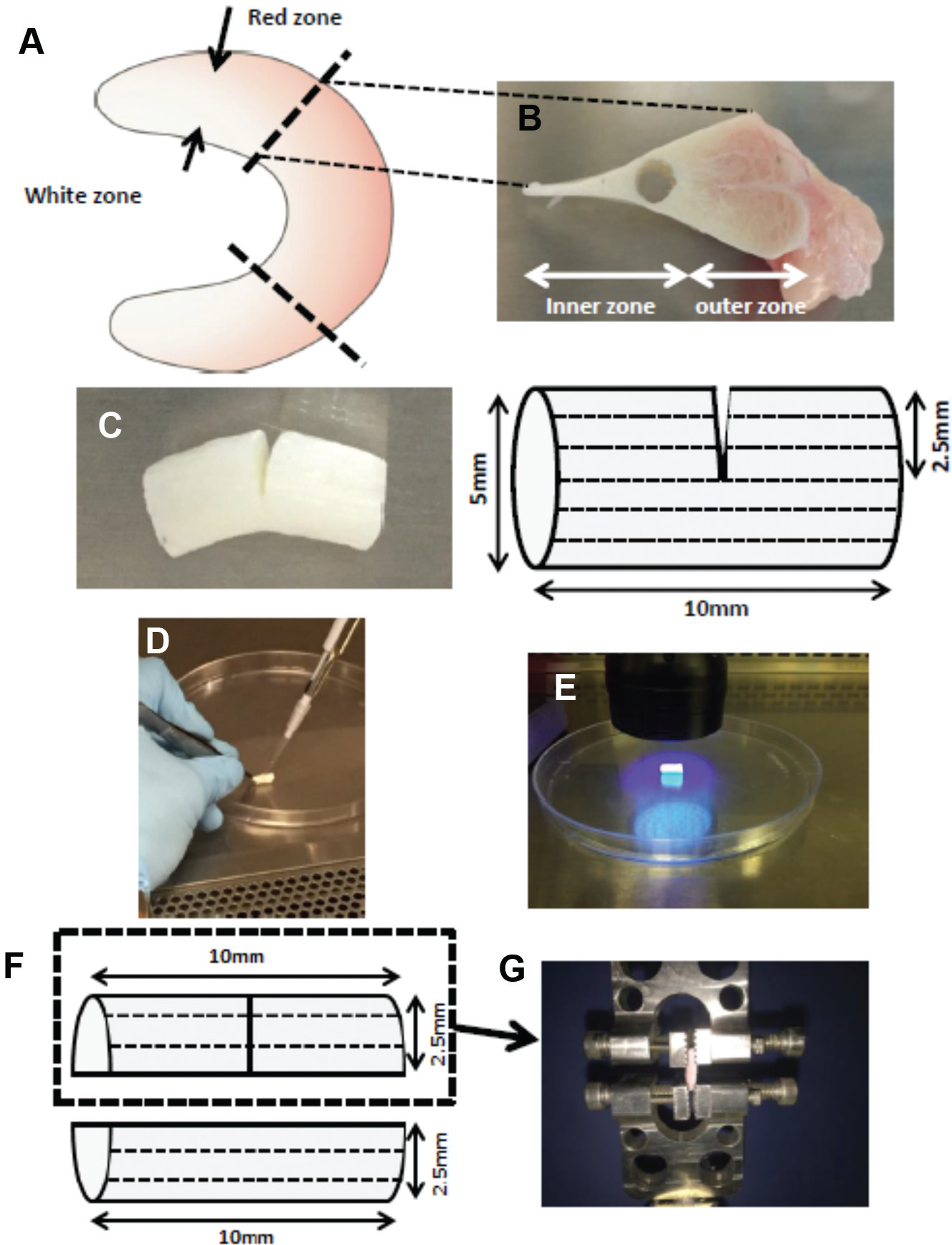
Schematic of the experimental setup. Preparation of composite constructs of meniscus and scaffold adapted from Shimomura et al. 41 (A, B) Cylinder-shaped explants (5-mm diameter, 10-mm height) were excised from the inner avascular region of the meniscus, with the long axis aligned along the direction of the meniscal circumferential fibers. (C) The adipose-derived stem cell–seeded or acellular hydrogels were (D) injected into the defect site and (E) polymerized by visible light. Dotted line in the meniscal construct represents meniscal fiber direction. For mechanical testing, only (F) the semicylinder containing the simulated radial tear was retained (to isolate the neotissue) and (G) tested under uniaxial tension.
Histology and Immunohistochemistry of In Vitro Model of Radial Tear
The meniscal explants were collected at 8 weeks of culture; washed twice in phosphate-buffered saline; fixed in 4% paraformaldehyde overnight at room temperature; immersed in 10%, 20%, and 30% sucrose solutions, each for 1 hour at room temperature; and then embedded in Tissue-Tek optimal cutting temperature compound (Sakura Finetek USA, Inc). The embedded samples were sectioned at 8-µm thickness and stained with picrosirius red. Repair of the radial tear was evaluated to obtain a “percentage histological repair” 41 (%HR) value; namely, 3 sections of the entire gap were evaluated from each of 3 explants per group (n = 9 per group). The entire length of the cut edge in the radial tear and the length of the adhesion site were outlined and measured by Image J (National Institutes of Health). The %HR was the length of the adhesion site, represented as a percentage of the total length of the cut edge.
The presence and distribution of procollagen types I and II were evaluated in the tear site at 8 weeks with standard fluorescence immunohistochemistry, counterstained with DAPI to identify cell nuclei. Briefly, sections were fixed in 4% paraformaldehyde for 5 minutes, treated with antigen retrieval solution (625-µg/mL hyaluronidase and 10-µL/mL chondroitinase in 0.02% bovine serum albumin), permeabilized for 15 minutes with 0.1% Triton X-100 (Sigma-Aldrich), blocked with 1% bovine serum albumin for 1 hour, and incubated with specific primary antibodies overnight at 4°C, followed by incubation with secondary antibodies for 1 hour. The primary antibodies included mouse monoclonal anti-collagen type I antibody (1:250, ab23446; Abcam) and mouse monoclonal anti-collagen type II (1:250, ab3092-500; Abcam), with Alexa 488–labeled polyclonal goat anti-mouse IgG antibodies (1:2000; Molecular Probes) as secondary antibodies. Negative controls for collagen type I or type II were incubated without the use of the primary antibody.
Mechanical Testing
Mechanical testing was performed with a Bose 3230 Mechanical Tester consisting of a 450-N load cell. At 4 and 8 weeks of culture, the constructs (n = 9 per group) were washed twice in phosphate-buffered saline, and a semicylindrical explant containing the repair site was harvested by precisely cutting with a plastic cutting guide after removal of the NFS (Figure 1F). As a control, a similar semicylindrical explant (5-mm diameter × 10-mm height) was harvested. The semicylindrical explant was fixed to the mechanical tester at both ends (Figure 1G). The samples were preloaded to remove slack (gauge length averaged 5.05 mm) and then subjected to 5-mm strain at an elongation rate of 0.0833 mm/s to obtain a load/displacement curve. For each construct, load to failure, stiffness, and Young modulus of the repaired site were calculated. Cross-sectional area was measured with a digital caliper (Fischer Scientific).
Statistical Analysis
Three independent trials of experiments were performed, with each condition tested in triplicate at minimum (n ≥ 9 per condition). The results are expressed as the mean ± SD. In experiments involving multiple groups and time points, a factorial analysis of variance was performed. Significant main effects and/or interactions prompted the performance of 1-way analysis of variance with Tukey post hoc test for multiple comparisons. Comparison between independent groups was achieved by a 2-tailed Student t test. The data were analyzed with SPSS (v 22.0; IBM), and significance was considered at P < .05.
Results
hASC Characterization
hASCs from 3 donors displayed comparable clonogenicity, forming 26 ± 3, 22 ± 2, and 24 ± 4 colonies per 100 p2 hASCs after 14 days of culture (Figure 2A). p2 hASCs underwent chondrogenic, osteogenic, and adipogenic differentiation, as shown by positive alcian blue, alizarin red, and oil red O staining, respectively (Figure 2B). Flow cytometry analysis of cell surface markers revealed the presence of the MSC-associated epitopes CD44, CD73, CD90, and CD105 and the absence of hematopoietic and endothelial markers CD31, CD34, and CD45 (Figure 2C), confirming an MSC-like phenotype.
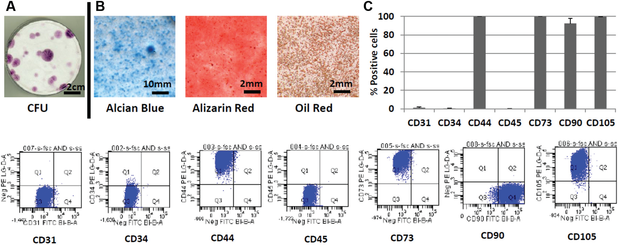
Characterization of infrapatellar fat pad–derived human adipose-derived stem cells. (A) Colony-forming unit (CFU) assay. (B) Multilineage differentiation potential (chondrogenesis, osteogenesis, and adipogenesis), assessed by histological staining with alcian blue, alizarin red, and oil red O, respectively. Day 21 cultures are shown. (C) Flow cytometric analysis of passage 2 human adipose-derived stem cells, confirming the presence of mesenchymal stem cell–associated surface markers (CD44, CD73, CD90, CD105) and absence of hematopoietic and endothelial markers (CD31, CD34, CD45). The data are expressed as mean ± SD.
In Vitro TGF-β3 Release Studies
In vitro release assays were performed to examine TGF-β3 release patterns (Figure 3). The results showed sustained release over 21 days; mGL hydrogels released 48.7% of total TGF-β3 in 7 days and 78.4% of total TGF-β3 in 21 days.
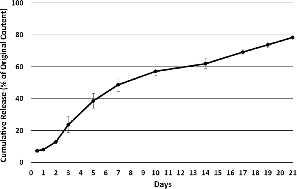
Transforming growth factor β3 (TGF-β3) release from TGF-β3-preloaded methacrylated gelatin hydrogels. Approximately 80% of total TGF-β3 was released over 3 weeks. The data are expressed as mean ± SD.
Effect of TGF-β3 on Chondrogenesis: Histology and Biochemical Composition
Minimal proteoglycan deposition was seen in ASC-seeded hydrogels cultured in the absence of TGF-β3 (Figure 4, left column). However, positive safranin O staining was found in ASC-seeded hydrogels preloaded with TGF-β3 (Figure 4, middle column) as well as ASC-seeded hydrogels cultured in differentiation medium supplemented with soluble TGF-β3 (Figure 4, right column). While the chondrogenic effect appeared qualitatively equivalent in the preloaded and medium-supplemented TGF-β3 groups at 3 weeks, superior sGAG deposition was noted in the latter group by 6 weeks. Alcian blue staining (Appendix Figure A1, available in the online version of this article) showed similar results.
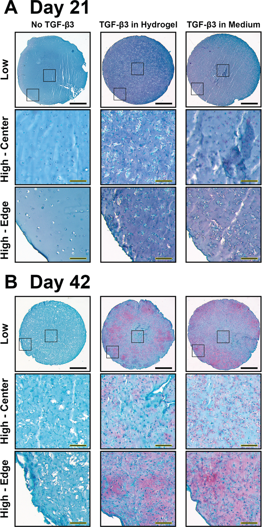
Histological assessment of chondrogenesis in adipose-derived stem cell–seeded hydrogel cultured for (A) 21 days and (B) 42 days. Safranin O (pink) staining demonstrates that the presence of transforming growth factor β3 (TGF-β3), either preloaded in the hydrogel or as a culture medium supplement, is required for adipose-derived stem cell chondrogenesis. Scale bars: 1 mm (low magnification), 200 μm (high magnification). Black boxes indicate regions of higher magnification in hydrogel center and edge.
Quantification of total sGAG production showed a similar result as histology. TGF-β3-preloaded hydrogels and hydrogels exposed to TGF-β3-supplemented medium showed equivalent increases in sGAG levels as compared with controls on day 21 (Figure 5). However, ASC hydrogels cultured in TGF-β3-supplemented medium possessed greater sGAG content on day 42 as compared with both control and preloaded TGF-β3 groups. Double-stranded DNA levels were also higher at days 21 and 42 in both TGF-β3 groups, but there was no significant difference between the preloaded and soluble TGF-β3 groups. As a result, normalized sGAG content (Figure 5C) showed the same trends as total GAG content (Figure 5A).
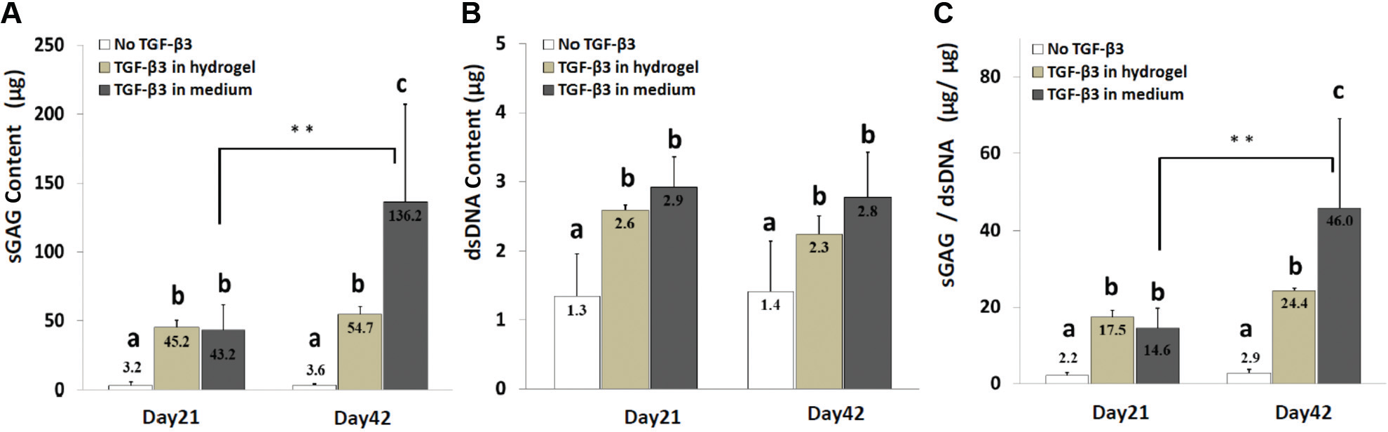
Sulfated glycosaminoglycan (sGAG) content of adipose-derived stem cell hydrogels after 21 and 42 days of culture. (A) Total sGAG content. (B) Total double-stranded DNA (dsDNA) content. (C) sGAG production normalized to DNA content. The results indicate the requirement of transforming growth factor β3 (TGF-β3) for production of sGAG and the highest sGAG content in cultures maintained in TGF-β3-supplemented medium. The data are expressed as mean ± SD. Letters correspond to statistically equivalent groups at a given time point, P < .05. Statistically significant differences between 3 and 6 weeks for a given group are shown by bars. **P < .01.
In Vitro Healing of Radial Meniscal Tear
Matrix staining with picrosirius red showed minimal in vitro healing mediated by acellular hydrogels, as represented by the low %HR, regardless of TGF-β3 supplementation (Figure 6). Conversely, ASC-seeded hydrogels promoted in vitro healing, as evidenced by increased neotissue formation. Healing was further improved by exposure of hASCs to TGF-β3, with no difference between ASC-seeded hydrogels preloaded with TGF-β3 and those supplemented with soluble TGF-β3. Notably, ASC-seeded hydrogels with preloaded TGF-β3 (%HR = 59.0) and those with soluble TGF-β3 (%HR = 66.7) showed significantly improved healing as compared with ASC-seeded hydrogels without TGF-β3 (%HR = 31.5) and with all acellular groups.
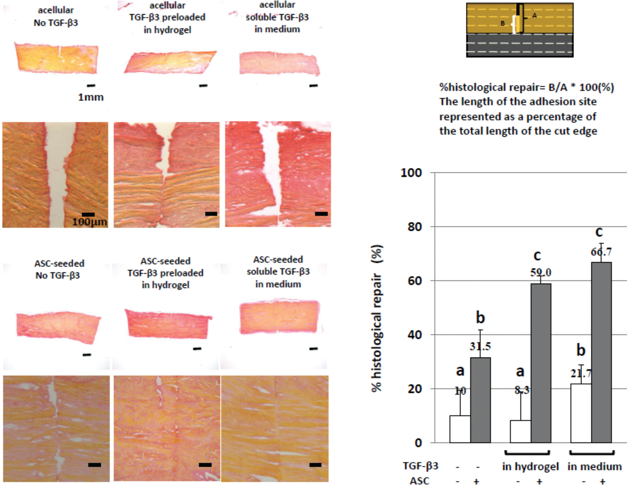
Histological analysis of meniscal radial gap repair in vitro. (Left) Histological picrosirius red staining of sections from the 6 experimental groups. (Right) Quantification of gap repair based on percentage histological repair. The results showed that radial tears in explants injected with adipose-derived stem cell (ASC)–seeded hydrogel preloaded with transforming growth factor β3 (TGF-β3) or cultured in soluble TGF-β3 exhibited a partial repair at 8 weeks in culture. The data are expressed as mean ± SD. Bar = 200 µm. Letters correspond to statistically equivalent groups, P < .05.
Immunohistochemical analysis of the tear site showed strong procollagen type I staining at the surface of the radial tear in all groups (Figure 7). However, procollagen type II was detected only in the ASC-seeded hydrogel group preloaded with TGF-β3 and that with soluble TGF-β3 (Figure 7), with positive cells found deep in the tear site.
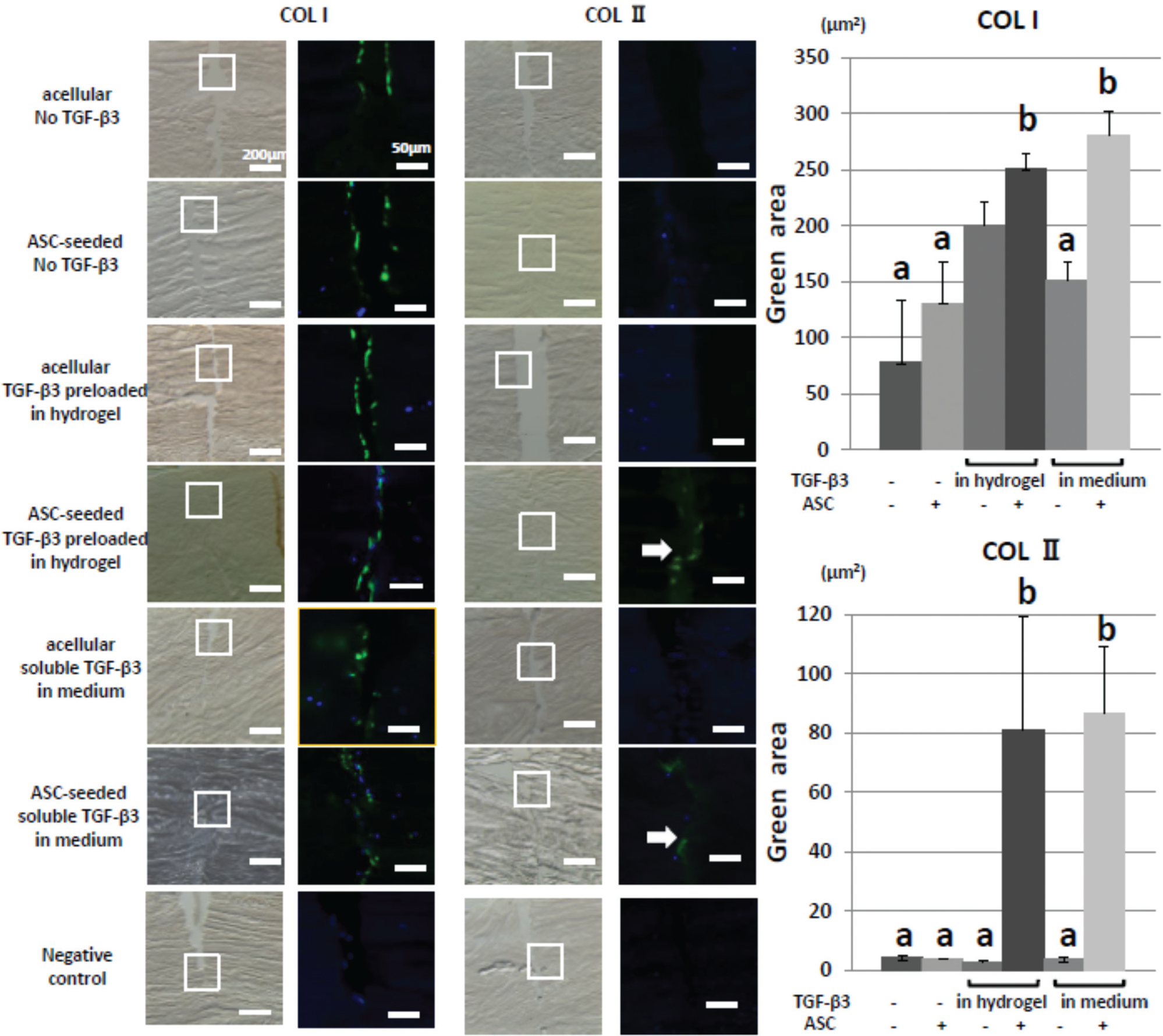
Immunohistochemical analysis of meniscal radial gap repair in vitro. Histological sections were immunostained for procollagen types I (COL I) and II (COL II). COL I staining was seen at the surface area of the radial tear in all groups, whereas procollagen type II staining was detected in the radial tear only in adipose-derived stem cell (ASC) hydrogels treated with transforming growth factor β3 (TGF-β3). Arrows indicate immunopositive cells. Bars: 200 µm (bright field), 50 µm (immunohistochemistry). The total area of positive (green) staining in the tear site was quantified for COL I and COL II stains. Letters correspond to statistically equivalent groups, P < .05.
Mechanical Properties
As shown in Figure 8, mechanical testing of the repaired meniscus explants showed that the values of load to failure (Figure 8A), stiffness (Figure 8B), and Young modulus (Figure 8C) in ASC-seeded hydrogels with preloaded TGF-β3 and those with soluble TGF-β3, at 4 and 8 weeks, were significantly higher than those in ASC-seeded hydrogels without TGF-β3. There was no significant difference between ASC-seeded hydrogels with preloaded TGF-β3 and those with soluble TGF-β3. Mechanical properties were also higher in TGF-β3-supplemented cellular groups as compared with the corresponding acellular hydrogels at 4 and 8 weeks. Conversely, there was no significant difference between ASC-seeded hydrogels without TGF-β3 and acellular hydrogels without TGF-β3 at both time points. Compared with week 4, mechanical properties at week 8 were significantly higher in the ASC hydrogels supplemented with preloaded TGF-β3 or TGF-β3-containing medium.
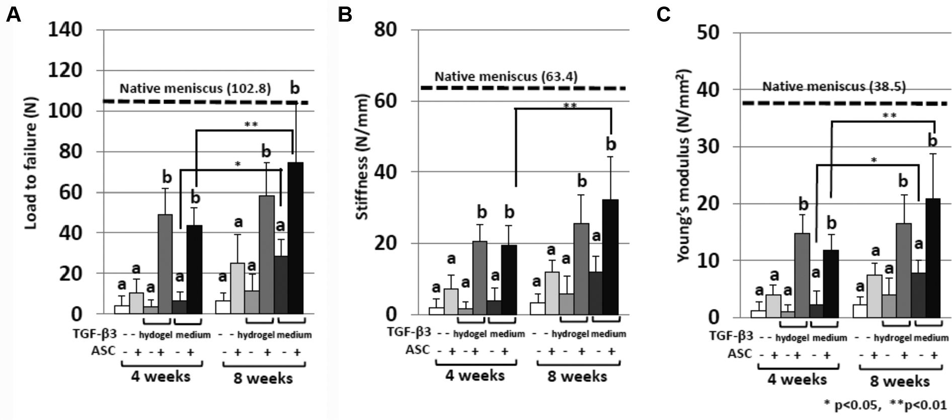
Mechanical testing of repaired meniscus at 4 or 8 weeks of culture. Mechanical testing of the repaired meniscus explants showed that the (A) load-to-failure, (B) stiffness, and (C) Young modulus values in adipose-derived stem cell (ASC)–seeded hydrogels preloaded with transforming growth factor β3 (TGF-β3) and ASC-seeded hydrogels with soluble TGF-β3 at 4 and 8 weeks were significantly higher than ASC-seeded hydrogels without TGF-β3. In addition, the requirement of ASCs for augmented healing is indicated by the significantly higher values in cellular groups as compared with the corresponding acellular groups. The data are expressed as mean ± SD. Letters correspond to statistically equivalent groups at a given time point, P < .05. Statistically significant differences between 4 and 8 weeks for a given group are shown by bars. *P < .05. **P < .01.
Discussion
This study found that (1) mGL hydrogels preloaded with TGF-β3 upregulated sGAG synthesis in ASCs at 3 weeks to a similar degree to mGL hydrogels cultured in full chondrogenic medium supplemented with TGF-β3 and (2) ASC-seeded hydrogels enhanced in vitro meniscal healing, with TGF-β3 supplementation providing a synergistic effect regardless of delivery method. As with other tissues, tissue-engineering strategies applied to enhance meniscal healing have utilized 3 principal elements: scaffolds, cells, and bioactive agents such as growth factors. As the avascular zone of menisci possesses a poor intrinsic healing capacity, several clinical studies explored the efficacy of fibrin clot or platelet-rich plasma as a source of autologous growth factors, with equivocal results. 28 It is likely that the hypocellularity of the meniscus, combined with the dense extracellular matrix that imparts its unique structure and function, prevents a sufficient number of endogenous cells from localizing to the tear site. 33 To that end, several preclinical studies showed improved healing of meniscal tears when augmented with exogenous chondrocytes.22,30 However, a seminal study by Port et al 32 found no improvement in healing when tears were augmented with fibrin clots seeded with autologous bone marrow–derived cells, suggesting that improved healing mediated by exogenous cells depends in part on the chondrogenicity of the cells.
While autologous chondrocytes are presently employed to facilitate repair of focal articular cartilage defects, there are several limitations with their use, including donor site morbidity, low tissue volume/hypocellularity necessitating ex vivo expansion, and their resulting propensity for rapid dedifferentiation.27,34 Consequently, MSCs derived from multiple tissues have been investigated as alternative sources of autologous chondrocytes. 27 Stimulation of MSCs by TGF-β3—whether supplied as a medium supplement or preloaded into a scaffold—appears essential to promote robust chondrogenic differentiation of MSCs.1,44 Lai et al 23 demonstrated that MSCs encapsulated within a hydrogel underwent chondrogenic differentiation when cultured in medium supplemented with TGF-β3. However, the use of soluble TGF-β3 as a medium supplement presents several challenges in terms of clinical translation, since multiple intra-articular injections of TGF-β3 would likely cause adverse side effects, such as arthrofibrosis by synovial hyperplasia.45,46 Conversely, localized release of TGF-β3 to exogenous MSCs delivered to a meniscal tear may provide the appropriate chondrogenic cues without off-target effects. However, it was not known to what extent a single but sustained exposure to TGF-β3—for example, preloaded into a biomaterial scaffold as described in this study—would be able to promote chondrogenesis as compared with standard chondrogenic differentiation medium supplemented with soluble TGF-β3. In this study, we found that hydrogels preloaded with TGF-β3 induced early chondrogenic differentiation (ie, by 3 weeks) of ASCs and promoted meniscal healing in an in vitro model of radial meniscal tear to a similar extent as ASC hydrogels cultured in TGF-β3-supplemented medium. Additionally, improved healing was seen only in ASC-seeded hydrogels, with little benefit observed when TGF-β3 was applied alone in acellular hydrogels.
Our findings reported here showed that p2 hASCs, isolated from the IPFP, represent a relatively homogeneous cell population that expresses mesenchymal cell markers (CD44, CD73, CD90, CD105) but not hematopoietic and endothelial markers (CD31, CD34, CD45). hASCs display multilineage differentiation potential, undergoing chondrogenic, osteogenic, and adipogenic differentiation, in accordance with other studies. 7 We utilized IPFP as the tissue source for the hASCs, as IPFP ASCs were shown to possess greater chondrogenicity than patient-matched ASCs isolated from subcutaneous fat. 26 In principle, IPFP-ASCs may be harvested intraoperatively from the same incisions used for arthroscopic meniscal repair, offering the prospect of a 1-stage procedure for cell-based augmentation of meniscal repair.
To localize the cells to the defect, a chondrosupportive carrier is needed. Hydrogels have been used effectively for soft tissue regeneration, given their ability to deliver high concentrations of homogeneously distributed cells while being amenable for in situ gelation within irregularly shaped defects. A method for VL-based photocrosslinking of an mGL hydrogel that permits live cell encapsulation was recently reported. 25 The VL-activated hydrogel had many properties desirable for articular cartilage repair, including cytocompatibility (ie, high cell viability), chondroconductivity, and tunable mechanical properties. In addition, mGL hydrogels can be rapidly photocrosslinked by VL, easily adaptable for arthroscopic procedures.
As expected, ASC-seeded hydrogels cultured in medium supplemented with soluble TGF-β3 showed robust proteoglycan deposition (Figures 4 and 5). Interestingly, ASC-seeded hydrogels preloaded with TGF-β3 possessed equivalent sGAG content as ASC hydrogels cultured in full chondrogenic medium (ie, soluble TGF-β3) at day 21, with continued GAG deposition through day 42. However, the preloaded hydrogels exhibited diminishing rates of proteoglycan deposition by day 42 as compared with ASC hydrogels cultured in TGF-β3-supplemented medium, likely because 80% of the preloaded TGF-β3 content is released by day 21. While ASC hydrogels cultured in full chondrogenic medium possessed greater GAG content than preloaded hydrogels at this later time point, the former group was exposed to substantially more total TGF-β3 (120 ng total, with twice weekly changes in chondrogenic medium containing 10-ng/mL soluble TGF-β3) than the latter (100 ng total, with TGF-β3 preloaded at 2 µg/mL before VL photocrosslinking), possibly contributing to the final quantitative difference in GAG content.
However, these groups improved healing equally well in the in vitro radial tear model, as evaluated by histology (Figure 6) and mechanical testing (Figure 8). Regardless of the means of TGF-β3 delivery, only hydrogels containing ASCs significantly improved healing. Specifically, cells expressing procollagen type I were seen in the defects of all groups, but only ASC hydrogels with preloaded TGF-β3 and those supplemented with soluble TGF-β3 showed positive expression of procollagen type II, detectable deep within the radial tear, at 8 weeks (Figure 6). The coexpression of collagen types I and II is notable, as the extracellular matrix of the inner avascular zone of the meniscus is principally composed of proteoglycan and collagen (70% by dry weight), consisting of collagen types II (60%) and I (40%). 4 While immunohistochemical staining of mature collagen types I and II would have permitted characterization of the biochemical composition of the neotissue, the use of antibodies for procollagen allowed the delineation between collagen produced by the hASCs and the existing collagen of the meniscus explant.
Beyond this limited examination of neotissue composition, this study had several other limitations. In vitro experiments exploring the effect of TGF-β3 delivery on sGAG deposition by hASCs were performed at 3 and 6 weeks, while experiments involving the in vitro repair model were performed at 4 and 8 weeks. In a preceding study that examined the use of mGL as a biomaterial capable of supporting fibrochondrogenic differentiation of human BM-MSCs, time points of 3 and 6 weeks were used. 36 However, in a recent study detailing the development of the utilized in vitro explant model, time points of 4 and 8 weeks were employed. 41 To allow comparison among the results in this study and the previous investigations, disparate time points were utilized.
Although the results of this study suggest that an ASC-seeded mGL hydrogel preloaded with TGF-β3 can promote neofibrocartilage formation, thereby enhancing “healing” in radial meniscal tear, the in vitro model utilized in this study has not yet been validated as being predictive of in vivo healing. To that point, the intra-articular microenvironment after meniscal injury presents several challenges to healing, some of which were not captured in the in vitro model. Namely, the synovial fluid of the joint contains numerous biological factors, including catabolic and inflammatory mediators, which may adversely affect ASC viability and/or chondrogenesis. Additionally, the forces exerted on the meniscus are not easily modeled given their complexity, changing with joint position and activity level. Furthermore, the ability to accurately deliver the ASC hydrogel through standard surgical approaches has yet to be explored. These limitations notwithstanding, the results described here suggest a promising new approach for biological augmentation of meniscal tears through a potentially single-step surgical procedure, although the degree of clinical utility must be further explored in a large animal model, which is being investigated in an ongoing study in our laboratory.
Conclusion
This study demonstrated that ASCs encapsulated in a photocrosslinkable mGL hydrogel scaffold preloaded with TGF-β3 at 2 µg/mL displayed robust chondrogenic differentiation and, when injected into a radial meniscal tear, resulted in significant repair, as examined by histology and mechanical testing. Photocrosslinked hydrogel seeded with ASCs and preloaded with TGF-β3 may represent an effective therapeutic option to repair meniscal radial tears and reduce the development of osteoarthritis.
Supplemental Material
DS_10.1177_0363546518782973 – Supplemental material for In Vitro Repair of Meniscal Radial Tear With Hydrogels Seeded With Adipose Stem Cells and TGF-β3
Supplemental material, DS_10.1177_0363546518782973 for In Vitro Repair of Meniscal Radial Tear With Hydrogels Seeded With Adipose Stem Cells and TGF-β3 by Hiroshi Sasaki, Benjamin B. Rothrauff, Peter G. Alexander, Hang Lin, Riccardo Gottardi, Freddie H. Fu and Rocky S. Tuan in The American Journal of Sports Medicine
Footnotes
Acknowledgements
The authors thank Dr Kazunori Shimomura for development of the in vitro meniscal tear model and Dr Jian Tan for ASC isolation and characterization.
One or more of the authors has declared the following potential conflict of interest or source of funding: This work was supported by the Department of Defense (grant W81XWH-15-1-0104). B.B.R. was an NIH Predoctoral Trainee (CATER training grant 5T32 EB001026).
References
Supplementary Material
Please find the following supplemental material available below.
For Open Access articles published under a Creative Commons License, all supplemental material carries the same license as the article it is associated with.
For non-Open Access articles published, all supplemental material carries a non-exclusive license, and permission requests for re-use of supplemental material or any part of supplemental material shall be sent directly to the copyright owner as specified in the copyright notice associated with the article.