





Abstract
In this study, the effects of a potent antioxidant, selenium, on apoptosis induced by acrolein, a cytotoxic and genotoxic environmental pollutant, were investigated by immunohistochemical and electron microscopic methods. One hundred adult male Wistar albino rats were used in the study. The rats were divided into four main groups: control, acrolein, selenium, and acrolein + selenium. The animals in the experimental groups were given 1 mg/kg/day selenium and 4 mg/kg/day acrolein daily for 7 days by gavage. After drug administration, each group was divided into subgroups according to the time they were to be euthanized: 12th hour, 1st, 2nd, 3rd, and 5th day. The rats in each group at the determined time were euthanized and their livers were removed. Routine histological procedures were performed for light and electron microscopy examinations. After applying the Terminal Deoxynucleotidyl Transferase dUTP nick end labeling assay on the liver sections, apoptotic index values were calculated. Comparing the liver sections of the rats in the acrolein group and the control group, acrolein was found to cause a significant increase in the apoptotic index. The apoptotic index values of the acrolein + selenium group decreased compared to the acrolein group. In the electron microscopic examinations, apoptotic findings were observed in the liver tissues of the rats given acrolein, such as chromatin condensation in the nucleus of hepatocytes, dilatations in the perinuclear space, and cytoplasmic vacuolization. These apoptotic findings were not observed in the acrolein + selenium group after the 12th hour. These findings show that selenium may potentially be useful as a protective agent for people exposed to acrolein.
Introduction
Acrolein is a highly electrophilic α, β-unsaturated aldehyde to which humans are exposed under industrial, environmental, and therapeutic conditions. This active aldehyde is produced during both synthetic and natural processes, such as the incomplete combustion of organic compounds and fuels such as petrol, coal, and wood, photochemical oxidation of airborne hydrocarbons, and cigarette smoke. In biological systems, acrolein is a metabolite of cyclophosphamide and allylamine, and has anticancer properties (Kehrer and Biswal, 2000). Acrolein has been determined to cause apoptosis in several cell types (Tanel and Averill-Bates, 2007).
Glutathione (GSH) is the main factor that controls the apoptosis and provides the association between acrolein and apoptosis (Kehrer and Biswal, 2000). Acrolein reacts with GSH (Calingasan et al., 1999) and causes GSH to rapidly decrease (Tanel and Averill-Bates, 2007). Acrolein hepatotoxicity is explained by the rapid decrease of liver GSH due to the formation of the conjugated form of acrolein and GSH (Sun et al., 2006). Acrolein increases reactive oxygen species (ROS) and decreases antioxidants (especially GSH) causing deterioration of mitochondrial functions, water intake and swelling of organelles, and release of cytochrome c, leading to cell apoptosis (Jia et al., 2007; Luo and Shi, 2005).
Today, people are exposed to various toxins in their environment that can be potent oxidants. Antioxidants are the most commonly used agents in the in vitro and in vivo studies of these toxins that form protective effects against various diseases and significantly reduce their toxicity (Faroon et al., 2008; Ratnam et al., 2006).
Selenium is considered one of the most powerful immunomodulators because of its low toxicity and beneficial effects on the immune system. Thus, intensive studies are carried out on selenium. Since enzymes related to selenium prevent oxidative damage by breaking down reactive oxygen radicals in humans, selenium is an important antioxidant in humans (Ramoutar and Brumaghim, 2007; Tapiero et al., 2003). In studies conducted on rats and other experimental animals, selenium has been suggested to reduce apoptosis through the antioxidant effect it provides by joining the structure of the GSH peroxidase that is responsible for the degradation of hydrogen peroxide (H2O2) (Burk, 2001).
The protective effect of selenium on hepatoxicity induced by various agents has been demonstrated in many studies (Sivaram et al., 2003; Soudani et al., 2011). Generally, seleno-enzymes are known to be involved in oxygen metabolism, detoxification events, inhibition of apoptosis in cancer cells, and control of cell division (Björnstedt and Fernandes, 2010).
Thus, the aim of this study was to investigate whether the antioxidant selenium could protect against cytotoxicity and apoptosis induced by acrolein, a toxic environmental pollutant humans are constantly exposed to.
Materials and methods
Animals
This study was carried out with permission from the Animal Experiments Local Ethics Committee in Ondokuz Mayis University. One hundred Wistar albino adult male rats (250–320 g) were used in the study. Animals were fed ad libitum with standard rat feed in plastic cages. A 12-h light/dark cycle was provided at 18–22°C room temperature. Before starting the experiment, the weights of the rats were determined, and mean rat weight in each group was ensured to be approximately the same.
Experimental design
In the study, four main groups of control (C), acrolein (Ac), selenium (Se), and acrolein + selenium (Ac + Se), and five subgroups of 12th hour, 1st day, 2nd day, 3rd day and 5th day were formed. A total of 100 rats were used, five in each group. No procedure was applied for the control group. The acrolein group was given 4-mg/kg/day acrolein (Yousefipour et al., 2005) (Kat No: 01679; Sigma-Aldrich, Germany) and the selenium group was given 1-mg/kg/day selenium (Kat No: S3132; Sigma-Aldrich) by gavage with a single daily dose for 7 days (Antunes et al., 2001; Newairy et al., 2007). The acrolein + selenium group was given selenium (1 mg/kg/day) by gavage 1 hour after administration of acrolein (4 mg/kg/day). The animals in this group (acrolein + selenium) were given selenium 1 day before the first administration of acrolein. In this study, the doses of the chemicals given to the rats were determined by considering the results of the previous experimental studies with the rats. In accordance with the hypothesis of the study, the dose of acrolein showing cytotoxic effect in liver and selenium showing hepatoprotective effect was selected.
After the last gavage procedure, intracardiac perfusion was performed on all rats under ketamine hydrochloride general anesthesia at the 12th hour and the 1st, 2nd, 3rd, and 5th days, and their liver tissues were extracted. The tissues were fixed in 10% buffered neutral formalin for light microscopy examinations and 2.5% glutaraldehyde for electron microscopy examinations.
Immunohistochemical analysis
Terminal Deoxynucleotidyl Transferase dUTP nick end labeling (TUNEL) assay
The sections were stained in accordance with the recommended standard procedure using the In Situ Cell Death Detection, POD (Roche, Manheim, Germany) apoptosis kit. The 4.5-µm thick sections were placed on adhesive slides. They were dried in an incubator at 37°C for one night. They were deparaffinized in xylol and dehydrated in serial alcohols. To eliminate the masking effect of formalin on tissue, the sections were boiled in a citrate buffer solution in a microwave oven at 300 W for 5 min. After washing, they were left in 3% H2O2 prepared in methanol for 10 min to eliminate endogenous peroxidase activity. After the sections were left in a mixture of terminal deoxynucleotidyl transferase enzyme and marking solution at 37°C for 1 h, they were held in a protein blocking serum for 10 min to prevent their nonspecific antigenic binding. They were incubated with peroxidase-conjugated anti-fluorescein isothiocyanate antibody at 37°C for 30 min. The sections were washed twice with phosphate-buffered saline for 5 min after all procedures except for the incubation with protein blocking serum. A 3-amino-9-ethylcarbazole (AEC; Zymed RED substrate kit, Paisley, OR, USA) chromogen was used to stain the sections. The staining was performed under a microscope for 10 min. The sections stained with AEC substrate and contrast stained with hematoxylin and sealed using a water-based adhesive (Shandon Immu-mount, Thermo Scientific™-9990402, Schwerte, Germany).
Apoptotic index measuring
In the tissue sections stained by the TUNEL method, the determination of apoptotic activity was performed under a light microscope (Nikon Eclipse E600W with Nikon DS-5M camera attachment, Tokyo, Japan). In 10 different areas, randomly selected in each section, red-brown-stained nuclei were considered positive, and blue-stained nuclei were considered negative, regardless of the staining intensity of the cells. At least 100 (positive and negative) nuclei were counted in each area. Then, the apoptotic index was calculated using the following formula
Electron microscopy analysis
The tissues that were fixated at +4°C in glutaraldehyde for 24 h were post-fixated with osmium tetroxide. Extracting the semi-thin and thin sections of the tissues blocked in Araldite resin by ultramicrotome, staining of the semi-thin sections, and obtaining images from the thin sections were performed in the Electron Microscopy and Image Analysis Unit (ESOGUSTEM) in Eskisehir Osmangazi University.
In the transmission electron microscopy evaluation, apoptotic findings such as chromatin condensation in the nucleus, jagged appearance of the nuclear membrane, expansion in the perinuclear space, swelling in the mitochondria and endoplasmic reticula, cytoplasmic vacuolization, and degeneration were taken into account.
Statistical analysis
For the statistical analyses, the Statistical Package for Social Sciences for Windows 15.0 program was used. The nonparametric Kruskal–Wallis test was used for the comparison of the data between groups. With a 95% confidence interval, the results were considered statistically significant at p < 0.05 and statistically insignificant at p > 0.05. The Bonferroni correction nonparametric Mann–Whitney U test was used for the paired comparison of the groups (Bonferroni correction p value: p = 0.05/number of groups).
Results
Immunohistochemical results
The apoptotic index values obtained from the livers of the control and experimental groups at all determined time periods were evaluated (Table 1). In the paired comparisons, the apoptotic index value of the acrolein group was higher compared to all the other groups (p = 0.007; Figure 1). Apoptotic index value of the acrolein + selenium group was lower than the acrolein group (p = 0.007) and higher than the control and selenium groups (p = 0.008). These differences were statistically significant. No significant difference was found between the control and selenium groups in terms of apoptotic index values (p > 0.05; Figure 2).

(a) Apoptotic nuclei in the liver tissue of the Ac group. Positive nucleus (white arrowhead), negative nucleus (white arrow) TUNEL ×200: (a) 12th hour, (b) 2nd day, (c) 3rd day, and (d) 5th day.
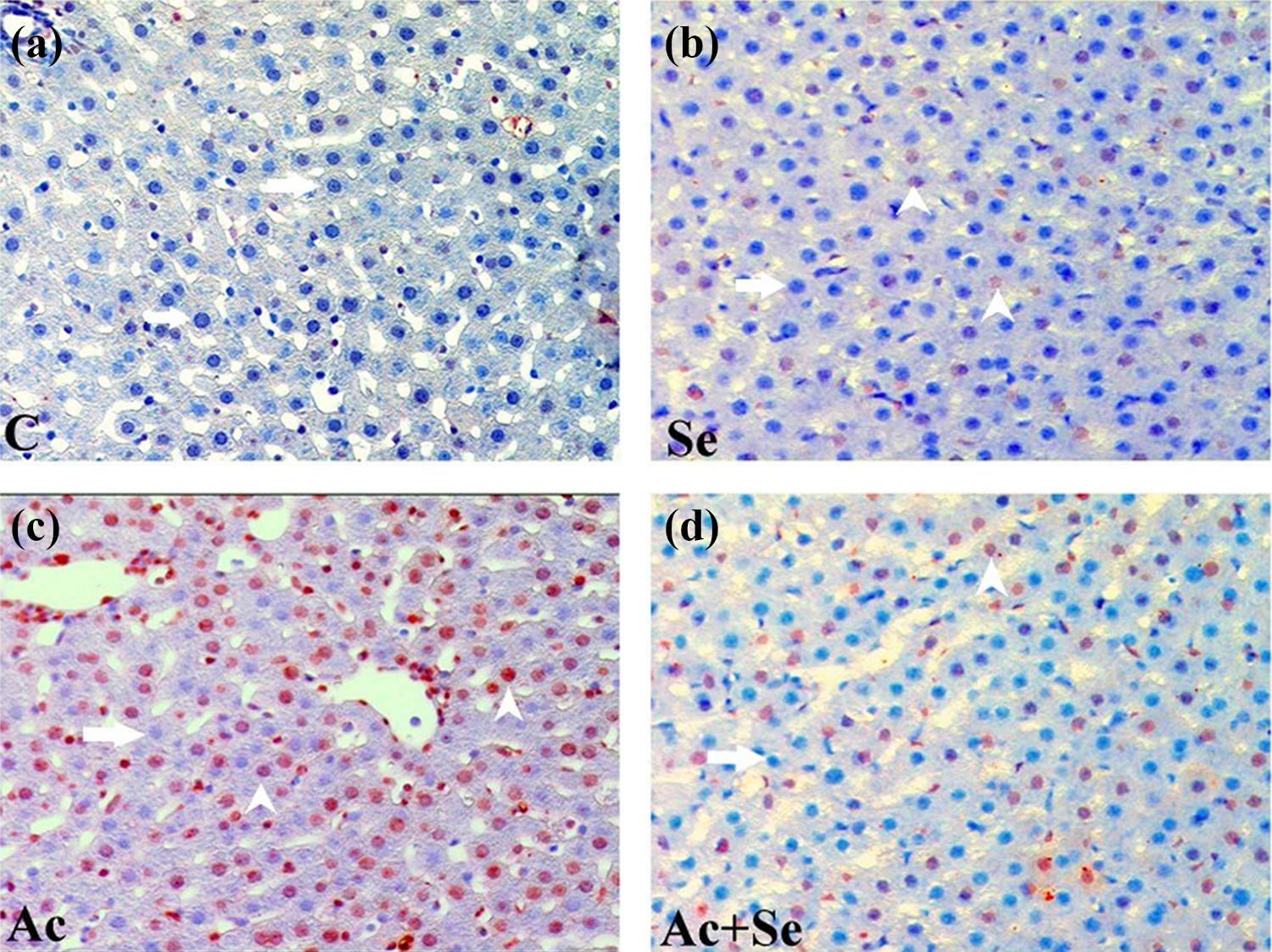
Apoptotic nuclei in liver tissues of (a) C, (b) Se, (c) Ac, and (d) Ac + Se groups. Positive nucleus (white arrowhead), negative nucleus (white arrow) TUNEL ×200.
The apoptotic index values and standard deviations obtained from the livers of the control and experimental groups at all determined time periods.
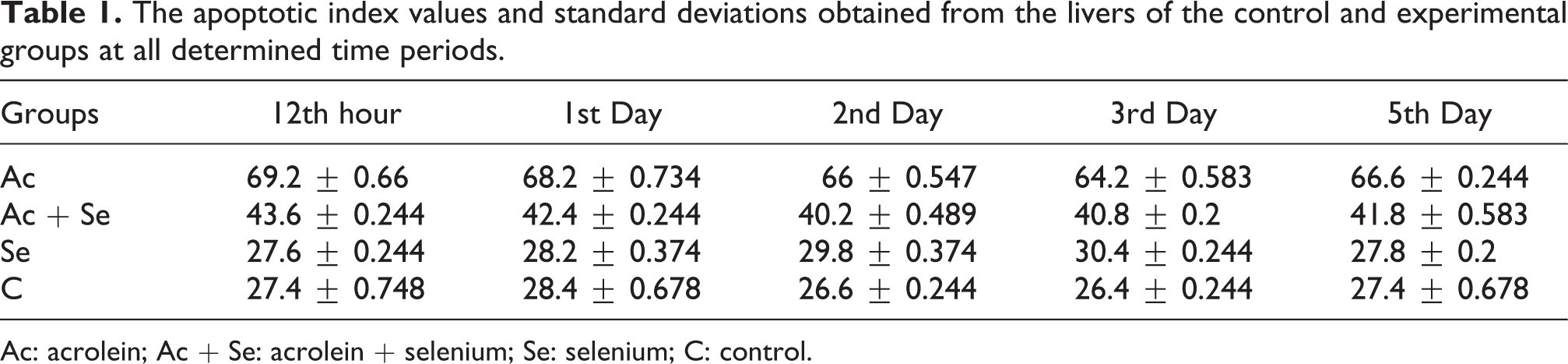
Ac: acrolein; Ac + Se: acrolein + selenium; Se: selenium; C: control.
The apoptotic index values obtained from the liver tissues of the hour and day subgroups of the C, Ac, Se, and acrolein + selenium experimental groups were evaluated. In the paired comparisons, no statistically significant difference was found between the 12th hour, 1st day, 2nd day, 3rd day, and 5th day subgroups of the main groups (p > 0.05).
Electron microscopy results
In transmission electron microscopy examinations, the hepatocytes in the livers of the rats in the control group were found to be normal. In the hepatocytes, the endoplasmic reticula and mitochondria abundant in the cytoplasm were observed to be normal. The nucleus generally located in the center of the cell was determined to be large, round, and euchromatic. Glycogen was observed to have a uniform distribution in the cell.
In the liver of the rats in the 12th hour subgroups of the acrolein group, apoptotic changes such as chromatin condensation in the nuclei of hepatocytes, dilatation in the endoplasmic reticulum and perinuclear space, cytoplasmic vacuolization, and changes in the plasma membrane were observed. In the nuclei of the 1st day subgroups, apoptotic findings such as dilatation in the perinuclear space and swelling in the mitochondria, deterioration in the matrix structure, and cytoplasmic vacuolization were observed. In the 2nd, 3rd, and 5th day subgroups, apoptotic properties such as the jagged appearance of the nucleus membrane, deterioration of the cytoplasm, and condensation in the mitochondrial matrix were determined (Table 2 and Figure 3).
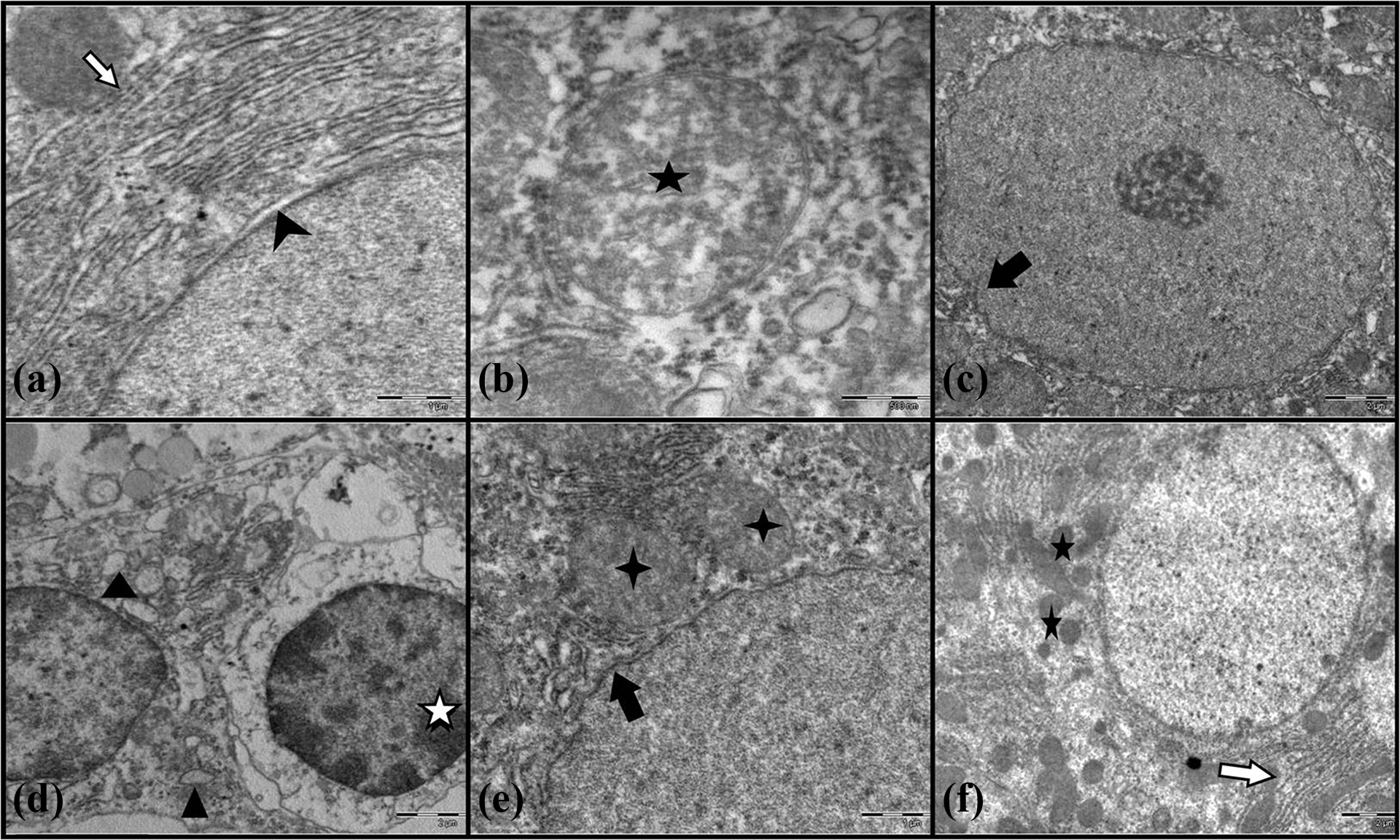
Transmission electron microscopy images of hepatocytes in liver sections of rats in acrolein groups. a) Dilatations in the perinuclear space (arrowhead) and lumen of the endoplasmic reticula (white arrow), b) Chromatin condensation in the nuclei of hepatocytes (black star), c) Jagged appearance of the nucleus membrane (black arrow), d) Cytoplasmic vacuolization (black triangle), deterioration in the matrix structure (white star), e) Swelling in the mitochondria (black rosette), jagged appearance of the nucleus membrane (black arrow), f) Lumen of the endoplasmic reticula (white arrow), condensation in the matrix of mitochondria (black star).
Semi-quantitative evaluation of apoptotic properties of hepatocytes in liver sections of rats in acrolein groups by transmission electron microscope.
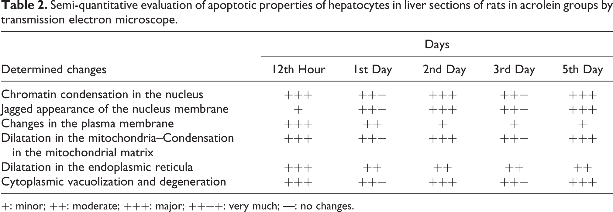
+: minor; ++: moderate; +++: major; ++++: very much; —: no changes.
The livers of the selenium 12th hour and 1st, 2nd, 3rd, and 5th day subgroups were found to be similar to the control group. In the 12th hour subgroup of the acrolein + selenium group, apoptotic findings such as chromatin condensation in the nucleus, dilatation in the perinuclear space, and cytoplasmic vacuolization were observed (Table 3 and Figure 4). However, not much significant changes were observed in the mitochondria, the endoplasmic reticula, or the chromatin structures and borders of the nuclei in the 1st, 2nd, 3rd, and 5th day subgroups.
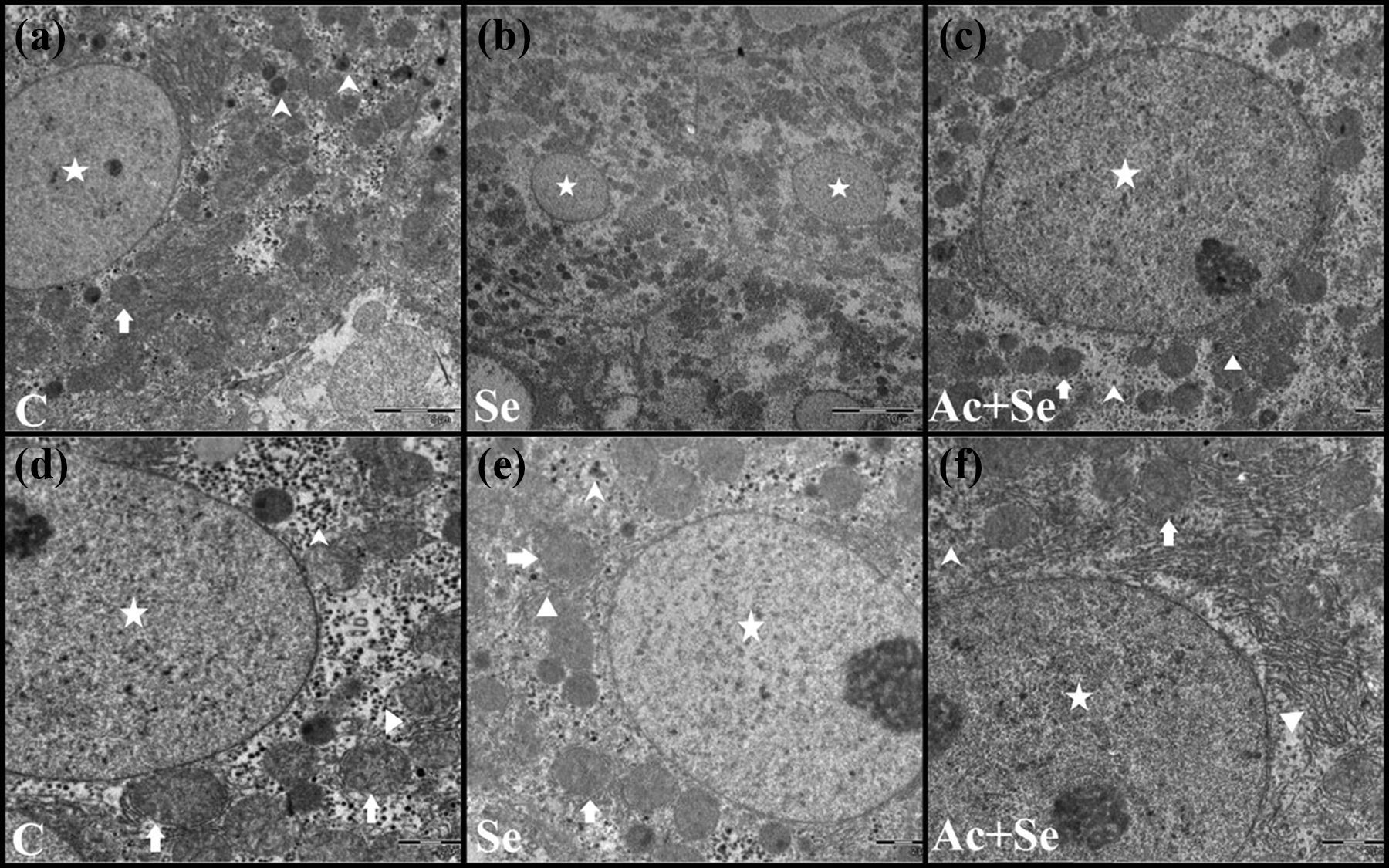
Transmission electron microscopy images of liver sections of rats in ((a) and (d)) C, ((b) and (e)) Se, and ((c) and (f)) Ac + Se groups. Nucleus (star), mitochondria (arrow), glycogen particles (arrowhead), and endoplasmic reticulum (triangle).
Semi-quantitative evaluation of apoptotic properties of hepatocytes in liver sections of rats in acrolein + selenium groups by transmission electron microscope.
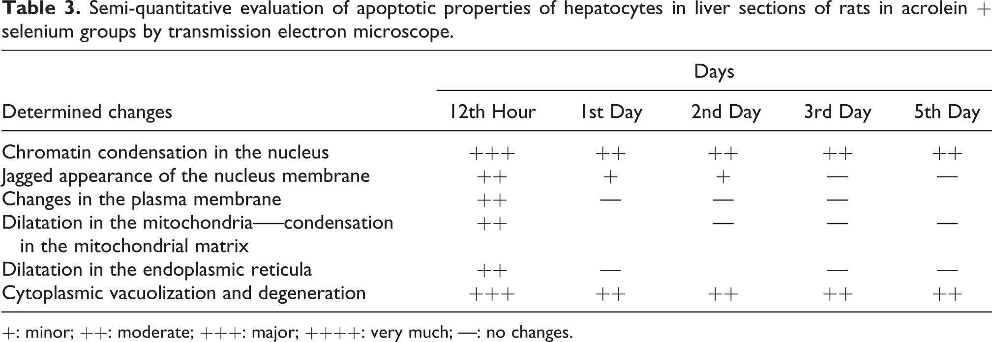
+: minor; ++: moderate; +++: major; ++++: very much; —: no changes.
Discussion
A cytotoxic and genotoxic environmental pollutant, acrolein, occurs during the combustion of nutrients and organic substances (Jia et al., 2007). It is found at any time and at any place, particularly in all smoke types such as cigarette smoke (Sun et al., 2006), in internal combustion engine exhausts, in overheated cooking oil vapors, and in polluted air as a result of photochemical reactions (Kehrer and Biswal, 2000).
The constant exposure of humans to the toxic environmental pollutant acrolein has required research into the effects of acrolein on tissues. This study was conducted to determine whether the cytotoxic and genotoxic environmental pollutant acrolein caused apoptosis in the hepatocytes in the livers of rats, and whether the potent antioxidant selenium could have a protective effect against this damage. To this end, we comparatively evaluated the immunohistochemical and electron microscopic changes of the liver tissues of rats given acrolein according to time. Aside from acrolein, we compared these effects with the rats given selenium, a potent antioxidant.
In the transmission electron microscopy examination of the hepatocytes of the liver sections of rats given acrolein, changes such as chromatin condensation in the nuclei of hepatocytes, dilatations in the perinuclear space and lumen of the endoplasmic reticula, cytoplasmic vacuolization, changes in the plasma membrane, swelling in the mitochondria, deterioration in the matrix structure, and jagged appearance of the nucleus membrane were observed in all time periods beginning from the 12th hour compared to the control group. These determined changes at the ultrastructural level reflected the characteristic apoptotic cell changes defined by many previous studies. In addition, the apoptotic index values of rats given acrolein measured by the TUNEL staining of the liver sections were higher than all the other groups, and this difference was statistically significant. These two important findings of the study suggest that acrolein causes apoptosis in hepatocytes.
GSH is the main factor that controls the apoptosis and provides the association between acrolein and apoptosis (Kehrer and Biswal, 2000). Luo and Shi (2005) showed acrolein to have many harmful effects associated with oxidative stress and ROS to play an important role in acrolein toxicity. The researchers suggested that acrolein performed its oxidative toxicity effect by reducing intracellular antioxidants (especially GSH), inhibiting several antioxidant enzymes that provide the detoxification of RNA, and increasing ROS production (Luo and Shi, 2005). It has been determined by many researchers that when oxidative stress increased, the cells died by necrosis or apoptosis (Kern and Kehrer, 2002; Luo and Shi, 2005; Tanel and Averill-Bates, 2007). ROS has been stated to show its effects by deteriorating mitochondrial permeability or transmembrane potential or by inducing the release of Ca+2 into the cytosol. Having a synergistic effect, ROS and Ca+2 cause the deterioration of mitochondrial functions, water intake and swelling of organelles, and release of cytochrome c to the cytosol, leading to the activation of the intrinsic pathway of apoptosis, caspase-9, and subsequently caspase-3 (Luo and Shi, 2005; Tanel and Averill-Bates, 2007). In light of this information, in our study, we can say that the changes such as swelling of the mitochondria and the dilatation of the endoplasmic reticula we determined in the electron microscopy images of the hepatocytes of the livers of the rats in the acrolein group resulted from oxidative stress caused by acrolein, thus inducing apoptosis in the cells.
Another interesting finding in this study was that intense apoptotic cells were observed in the liver sections of all the time subgroups (12th hour, 1st, 2nd, 3rd, and 5th days) of rats given acrolein, and there was no statistically significant decrease in apoptotic index level according to time. Parent et al. (1998) found similar levels of acrolein [2, 3-14C] in the kidney, spleen, lung, blood, liver, fat, adrenal glands, and ovaries of rats euthanized 17 days after the administration of acrolein. Draminski et al. (1983) found that the acrolein conjugate metabolite S-carboxyethyl-mercapturic acid occurred in the urea of rats given single oral dose of 10 mg/kg acrolein. In this study, the fact that the apoptotic index value of rats given acrolein did not decrease according to time is an indicator of the fact that acrolein itself or its metabolites that occur can remain in the liver for a long period of time, and its presence for this period leads to apoptosis of the hepatocytes.
One of the important findings of this study was that selenium, which affects the GSH metabolism, a major antioxidant for the cell, had the ability to prevent acrolein-induced apoptosis in hepatocytes.
In the present study, while the electron microscopy appearances of the hepatocytes of the liver sections of rats given selenium were similar to the control group, the apoptotic findings determined in the acrolein group after the 12th hour were not observed in the groups that were given selenium together with acrolein. In addition, it was determined that the apoptotic index values measured from the liver sections of rats given selenium did not differ compared to the control group, and they were significantly lower in the groups that were given selenium together with acrolein than the acrolein group. The selenium-dependent GSH peroxidase in the cell metabolizes both H2O2 and lipid peroxides. Frisk et al. (2003) found that selenium reduced apoptosis by its antioxidant effect by joining the structure of GSH peroxidase that degrades H2O2 (Frisk et al., 2003), and Björnstedt and Fernandes (2010) showed that seleno-enzymes were generally involved in oxygen metabolism, detoxification events, inhibition of apoptosis in cancer cells, and control of cell division (Björnstedt and Fernandes, 2010).
Conclusion
We concluded that environmental exposure to acrolein has a toxic effect even at low dose and that selenium has a beneficial effect in protecting cells against acrolein-induced apoptosis. In addition, we believe that these findings will contribute to the understanding of the environmental exposure toxicity of acrolein, as well as of the normal tissue toxicity of cyclophosphamide, which produces acrolein as one of its metabolites.
Footnotes
Declaration of conflicting interests
The author(s) declared no potential conflicts of interest with respect to the research, authorship, and/or publication of this article.
Funding
The author(s) disclosed receipt of the following financial support for the research, authorship and/or publication of this article: This study was supported by Ondokuz Mayıs University Scientific Research Projects Unit. No financial support was received outside the university.
