





Abstract
Aims:
To assess the long-term outcome of childhood tuberculous meningitis treated with modern 4-drug antitubercular regimens and to determine predictors of survival and morbidity.
Methods:
In this single-center prospective cohort, outcome of children with tuberculous meningitis treated with standard regimens was assessed at 6 months and 12 months after discharge using the Pediatric Cerebral Performance Category Scale.
Results:
Of 130 children, 38 died in hospital and 34 were either severely disabled or comatose/vegetative at discharge. At 6 and 12 months, 87% of the survivors were either normal (n = 62) or mildly disabled (n = 17, on the Pediatric Cerebral Performance Category scale). On multivariate analysis, the factors associated with poor outcome at 12 months were stage III at admission (adjusted odds ratio 4.4, 95% confidence interval, 1.7-11.2, P = .002) and presence of infarcts on neuroimaging (adjusted odds ratio 2.6, 95% confidence interval, 1.1-6.6, P = .037).
Conclusions:
Despite the high in-hospital mortality, in resource-constraint settings, the survivors showed remarkable improvement, with two-thirds returning to a normal functional status at 6 months’ follow-up.
Keywords
Tuberculous meningitis is the most severe extrapulmonary complication of tuberculosis. Nearly 80% of tuberculous meningitis occurs in children younger than 5 years. With the decline of bacterial meningitis because of effective conjugate vaccines in many areas of the world, Mycobacterium tuberculosis becomes a relatively more important, treatable, and preventable cause of death and disability due to childhood meningitis. 1 In a recent meta-analysis and systematic review of 19 studies with 1636 children, the overall risk of death was 19.3% (95% confidence interval 14.0-26.1), and the probability of survival without neurologic sequelae was 36.7% (27.9-46.4). 2 Among survivors, the risk of neurologic sequelae was 53.9% (95% confidence interval 42.6-64.9). The authors emphasize the need for implementation of consensus definitions, standardized reporting of data, and high-quality clinical trials to improve outcomes. The review includes studies with considerable heterogeneity and includes studies over a period when the treatment regimens and supportive care standards have changed considerably. 2 Additionally, most of the prospective studies in childhood tuberculous meningitis have been done in an era before the modern drug regimens came to use. 3 –5 Despite being an endemic country, very few prospective studies on childhood tuberculous meningitis have been published from India. 5 Hence, we planned this cohort study with an objective to assess the long-term outcome of childhood tuberculous meningitis treated with modern 4-drug antitubercular regimens along with steroids and to determine predictors of survival and morbidity. We followed the recommended case definitions to diagnose tuberculous meningitis and used outcome scales to track recovery. 6
Method
This prospective, cohort study was conducted at a single children’s hospital, teaching, and research center in North India from October 2010 to June 2013. The institute ethics committee approved the study, and informed consent was obtained from parents or primary caregivers. Children were enrolled from October 2010 to June 2012 and followed up for at least 12 months. All children presenting with symptoms of fever with at least one of the following: headache, seizure, or encephalopathy, of more than 5 days’ duration were evaluated for central nervous system tuberculosis. All children underwent investigations to exclude alternative diagnoses such as enteric fever, rickettsial infection, leptospirosis, dengue, cerebral malaria, Brucella meningitis, cryptococcal meningitis, Acanthamoeba meningitis as per the treating unit’s protocol. Children who were meeting the criteria for possible, probable or definite tuberculous meningitis as per scoring system described by Marais et al were enrolled for the study. 6 Children diagnosed with HIV (n = 3), those on antitubercular therapy of more than 1 month duration at presentation (n = 2) were excluded from the study. All children underwent cerebrospinal fluid analysis, neuroimaging, and chest radiography. A structured proforma was used to record the clinical features, investigation results and follow up. Baseline information regarding demographic variables, duration of symptoms before admission, presenting symptoms, nutritional state, Modified Glasgow Coma Scale (Modified GCS) score, motor function, the presence of neurologic signs such as meningeal irritation, signs of raised intracranial pressure, signs of brainstem dysfunction, and cranial nerve palsies was recorded. Details of investigations including cerebrospinal fluid study, neuroimaging (computed tomographic [CT] scan or magnetic resonance imaging [MRI]), chest radiograph findings, ultrasonography head or abdomen, gastric aspirate for acid-fast bacilli, tuberculin skin test, etc were also recorded. Neuroimaging (computed tomography or MRI) was done in all patients. Neuroimaging findings, especially basal exudates, hydrocephalus, trans-ependymal flow, tuberculomas, and infarcts were noted. Chest radiography findings such as hilar or mediastinal lymphadenopathy, collapse, consolidation, effusion, cavitation, or military mottling were noted. Tuberculous meningitis was staged using the modified criteria of the British Medical Research Council: stage I tuberculous meningitis (GCS 15 with no focal neurologic signs), stage II tuberculous meningitis (GCS 11–14 or GCS of 15 with focal neurologic deficit), and stage III tuberculous meningitis (GCS <11). 7
All children were given daily intensive therapy comprising 4 drugs for 2 months; isoniazid (10 mg/kg), rifampicin (15-20 mg/kg), pyrazinamide (35-40 mg/kg), and ethambutol (20-25 mg/kg). The maintenance therapy was given for 10 months or more using isoniazid (10 mg/kg) and rifampicin (15-20 mg/kg). Steroids were given for the initial period of 6 to 8 weeks. Antiepileptics were given whenever indicated. Ventriculoperitoneal (VP) shunt was done in all children with a hydrocephalus and transependymal flow, papilledema, or with clinical signs of raised intracranial pressure not resolving with medical therapy. Details of complications including nosocomial complications, complications of neurosurgical procedures, electrolyte abnormalities, paradoxical tuberculomas, treatment failure, seizures, vision and hearing impairment and new motor deficits were noted. A repeat neuroimaging was done for children who developed neurologic worsening during follow-up. All children were followed up at 6 months and 12 months after discharge. During these follow-up visits, history and clinical examination were done, compliance to medication was assessed, hearing and vision assessment was done (if not done earlier after discharge), and functional performance and recovery were assessed using the PCPC (Pediatric Cerebral Performance Category). Children who had persistent or recurrent symptoms of meningitis were subjected to repeat neuroimaging and cerebrospinal fluid examination. The neurologic outcome was categorized as normal; mild, moderate, or severe disability; coma or vegetative state; and death. Children with no deficits, mild, and moderate deficits were categorized to have “Good Outcome,” whereas severe disability, vegetative state, and death were categorized to have “Poor Outcome.” Hearing and visual screening was done in all patients at 3 months or later after discharge.
Data were summarized using descriptive statistics. Fisher exact test was used to determine the significance of categorical variables. Test of normalcy was applied to quantitative variables. Student t test was used to determine the significance of normally distributed continuous variables. Nonparametrically distributed numerical variables were assessed using Mann-Whitney U test. For univariate analysis, the statistical significance was placed at the 1% level, taking into account the large number of variables and multiple comparisons. To study the independent effect of predictors, a multivariate logistic regression analysis was performed. All variables with P value <.05 were subjected to binary regression analysis. Variables with incomplete data were removed, and all variables were simultaneously analyzed in the regression model.
Results
A total of 24 232 children were admitted in the hospital during the enrollment period. A total of 130 children meeting study criteria were enrolled in the study.
Clinical Data (Table 1)
Boys predominated (61.5%), and almost one-third of children were below 2 years of age (Table 1). Forty-four percent of children had a history of contact with an adult with tuberculosis. However, none of them had received prophylaxis. Only 45% of the children presented within 15 days of onset of symptoms (median 19 days, interquartile range IQR 6.25-30).
Comparison of Clinical Profile of Children With Tuberculous Meningitis in Relation to Long-Term Outcome.

Abbreviations: BCG, Bacillus Calmette-Guérin; CI, confidence interval.
aDefined as children either with hypertension and bradycardia OR irregular breathing OR abnormal tonic flexor/ extensor posturing OR papilledema on funduscopy OR bulging anterior fontanelle OR poorly reactive asymmetric pupils.
Diagnostic Data
Children were classified as definite, probable or possible tuberculous meningitis as per the scoring system by Marais et al. 6 Eighty-two percent children had probable tuberculous meningitis (n = 107), 16.2% had possible tuberculous meningitis (n = 21), and 2 children had definite tuberculous meningitis. Acid-fast bacilli were isolated in 2 children each from sputum, gastric aspirates, and fine needle aspirate of the lymph node. Only a quarter (n = 32) of children had an abnormal chest radiograph. Twenty-nine children had only ventricular cerebrospinal fluid drawn, and the rest had lumbar cerebrospinal fluid examination. The lumbar cerebrospinal fluid cytology revealed a median cell count of 40/mm3 (interquartile range 10-150), a median protein content of 157 mg/dL (interquartile range 90-225), glucose 41 mg/dL (interquartile range 29-60), and adenosine deaminase of 9 U/L (interquartile range 7-16) (n = 88) (Table 2). Seven percent (n = 8) children had an entirely normal cerebrospinal fluid examination. The diagnosis of the children with normal cerebrospinal fluid examination was made on the following grounds: basal exudates and hydrocephalus (n = 4); tuberculomas with abnormal chest radiology (n = 3; 2 had hilar lymphadenopathy, and 1 had miliary tuberculosis); and 1 child had lacunar infarct, mesenteric lymphadenopathy, and contact with mother suffering from sputum smear–positive pulmonary tuberculosis.
Comparison of Investigations in Children With Tuberculous Meningitis in Relation to Long-Term Outcome.

Abbreviation: CI, confidence interval; CSF, cerebrospinal fluid.
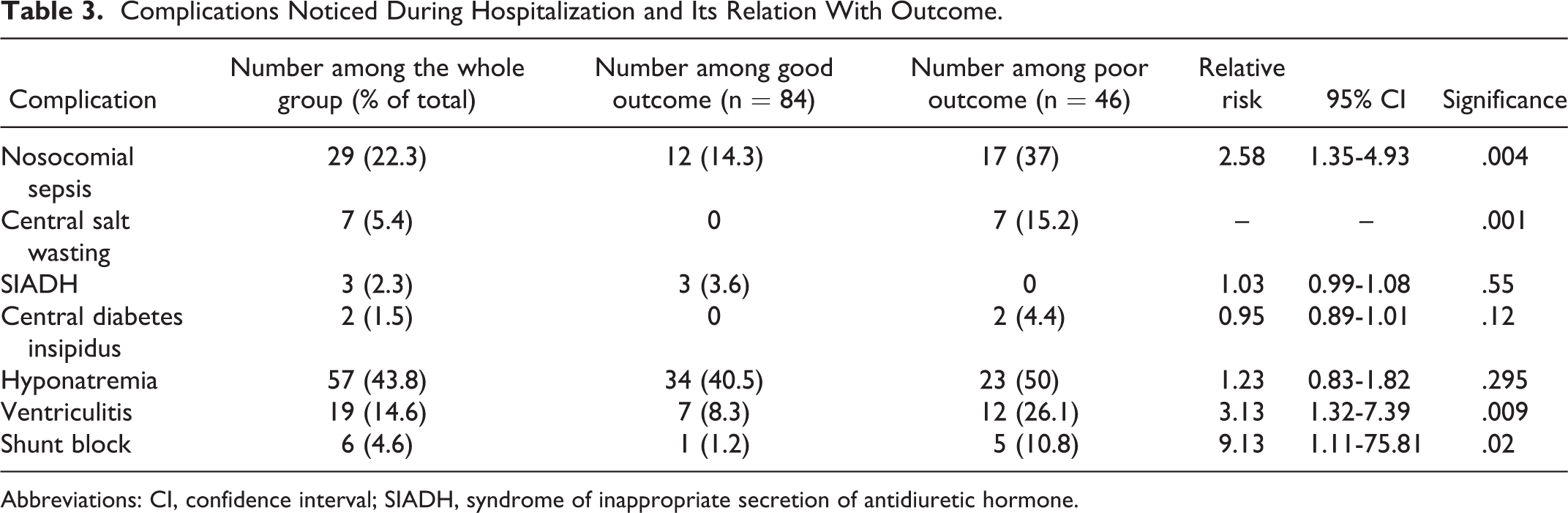
Seven children had evidence of cerebral salt wasting; 3 had the syndrome of inappropriate antidiuretic hormone secretion, and 2 had central diabetes insipidus during hospital stay (Table 3). Three children had cortical venous thrombosis. Antitubercular therapy–induced hepatitis was seen in 7 children. Six children developed shunt block on follow-up requiring shunt revision.
Complications Noticed During Hospitalization and Its Relation With Outcome.
Abbreviations: CI, confidence interval; SIADH, syndrome of inappropriate secretion of antidiuretic hormone.
Neuroimaging
The major findings on neuroimaging were hydrocephalus (n = 112, 86%), basal exudates (n = 88, 68%), infarcts (n = 39, 30%) and tuberculomas (n = 33, 25.4%). Twenty-one children had one of the above 4 findings, 27 had 2 findings, 54 had 3 findings, and 22 had all the 4 findings. Thirty children underwent neuroimaging on follow-up 12 months or later after diagnosis. Sixteen of these children had persistent neuroimaging findings either in the form of exudates or tuberculomas.
Treatment and Follow-up
Ninety-eight children (87.5%) with hydrocephalus (n = 112) underwent a neurosurgical drainage procedure. Eighty-eight underwent ventriculo-peritoneal shunt surgery; 8 underwent external ventricular drainage; and 2 underwent omaya chamber insertion. Five children developed symptomatic paradoxical tuberculomas while on antitubercular therapy. In 12.3% [n = 16] children, anti-tubercular therapy was extended beyond 1 year because of persisting tuberculomas or exudates. Eight (6%) children developed epilepsy and another 8 developed new focal deficits during treatment.
Outcome
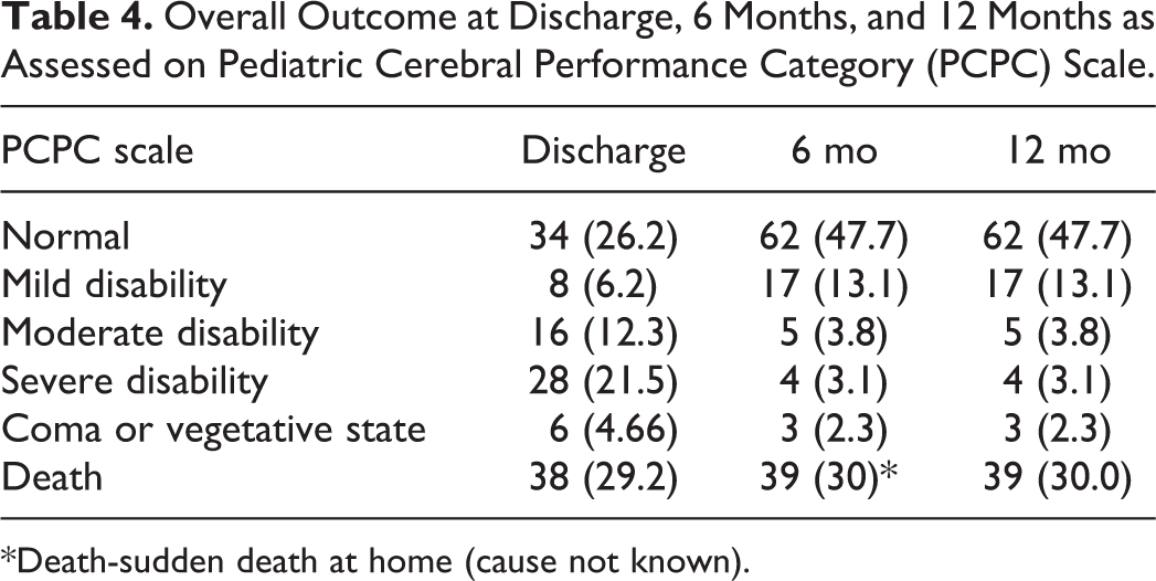
Thirty-eight (29.2%, 95% confidence interval 21.4-37) children died in hospital. Of the 92 survivors, 1 child died 2 months after discharge. At discharge 34 children were either severely disabled (n = 28), or comatose/vegetative (n = 6). At 6 months’ follow-up only 7 children remained severely disabled (n = 4), or comatose/vegetative (n = 3). These numbers remained the same at 12 months’ follow-up. At 6 months, 87% of the survivors were either normal (n = 62) or mildly disabled (n = 17) on PCPC scale (Table 4). Among survivors 7 (8%) children had a hearing impairment, 11 (12%) had a visual impairment, 12 (13%) had epilepsy and 17 (19%) had motor deficits. Two children had persistent meningitis after 3 months of 4 drug anti-tubercular therapy; they were treated as drug-resistant tuberculosis. No child had a relapse of meningitis during the follow-up period. The overall risk of neurologic sequelae in survivors was 32.6% (95% confidence interval 23-42.2) and the risk of severe sequelae was 7.6% (95% confidence interval, 2.2-13). The stage of tuberculous meningitis at admission had a strong influence on the outcome; children in stage III at admission had the maximal mortality and least chance of outcome without deficits. Beyond 6 months, the outcome remained the same in children in various stages of disease severity.
Overall Outcome at Discharge, 6 Months, and 12 Months as Assessed on Pediatric Cerebral Performance Category (PCPC) Scale.
*Death-sudden death at home (cause not known).
Predictors of Poor Outcome
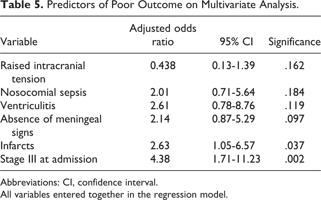
On univariate analysis, none of the presenting clinical features (fever, vomiting, seizures, altered sensorium, headache, weight loss, cough, or focal deficits) predicted a poor outcome. Factors associated with poor outcome were clinical stage III at admission (relative risk 2.78, 95% confidence interval 1.77-4.38, P < .001), infarcts on neuroimaging (relative risk 2.36, 95% confidence interval 1.4-3.97, P = .001), and cerebrospinal fluid glucose <20 mg/dL (relative risk 2.33, 95% confidence interval 1.3-4.19, P = .01). Complications during hospitalization associated with a poor outcome included nosocomial sepsis (relative risk 2.58, 95% confidence interval 1.35-4.93, P = .004), cerebral salt wasting (15.2% vs 0%, P < .001), and ventriculitis after shunt surgery (relative risk 3.13, 95% confidence interval 1.32-7.39, P = .009) (Tables 1 –3). On multivariate analysis, tuberculous meningitis stage III at admission (adjusted odds ratio 4.38, 95% confidence interval 1.71-11.23, P = .002) and infarcts on neuroimaging (adjusted odds ratio 2.6, 95% confidence interval, 1.1-6.5, P = .037) were associated with a poor outcome in tuberculous meningitis (Table 5).
Predictors of Poor Outcome on Multivariate Analysis.
Abbreviations: CI, confidence interval.
All variables entered together in the regression model.
Discussion
Figures of mortality and morbidity from childhood tuberculous meningitis vary vastly across studies. This heterogeneity has resulted in a lack of uniformly accepted data for mortality and morbidity from this disease. The need to have multisite data from prospective cohorts using standard definitions and treatment regimens has been emphasized. 2 Recognizing this need, we planned this study. We used the recommended criteria for defining tuberculous meningitis. 6 In keeping with the observation from a recent review, we have provided clear information on the treatment regimens and use of surgical interventions. 2 In this prospective cohort of 130 children with tuberculous meningitis, we found dismal short-term outcomes. Nearly 30% of children died in the hospital. The mortality rate observed in this study exceeds the recently published rates and the 95% confidence interval limits. 2 There could be several reasons why we encountered a higher mortality. This study was conducted in a referral center and the children admitted were frequently referred from other health care centers due to advanced disease, nonimprovement, or due to complications like hydrocephalus. Overall, three-fourths of the children required neurosurgical cerebrospinal fluid drainage procedure. Second, we cared for children in resource-constrained settings with problems of overcrowding, delayed treatment, and high rates of hospital-acquired infections.
In our study, only 20% children presented in stage 1 and only 45% children presented within 15 days of onset of symptoms. The median duration of symptoms before presentation was 20 days compared to 9 days in another large cohort. 8 None of children younger than 2 years presented in stage 1 compared with 79% above 5 years of age who were in stage 1, suggesting a delay in diagnosis in younger children. Critically ill children with tuberculous meningitis are at risk from their primary disease as well as from interventions, complications of interventions, and hospital stay–related problems. In our cohort, the rate of hospital-acquired bloodstream infections was 22.3%, and 19.4% of children undergoing a cerebrospinal fluid drainage procedure developed ventriculitis. In previous reports from our center, the rates of bloodstream infections in intensive care unit settings have been reported to be around 30% to 33.6%. 9,10 The shunt infection rates in children with tuberculous meningitis have been reported to be high (16%) in reports from India. 11 It is clear that efforts to reduce in-hospital mortality from tuberculous meningitis would need improving supportive care and infection control strategies. But the provision of these interventions in resource-constrained settings remains a challenge.
Very few studies in the literature have described follow-up of tuberculous meningitis survivors at uniform intervals. We examined all children after 6 months of discharge and again after 12 months. This approach allowed a better understanding of the timeline of recovery. The long-term outcome in the survivors was, fortunately, better than the short-term outcome. Additionally, nearly all the neurologic recovery occurred by 6 months. The pooled risk of any neurologic sequelae was 32.6% (95% confidence interval 23-42.2), comparable to the risk described in a recent systematic review, but the risk of severe sequelae was only 7.6% (95% confidence interval, 2.2-13). Intuitively, it seems that children with a severe disease died in the acute phase, and those with less severe disease survived with a good outcome. However, countering this assumption is the fact that a significant proportion of children with severe disability at discharge showed improvement to lower disability states (Table 4).
Antitubercular chemoprophylaxis is highly effective in preventing progression to active tuberculosis in children infected with M tuberculosis, with a protection of up to 90%. 12 Regrettably, none of the children with adult contacts in our cohort received preventive antitubercular treatment. Our study and previous reports emphasize that there is a significant gap between policy and practice in the developing world on this aspect. 13
Neuroimaging, especially MRI, has improved the diagnosis of tuberculous meningitis. In our cohort, all children underwent neuroimaging, and hydrocephalus was the most common finding on neuroimaging (86%). Furthermore, 65% with communicating hydrocephalus needed cerebrospinal fluid diversion procedures, possibly indicating more advanced disease. In a study by van Well et al, 75% of children with communicating hydrocephalus were managed with diuretic alone, and only 18% required ventriculoperitoneal shunt. 8
Several studies have indicated that age and stage of disease at diagnosis are most closely related to the outcome of childhood tuberculous meningitis. 5,14,15 The stage of tuberculous meningitis defined using the refined’ Medical Research Council scale at 1 week has been suggested as a better indicator for prediction of outcome. 16 In keeping with these observations, on multivariate regression, stage III tuberculous meningitis of disease at admission emerged as the most important factor independently predicting a poor outcome in our cohort.
The findings of the current study have to be viewed in the context of its limitations. There were only 2 children with confirmed tuberculous meningitis in our cohort. However, the use of predefined, rigorous clinical criteria should have minimized the possibility that those with an alternative diagnosis may have been included. The lack of microbiological isolation prevented us from identifying drug resistance (reported to increase odds of death 64 times among children with tuberculous meningitis). 17 Hence, it is hard to study the effect of this on the outcome in our cohort. We defined outcome in survivors using established scales; however, we did not evaluate the cognitive or behavioral outcomes in detail. Tuberculous meningitis is known to affect these spheres adversely, and our figures of good outcomes may not necessarily translate into good school performance or good adjustments as adults in society. 18
In conclusion, the outcome of tuberculous meningitis in childhood remains poor despite multi-drug anti-tubercular therapy. Early diagnosis of tuberculous meningitis has remained elusive, and the situation is likely to remain so. More emphasis on Bacillus Calmette-Guérin vaccination and active search for child contacts of adults with tuberculosis and preventive therapy are the only hope to prevent what remains a dreaded disease. These strategies are likely to be financially sensible too.
Footnotes
Author Note
The manuscript was presented as an Oral Abstract in the 9th World Society for Pediatric Infectious Diseases at Rio De Janeiro in November 2015.
Author Contributions
SD was involved in data collection and analysis and prepared the initial draft of the manuscript; AG and NS supervised the patient care, did the statistical analysis, and edited the final draft of manuscript; PM supervised the development assessment and edited the manuscript. NK interpreted the radiologic data and edited the manuscript. PDS supervised the entire process, did the critical review, finalized the draft of manuscript, and will act as guarantor.
Declaration of Conflicting Interests
The authors declared no potential conflicts of interest with respect to the research, authorship, and/or publication of this article.
Funding
The authors received no financial support for the research, authorship, and/or publication of this article.
Ethical Approval
The study was approved by the Institutional Ethics Committee (Reference Memo No 8359/PG-2Trg/2010/4961 dated 29.3.2011).