






Abstract
Brachial plexus nerve damage following childbirth is the main cause of limb dysfunction in neonates. For infants with a history of dystocia, clinicians will use physical examination to diagnose brachial plexus injury. But the accurate assessment of the degree of injury needs to depend on auxiliary examination. Moreover, the brachial plexus anatomy is complex, and there are many different injury types that are simple to combined injuries, and easy to be combined. A single auxiliary examination method is easy to miss the diagnosis. Electromyography has difficulties in the assessment of combined injuries (particularly preganglionic injury paired with postganglionic injury). And when clavicular fracture or significant edema are present, ultrasonography / magnetic resonance imaging (MRI) have limits in the detection of plexus nerve injury. Some scholars believe that multiple auxiliary examinations is helpful to reduce the missed diagnosis and accurate evaluation. High-field strength MRI with its excellent soft tissue contrast and multiplanar capabilities allows good delineation of the brachial plexus. Three-dimensional-short T1 inversion recovery (3D-STIR) is a novel sequence, which is a heavy T2-weighted image, can reconstruct the brachial plexus with an arbitrary curved surface. A STIR fat suppression sequence can suppress the surrounding fat and other soft tissue signals, and has a good tissue contrast. It can display brachial plexus injury of adults clearly. The brachial plexus of infants is thinner than that of adults, and the contrast of surrounding soft tissues is poor, so the imaging effect of the brachial plexus on infants is not satisfactory. In recent years, three D-SHINKEI (three-dimensional nerve-sheath signal increased with inked rest-tissue rapid acquisition of relaxation imaging) sequence has been used in adult brachial plexus and provides excellent imaging, 1 but is rarely reported in infants. 3D-NERVE is a sequence based on 3D-SHINKEI technology. The purpose of this article is to explore diagnostic performance of 3D-NERVE as an adjunct to electromyography for the assessment of brachial plexus injury in infants.
Materials and Methods
Written informed consent was obtained from all parents of infants. Ethics approval for the study was obtained from the Ethical Committee of our hospital (SDFE-IRB/T-2023001).
Patients
This study included 37 infants referred for a clinical diagnosis of brachial plexus injury between January 2019 and November 2022. There were 21 (90.4% boys; age: 1.59 ± 2.42 months) infants who underwent 3D-NERVE sequence scanning, and 16 (43.7% boys; age: 2.28 ± 2.03 months) infants who underwent 3D-NERVE and 3D-STIR sequences scanning.
Imaging Technique
All study participants underwent MR imaging with a 3.0-tesla (T) system (Ingenia; Philips, the Netherlands) using a head and neck coil, supine position, shoulder straightened, and arms dropped down. All infants were scanned while asleep or after sedation with a chloral hydrate enema (0.5–1.0 mL/kg).
The 3D-NERVE sequence parameters were TR/TE (repetition time / echo time) = 2200/76 ms, FOV (field of view) = 150 × 225 mm, echo train length = 20, slice gap = 0; iMSDE duration = 40 ms, whole body–specific absorption rate < 25%. 3D-STIR sequence parameters TR/TE = 2200/76 ms, FOV = 150 × 225 mm, echo train length = 20, slice gap = 0, slice thickness = 2.3 mm, b = 200 s/mm2.
Analysis
All MRI scans were transferred to a postprocessing system (EWS Philips), and NERVE and STIR images of the coronal plane were displayed using partial maximum intensity projection (MIP). MRI scans were independently evaluated by 2 experienced radiologists. Any information regarding the applied MR sequence was removed from all images, and images were randomly evaluated. The images were assessed for the brachial plexus injury, homogeneity of fat suppression (dichotomous grading, yes/no), pulsation artifacts, and the displaying rate of trunks, bundles, and branches of the brachial plexus. The nerve signal-to-muscle signal ratio, nerve signal-to-fat signal ratio, and muscle-to-fat signal ratio (degree of fat suppression) were assessed and compared in both sequences. The level of the brachial plexus nerve root was selected as the measurement level (region of interest [ROI] = 0.5-1 mm2).
Statistical Analysis
A paired Student t test was used to assess the differences in signal intensity ratios between both imaging sequences in the same patient. Fisher exact test was used to assess the differences in the homogeneity of fat suppression and display rates of trunks, bundles, and branches of the brachial plexus between both imaging sequences in the same patient. Statistical significance was set at P <.05, and all data were stored on a spreadsheet and analyzed using the SPSS, version 17.0, software.
Results
Brachial plexus injury was identified in 97.3% (36/37) of infants, of which 58.33% (21/36) occurred on the left side, 38.89% (14/36) occurred on the right side, 2.78% (1/36) occurred on both the sides. Because of soft tissue edema, 2.70% (1/37) of infants could not be evaluated.
Thirty-two cases of brachial plexus injuries were nerve degeneration or edema; MRI demonstrates thickening and hyperintensity of brachial plexus (Figure 1a). In the 2 cases of nerve root avulsion injuries, MRI demonstrates preganglionic root avulsion injury with extradural pseudomeningocele (Figure 1b). One case had compound injury (Figure 1c). In advanced phases, fibrosis may present as thickening of the plexus in 1 case (Figure 1d). The results of examination were consistent with electromyography, and MRI is more accurate and visualized.
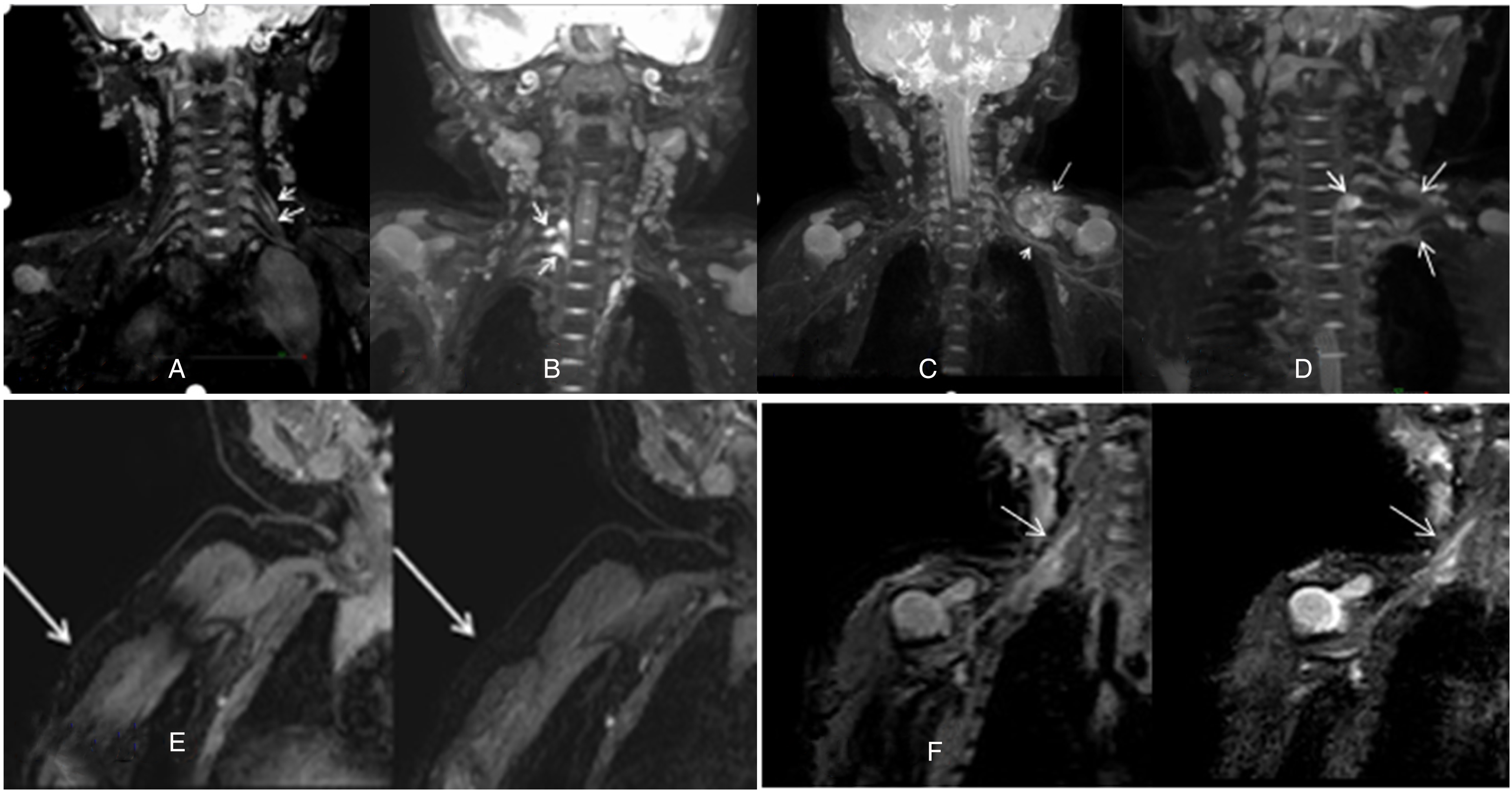
(A) 3D-NERVE MRN image demonstrates thickening and hyperintensity of the left C5,6 nerve roots. (B) 3D-NERVE MRI scan demonstrates preganglionic injury (right C7,8) with extradural pseudomeningocele. (C) 3D-NERVE MRI scan demonstrates thickening and hyperintensity of the left brachial plexus (root, trunk, bundle, branch). The nerves were aggregated and indistinct. A clavicular fracture is associated with surrounding hematoma. (D) 3D-NERVE MRI scan demonstrates preganglionic root avulsion injury with extradural pseudomeningocele (left C7), and fibrosis of brachial plexus (upper, middle, lower trunk and medial, lateral bundles of the brachial plexus). (E, F) 3D-NERVE (left) demonstrates better fat suppression and nerve highlight effects than 3D-STIR(right).
The 2 sequences were compared, yielding the following results (Table 1). Pulsation artifacts were not observed on 3D-STIR (0/16) and 3D-NERVE (0/16); heterogeneous fat saturation was observed in 5 (5/16) infants on 3D-STIR; however, use of 3D-NERVE revealed heterogeneous fat saturation in 1 (1/16) infant. The arterial signal was equally suppressed in both sequences. Venous signal suppression was better with 3D-NERVE. 3D-NERVE revealed a higher nerve-to-fat ratio (P < .05), nerve-to-muscle ratio (P < .05) but revealed insignificant difference on muscle-to-fat ratio (P > .05) (Table 1). 3D-NERVE and STIR showed 100% (16/16) of the brachial roots and brachial plexus trunks. Brachial plexus bundles and brachial plexus branches were seen in 93.75% (15/16) and 68.75% (11/16) of 3D-NERVE-derived images, respectively. Brachial plexus bundles and brachial plexus branches were seen in 93.75% (15/16) and 62% (10/16) of STIR images. The differences were not statistically significant (P > .05).
Comparison of Image Characteristics Between 3D-NERVE and 3D-STIR Sequences.

Discussion
Neonatal brachial plexus injury presents as a weakness or paralysis of the upper extremities at birth, and is most often related to stretching of the brachial plexus in the perinatal period. In recent years, the incidence of brachial plexus injury has increased because of the increasing birth rate of macrosomia. Preganglionic injury can present as avulsion injury with or without pseudomeningocele. Pseudomeningocele is an indirect sign of preganglionic injury and is due to a tear in the meningeal sheath surrounding the nerve roots with extravasation of cerebrospinal fluid into the adjacent tissues and is easily identified on T2-weighted MRI scans with attenuated or disrupted proximal roots within or immediately distal to the meningocele. Postganglionic injuries are either stretched with nerve continuity or avulsed with nerve disruption. In stretch injury, nerves are hyperintense on T2-weighted images. This reflects oedema with nerve continuity. In avulsion injury, there is nerve discontinuity with distal nerve retraction. Assessment of the brachial plexus is sometimes difficult when there is distortion due to severe trauma or surgical intervention. In advanced stages, fibrosis may manifest as thickening of the plexus. 2 Nerve rupture is a more severe form of injury, with limited recovery related to scar tissue that forms a neuroma and blocks axon regeneration, and surgery is required.3,4 The first 3 months are the “golden period” because of their significant impact on outcomes. 5 Incomplete nerve injury is usually treated conservatively, including drug therapy, functional training, physical therapy, and traditional Chinese medicine. Therefore, it is important to accurately evaluate the location and degree of injury and choose a reasonable treatment for the recovery of limb function in infants.
Brachial plexus injuries are diagnosed clinically based on the history and physical examination findings, and one can accurately diagnose the type of brachial plexus injury by electromyography and ultrasonography, but it cannot show the specific position, numbers, and degree nonintuitively. 2 The high-resolution ultrasonographic can identify and locate the postganglionic region associated with the upper and middle trunks. The ability of ultrasonography to evaluate pre- and postganglionic injuries associated with the lower trunk is quite limited. 6 MRI is not the first choice to evaluate these types of injuries in the past. With 3.0-T MRI coming into being, studies on brachial plexus of MRN have increased. 3D-STIR and 3D-NERVE sequences offer high-resolution MR imaging of postganglionic brachial plexus lesions. It is a valuable tool for the diagnosis of a variety of plexus lesions in adults, as well as for the planning of surgical interventions. The brachial plexus in infants is slender and has a poor contrast with the surrounding tissue, and rarely report on imaging brachial plexus injuries in infants. In our study, 36 of 37 infants were diagnosed with brachial plexus injury by MRI, and the agreement was good with electromyography.
This study aimed to study diagnostic performance of 3D-NERVE as an adjunct to electromyography for the assessment of brachial plexus injury in infants. 3D-STIR is a heavy-T2-based and stir-based technique; the former shows the lesions of nerve bundles based on its ability to sensitively distinguish the changes in fluid content in the endoneurium; the latter can sufficiently suppress the surrounding fat signal and highlight the neural hyperintensity.7, 8 3D-nerve is a sequence based on the 3D-SHINKEI technology, which is a new sequence that utilizes both spectral adiabatic inversion recovery type fat suppression and an improved Motion Sensitized Driven Equilibrium (iMSDE) pulse. 3D iMSDE encompasses repeated opposite-direction pulses for vascular signal suppression before fat suppression and variable flip angles for T2-weighted images are applied. Vessel signal-suppressed and fat-suppressed images have significant potential to produce high-resolution and volumetric neurographic images.1, 9
This study found that both 3D-NERVE and 3D-STIR sequences had good fat suppression and nerve highlight effects. They can not only depict the anatomy and lesions of the brachial plexus and identify the number of lesions, but also analyze their spatial extension within and along the nerve tracts. These sequences can be used as supplements to neuroelectromyography and US for visualization of the brachial plexus. 3D-nerve demonstrates better capacity than 3D-STIR (Table 1).
In the STIR technique, an initial 180° inverting radiofrequency pulse is followed by a standard 90°–180° spin-echo sequence. The time allowed to elapse between the inversion pulse and the 90° pulse is chosen to approximate the null point of fat, resulting in suppression of the signal intensity of fat. The primary advantage of the STIR pulse sequence lies in its ability to produce uniform fat suppression. 9 Even in difficult areas of the body, the STIR sequence has proven to be extremely reliable, with strong insensitivity to B0 inhomogeneities. The 3D-nerve sequence comprises high-bandwidth STIR pulse, and is thus slightly superior in fat suppression compared with STIR (Figure 1e). 10
Both 3D-NERVE and 3D-STIR sequences are able to depict the roots, trunks, bundles, and most of the nerve branches of the brachial plexus in infants, and the 3D-nerve sequence has proven to be better than the 3D-nerve sequence. Contrapose the eddy currents of the preparation pulse of MSDE, iMSDE introduces 2 refocusing pulses, and optimize the B0/B1 characteristics. 11 The iMSDE technique dynamically adjusts the order moment of the gradient, set a pair of bipolar gradient to dephase the blood flow to suppress the blood flow, and further improved the eddy current, to obtain a clearer image of the vessel suppress. 11 The iMDSE prepulse results in suppression of the arterial and venous vessel signals, enhancing the visualization of nerve structures, which becomes particularly obvious in regions with immediate proximity of blood vessels to the plexus nerves, for example, the clavicular region. 12 In addition, the high-bandwidth STIR pulse shows superior fat suppression, and the 3D-NERVE presents a higher nerve signal and clearer delineation of the nerve (Figure 1f).
In conclusion, compared with the 3D-STIR technique, 3D-NERVE presents a superior fat suppression, higher nerve signal, and clearer delineation of the nerve, which can accurately locate the injured nerve and clearly display the type and number of injured nerves. It is an effective adjunct to electromyography for doctors to assess brachial plexus injury. But this study has limitations in that it only investigated the accuracy of the 2 sequences in the diagnosis of brachial plexus injuries and did not investigate in detail the role of MRI as a guide to clinical management and prognosis. In the future, we will investigate the relationship between the comprehensive score (clinical, electrophysiologic, MRI, ultrasonography) and clinical treatment, prognosis. 13
Footnotes
Acknowledgements
I would like to express my gratitude to all those who helped me during the writing of this thesis. A special acknowledgment should be shown to Professor Li Min, from whose lectures I benefited greatly. I am particularly indebted to family members, who gave me kind encouragement all through my writing. Finally I wish to extend my thanks to the library assistants who supplied me with reference materials of great value.
Author Contributions
All authors contributed to the study's conception and design. Changyou Ma and Yi Lu obtained the images, while Chunhua Dong and Jianshe Zhao evaluated the MR images. Leilei Zhao collected and analyzed the data, and drafted the original manuscript. Jianshe Zhao revised the original draft of the manuscript. All authors have provided comments on previous iterations of manuscript. The final version of the manuscript underwent review and received approval from all authors.
Declaration of Conflicting Interests
The authors declared no potential conflicts of interest with respect to the research, authorship, and/or publication of this article.
Funding
The authors received no financial support for the research, authorship, and/or publication of this article.
Ethics Approval
This retrospective study was approved by the Ethics Committee of Jinan Children's Hospital :Qilu Children's Hospital of Shandong University (SDFE-IRB/T-2023001). The study was performed in accordance with the ethical standards as laid down in the 1964 Declaration of Helsinki and its later amendments or comparable ethical standards. The human research participants provided informed consent for publication of the images in  .
.