






Abstract
Polymeric hydrogel systems based on poly(2-hydroxyethyl methacrylate), poly(ε-caprolactone), and poly(ethylene glycol) were prepared by photopolymerization using 2,4,6-trimethyl benzoyl diphenyl phosphine oxide as the photoinitiator. The structural details, morphology, and crystallinity were evaluated by Fourier transform infrared, scanning electron microscope, and X-ray diffraction analysis. The fluid uptake of the hydrogels was measured using swelling analysis. Based on a hemolysis assay, the prepared hydrogels were non-hemolytic. No attachment of fibroblasts to the hydrogel systems was observed. Stress–strain results indicated that the poly(2-hydroxyethyl methacrylate), poly(ε-caprolactone), and poly(ethylene glycol) hydrogel system (85:10:5) possessed better mechanical properties. Cytotoxic assessment by direct contact method of 85:10:5 systems found that the hydrogel was non-cytotoxic to L929 fibroblasts. This hydrophilic polymer system has a potential for wound dressing applications.
Keywords
Introduction
Skin plays an important role in physiological functions such as immune surveillance, self-healing, thermoregulation, and sensory detection. 1 Loss of structural integrity of skin by cutaneous wounds may impair skin functions at various levels ranging from disabilities to even death. Cutaneous wounds often occur due to mechanical trauma, burns, surgical procedures, and reduced blood circulation. Proper wound management should be carried out to prevent excessive fluid loss and bacterial invasion, and can be achieved only by covering the wound using wound dressing material.2–4 An ideal dressing should provide a moist wound environment, absorb exudates, eliminate dead space, should not harm the wound, and provide a bacterial barrier while promoting wound healing. As reported, wounds in moisture environment heal faster than in dry environment.5,6
Hydrogels have the capacity to absorb wound exudates and retain them within the gel, providing a microclimate that stimulates and regulates most of the cellular functions and nutritional processes during the individual phases of wound healing. Hydrogels are easily removed from the wound surface because of their non-adherent nature. 7 A number of natural and synthetic polymers, including chitosan, collagen, alginate, poly(N-vinylpyrrolidone), and poly(2-hydroxyethyl methacrylate) (PHEMA), have been used as wound dressings.8–12
PHEMA hydrogel possesses high biocompatibility, excellent blood compatibility and good swelling characteristics. Hydrophilic nature of the polymer imparts high degree of swelling to the hydrogel systems.13–15 Main disadvantages of PHEMA hydrogels are their low mechanical strength and tear resistance that can be resolved, for instance, by incorporating other polymer systems, adding cross-linking agents, or copolymerization. Numerous reports have shown that incorporation of polycaprolactone improves the mechanical property of hydrogel systems such as poly(vinyl alcohol) and PHEMA.16,17 Biocompatibility and blood compatibility of hydrogel systems can also be improved using poly(ethylene glycol) (PEG). 18 PHEMA hydrogel tethered with PEG sulfonate was found to be non-thrombogenic with a superior swelling capacity compared to the homopolymeric PHEMA hydrogel. 19 Hydrogels based on PHEMA and PHEMA-co-PEG have earlier been investigated as wound dressing materials.20,21
Although hydrogels can be prepared using different polymerization techniques, the photopolymerization has special advantages such as spatial as well as temporal control over polymerization, faster rate (less than a second to few minutes) at room or physiological or even higher temperatures, and minimum heat of polymerization. 22 Photopolymerization is usually achieved using organic photoinitiators sensitive to ultraviolet (UV) or visible lights. Major problem associated with the commonly used photoinitiators is that they tend to impart color to the polymer system. In our study, we used a novel photoinitiator, 2,4,6-trimethyl benzoyl diphenyl phosphine oxide (TPO), which facilitates polymerization at a faster rate without coloring the polymer system.
The aim of this study was to prepare hydrogel systems composed of PHEMA–poly(ε-caprolactone) (PCL)–PEG by photo-curing technique using TPO for potential wound dressing application and to investigate their physical, chemical, mechanical, and morphological characteristics. Furthermore, the hydrogels were subjected to hemolysis, in vitro cytotoxicity, and cell adhesion studies to evaluate their potential for wound dressing applications.
Materials and methods
Materials
HEMA, TPO, and PCL (molecular weight (Mw) 14,000) were all purchased from Sigma–Aldrich Chemical Company Inc. (USA). PEG (MW 200) and formaldehyde were procured from Merck (Germany). Iscove’s modified Dulbecco’s Minimal Essential Medium − F12, antibiotic–antimycotic solution, fetal bovine calf serum, and trypsin–EDTA (all cell culture grade) were obtained from Invitrogen (USA). Texas red–phalloidin was obtained from Molecular Probes (USA). All reagents used were of analytical or equivalent grade. Phosphate buffered saline (PBS; pH 7.4) was prepared by dissolving 17.9 g of di-sodium hydrogen phosphate, 5.73 g of monosodium hydrogen phosphate, and 9 g of sodium chloride in 1 L distilled water. Photopolymerization was carried out using a dental light cure unit containing a halogen bulb (Model 301, Prolite, Caulk/Dentsply; Melfork, DE, USA) at 380–480 nm wavelength.
Preparation of hydrogel
HEMA monomer was vacuo distilled before use to remove the inhibitor. PCL (Mw 14,000) and PEG (Mw 200) were added to the monomer and heated up to 70–75°C to obtain a homogenous mix. TPO photoinitiator (1 wt%) was added to this mix under constant stirring. The solution was then poured into a Teflon mold (60 mm × 10 mm × 1 mm) and exposed to a dental light source to initiate the polymerization for a maximum of 2–3 min. After completing the polymerization, the mold containing the cured polymer was kept in distilled water till the polymer peeled away. Three hydrogel systems were prepared accordingly based on the formulations listed in Table 1. The sheets were stored in distilled water at 37°C (Figure 1; Scheme 1).
Sample code and composition of polymeric hydrogels.

HEMA: 2-hydroxyethyl methacrylate; PCL: poly(ε-caprolactone); PEG: poly(ethylene glycol).

Hydrogel sheet prepared by photopolymerization.

Production scheme of hydrogel systems.
Characterization of PHEMA-based hydrogels
Fourier transform infrared spectroscopy
Fourier transform infrared (FT-IR) spectrum of hydrogels was obtained (Jasco FT-IR 6300, Japan) in the range of 4000–400 cm−1. Lyophilized polymer samples were ground with KBr powder and spectra recorded on pellets.
X-ray diffraction studies
Hydrogel samples prepared were freeze dried using a lyophilizer (Alpha 1-4 LD; Christ, Germany), and X-ray diffraction (XRD) diffractogram was recorded using a X-ray diffractometer (Siemens D 5005, Germany) at 40 kV and 30 mA over a diffraction angle (2θ) in the range of 10°–80°.
Swelling studies
Pre-weighed dried circular disk samples (8 mm diameter × 1 mm thickness) were immersed in de-ionized water at 37°C for 24 h. After removing the surface water, the swollen samples were weighed at specific time intervals using an analytical balance (Model CP224S; Sartorius, Germany) to the nearest of 0.1 mg. The swelling percentage of the samples was determined according to the following equation
where Ws and Wo are the masses of the swollen and dry samples, respectively.
Mechanical properties
Evaluation of tensile strength and strain of hydrogels was carried out using a Universal Testing Machine (INSTRON Model 3365, UK) at 22°C. Swollen hydrogel samples with 30 mm length, 10 mm width, and 1 mm thickness were pre-conditioned before testing. A crosshead speed of 50 mm/min was maintained during the testing using a 100-N load cell. Tensile strength of materials was calculated using the equation:
Sample size was six per group, and the mean and standard deviation were calculated. Statistical analysis (analysis of variance (ANOVA) single factor) was used to determine significant changes in stress.
Surface morphology
Surface morphology of hydrogel samples was studied using a scanning electron microscope (SEM; Hitachi S2400, Japan). Dried hydrogel samples were gold coated and placed on the aluminum stub and observed under vacuum in SEM.
Tests for cytotoxicity
Presence of water in hydrogel makes the hydrogel flexible and soft providing minimum frictional irritation as well as soothing effect in contact with physiological systems. Cytotoxicity study of the 85:10:5 system was carried out by direct contact test and MTT [3-(4,5-dimethylthiazol-2-yl)-2,5-diphenyl tetrazolium bromide] assay per established standard procedures.
Direct contact test
The in vitro cytotoxicity test using direct contact method was performed according to the ISO 10993-5. The culture medium from the L929 monolayer was replaced with fresh medium. The EtO-sterilized samples, negative control high-density polyethylene (HDPE), and positive control poly(vinyl chloride) (PVC disk) in triplicate were placed gently on top of a monolayer L929 fibroblast cells. After incubation at 37 ± 1°C for 24–26 h, the cell monolayer was examined microscopically for the response around the test samples. Morphology of the cells was assessed with respect to positive and negative control samples. The cellular responses of the test samples were scored as 0, 1, 2, 3, and 4 for non-cytotoxic, mildly cytotoxic, moderately cytotoxic, and severely cytotoxic, respectively.
Viability test on extract by MTT assay
MTT assay was used to measure the metabolic activity of cells by monitoring the reduction in yellow-colored tetrazolium salt to purple-colored formazan. Extract was prepared by incubating 3 cm2 of presoaked test material in 1-mL culture medium containing serum at 37 ± 1°C for 24–26 h. After 24 h, the extract was diluted with culture medium to 50%, 25%, and 12.5% concentrations. Negative control was prepared by incubating 12.5-cm2 ultra-high-molecular-weight polyethylene with 1-mL culture medium containing serum at 37 ± 1°C for 24–26 h. A dilute solution of phenol (13 mg/mL to 1.3 mg/mL with culture medium containing serum) was used as positive control. Equal volumes (100 µL) of various diluted test samples and positive control were placed on subconfluent monolayer of L929 cells. After incubation of cells with various concentration of test samples and controls at 37 ± 1 for 24 ± 2 h, extract and control medium were replaced with 50 µL MTT solution (1 mg/mL in medium without supplements), wrapped with aluminum foil, and were incubated at 37± 1°C for 2 h. After discarding the MTT solution, 100 µL of isopropanol was added to all wells and swayed the plates. The color developed was quantified by measuring absorbance at 570 nm using UV spectrophotometer (MQX200R; BioTek, USA).
Percent hemolysis
Hemocompatibility test of hydrogel systems was carried out broadly on the basis of ISO 10993-4:2002 (E) selection of tests for interaction of materials with blood. The test is mainly aimed at finding the extent of hemolysis caused by the sample. Blood from healthy human volunteer was collected into the anticoagulant, citrate-phosphate-dextrose-adenine[CPD-A]. Samples kept in PBS were taken out and placed in polystyrene plates. Blood (2 mL) was added to each sample; 1 mL blood was taken immediately for initial analysis and another 1 mL blood was incubated with the samples for 30 min under agitation at 70 ± 5 r/min using an Environ shaker (Labline Instruments Inc., Melrose Park, USA) thermostated at 35 ± 2°C. Four empty polystyrene culture dishes were exposed with blood as reference. The total hemoglobin in the whole blood samples was measured using automatic hematology analyzer (Sysmex-K 4500, Sysmex Corporation, Japan). The free hemoglobin liberated into the plasma after exposure to materials was measured using diode array spectrophotometer (HP 8453; Hewlett-Packard GmbH, Germany) and the hemolysis percentage was calculated using the formula
Cell adhesion
Human dermal fibroblast cells were used for the cell adhesion study. Hydrogel samples were seeded with 70,000 cells/cm2, allowed to attach for 72 h, fixed with 3.7% formaldehyde for 20 min, and then permeabilized with 0.2% triton X 100 (Molecular Probes). Texas red–conjugated antibodies (Santa Cruz Biotechnology, USA) against actin cytoskeleton were added and kept at 37°C for 30 min, washed thoroughly using PBS, and observed through a fluorescent microscope (Leica DMIRB, Germany).
Results and discussion
Hydrogel systems based on PHEMA–PCL–PEG were prepared using photopolymerization method, as shown in Scheme 2. Conversion of HEMA monomer to PHEMA was confirmed by the disappearance of the unsaturated group at 1636 cm−1 in the FT-IR spectrum of all hydrogels studied (Figure 2). Characteristic absorption of PCL, PHEMA, and PEG is prominent in the spectrum. The absorption at 1725–1730 cm−1 corresponds to characteristic –C = O stretching frequency of PCL and PHEMA. Peaks at 1163 and 1400 cm−1 are indicative of the C–O–C stretching and –OH bending frequency of PEG, respectively. The absorption band at 3533 cm−1 indicates the free hydroxyl (–OH) group, a characteristic peak of PHEMA. The broad nature of these peaks suggests that hydroxyl groups are extensively hydrogen bonded.

Schematic representation of hydrogel formation.

(a) FT-IR spectra of HEMA, HI-1, HI-3, and HI-5. (b) FT-IR spectrum of HI-1 indicates characteristic peaks of PEG.
The X-ray diffractograms of H I-1, H I-3, and H I-5 and PHEMA alone are shown in Figure 3. PHEMA had an amorphous nature while the three blended polymer systems had semi-crystalline character. Sharp peaks at 21.40° and 23.71°, specific to PCL, confirmed the introduction of crystalline character to the hydrogel system.

XRD patterns of PHEMA (H), HI-1, HI-3, and HI-5.
Swelling characteristic of hydrogel network is important especially for wound dressing applications as it may affect mechanical and surface properties.23–25 Polymer swelling property is a complex phenomenon as it depends on the nature of the polymer, polymer–solvent compatibility, and degree of cross-linking. In case of ionic networks, swelling behavior depends on mass transfer limitations, ion exchange, and ionic interactions.26,27 Maintaining a moist environment over the wound bed is one of the most important criteria for wound dressings. A matrix with good swelling capacity is expected to possess a cleansing effect upon the wound, as it facilitates absorption of wound exudates, which in turn tend to protect the wound from infection. Prepared hydrogel samples contain both hydrophilic and hydrophobic units. When we compare HI-1 and HI-5, both systems contain 95 parts of hydrophilic component (HEMA). HI-1 contains 2.5 parts of hydrophobic component (PCL) and 2.5 parts of hydrophilic component (PEG). HI-1 possesses significantly greater swelling capacity compared to HI-5, which indicates HI-1 is more hydrophilic. This indicates that PEG plays an important role in the swelling property and hydrophilic nature of the systems. HI-3 displayed greater swelling than HI-1 due to the presence of PEG and HEMA. The swelling characteristics of all hydrogel systems are shown in Figure 4. The results for all hydrogel systems are significantly different (p < 0.05).

Swelling capacity of hydrogels.
Scanning electron micrographs of the outer surface and cross section of hydrogels (Figures 5(a) and (b)) reveal a dense outer skin layer and a porous cross-sectional layer. The dense layer tends to provide the hydrogel a capacity to prevent excessive loss of body fluids, control of water loss through evaporation, as well as protection of wound from external environment. The porous nature is expected to help in promotion of drainage, prevention of exudates build-up, and preparation of an optimal wound bed for autografting.

SEM micrographs of the HI-3 (a) cross-section and (b) surface.
Hydrogels are mechanically weak in their swollen state. The PCL was incorporated to the hydrogel systems to enhance hydrophobicity and to improve the mechanical properties to a desirable extent. Moreover, PCL is a Food and Drug Administration (FDA)–approved polymer used for many biocompatible applications. The tensile characteristics of the hydrogel systems are shown in Figures 6(a) to (c), where HI-3 had the highest tensile strength (0.23 ± 0.03 MPa) and an elongation (111 ± 17%). 28 Although reported values of tensile strength of human skin are ~5–30 MPa, skin grafts are rarely subjected to high tensile strengths at wound sites, and hence, substitute matrices with optimum elongation characteristics (around 105%) may be preferred. The inferior tensile strength does not hinder application of scaffolds for skin tissue engineering. 29 Hydrogel HI-3 was found to have good mechanical and swelling properties that are suitable for wound dressing applications, which is instrumental for further biocompatibility studies.

(a) Tensile strength of hydrogels. (b) Tensile strain of hydrogels. (c) A sample stress–strain curve of HI-3.
The results of direct contact test between fibroblast cells and HI-3 hydrogel sample are summarized in Table 2. In general, HI-3 was shown to be non-cytotoxic when compared to the negative reference material (HDPE) (Figure 7(a)) and retained its characteristic spindle shape. Hemolysis estimations have been used as a simple and reliable technique for estimating blood compatibility of materials. 30 The percentage hemolysis of the samples was evaluated (shown in Figure 9) to determine the hemolytic property of the material for wound dressing. In this study, empty polystyrene dishes were used as a reference generating negligible hemolysis <0.1%. As per ISO 10993-4:2002 (E), for material to be non-hemolytic, the percentage hemolysis should be less than 0.1%. All tested samples displayed less than 0.1% hemolysis.
Direct contact cytotoxic tests with hydrogel samples.

HDPE: high-density polyethylene; PVC: poly(vinyl chloride).

Microscopic images of L929 cells in direct contact with (a) HDPE-negative control, (b) hydrogel HI-3, and (c) PVC-positive control.

MTT assay; % viability of cells with respect to the controls.

Percentage hemolysis of samples with control.
An ideal wound covering material should not possess cell adhesion since it may cause pain upon the removal of dressing from the wound. We observed that incubation of our hydrogel samples on the subconfluent monolayer of fibroblasts for 72 h did not produce any unfavorable effect on cell spreading as evidenced by actin organization and network formation in cells lying underneath the gel (Figures 10(b), (e), and (h)). Microscopic observation of surface in contact with cells indicated non-adherence of the cells to the material (Figures 10(a), (d), and (g)). Cell morphology as well as spreading in the medium underneath the gel was found to be comparable to the bare medium (Figures 10(c), (f), and (i)). Spindle-shaped morphology was also prominent for the cells validating the earlier non-cytotoxic character observed for the hydrogel.
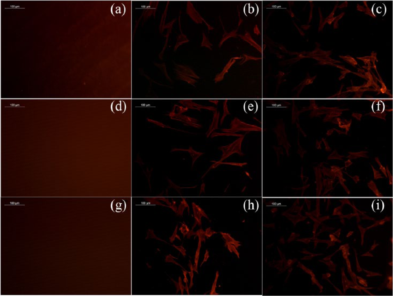
Microscopic images of actin staining on (a) HI-1, (b) fibroblast cells in contact with HI-1, (c) control cells, (d) HI-3, (e) fibroblast cells in contact with HI-3, (f) control cells, (g) HI-5, (h) fibroblast cells in contact with HI-5, and (i) control cells.
Conclusion
The formation of a semi-crystalline hydrogel by polymerizing HEMA monomer via photopolymerization at 380–480 nm was confirmed by FT-IR and XRD. With an optimum swelling and elongation properties, the HI-3 hydrogel system was found to be a potential candidate for further biological studies. Non-cytotoxic character, non-hemolytic behavior, and non-adherence to fibroblast cells support the prospective potential use for this hydrogel system in wound covering applications.
Footnotes
Acknowledgements
The authors acknowledge the Director and the Head, BMT Wing, SCTIMST, for the facilities provided. The authors wish to thank Dr Lissy K. Krishnan for providing facility for cell adhesion studies. They are also grateful to Dr S. Unnikrishnan and Dr Renjith P. Nair for their technical help during cell culture studies.
Declaration of conflicting interests
The authors declared no potential conflicts of interest with respect to the research, authorship, and/or publication of this article.
Funding
R.P.K. is grateful to Kerala State Council for Science, Technology and Environment, Thiruvananthapuram, India, for fellowship support and part funding (KSCSTE, Burn project 8042). The work forms part of PhD work of R.P.K.