
Abstract
pH-Sensitive hydrogels of 2-hydroxyethyl methacrylate/citraconic anhydride–modified collagen were obtained by free radical copolymerization/crosslinking in the presence of ammonium persulfate/N,N,N′,N′-tetramethylethylenediamine redox initiator system. Their pH-responsiveness was demonstrated by swelling behavior and ciprofloxacin release tests. Both unloaded and loaded hydrogels were characterized by Fourier transform infrared, scanning electron microscopy, and biocompatibility tests. The enzymatic degradation in the presence of Clostridium histolyticum mainly depends on initiator content. In vivo biocompatibility tests involving intraperitoneal hydrogels’ implantation in rats following the analysis by the granuloma test, leukocyte formula, immune parameters, and hepatic transaminases demonstrated their non-toxicity and biocompatibility with living tissues. The in vitro antimicrobial activity, in vivo biocompatibility, and in vitro biodegradability tests attest the possibility to use these new polymeric hydrogels with tailored properties as matrices for bioactive products in medical and pharmaceutical applications as wound care and targeting drug delivery systems. The ciprofloxacin release studies proved their potential as materials for wound dressings.
Keywords
Introduction
Poly(2-hydroxyethyl methacrylate) (PHEMA) is one of the most extensively studied material in tissue engineering, that is, in contact lens, because of its excellent biocompatibility and physicochemical properties similar to those of living tissues. It also exhibits good chemical and hydrolytic stability and good tolerance for entrapped cells. 1 PHEMA sponges are promising materials for implants and they promote fibroblast/collagen attachment. 2 2-hydroxyethyl methacrylate (HEMA)-based (co)polymers showed soluble to insoluble thermoresponses, 3 being widely used as the backbone for synthesizing stimuli-responsive hydrogels. 4 Gamma radiation was used to prepare new poly(oligo(propylene glycol) methacrylate)–HEMA copolymeric hydrogels having wide diversity in thermoresponsive properties which strongly depend on their composition. 5 Tomic et al. 6 coupled temperature-responsive polymers with pH-sensitive components and obtained copolymers based on HEMA and itaconic acid with potential for biomedical applications, especially for skin treatment. These copolymers show a swelling and drug release behavior dependent on both stimuli.
Due to their hydrophilic nature, porosity, and soft tissue–like properties, hydrogels emerge over different types of biomaterials as prime candidates for the biomedical applications especially for wound dressings.7,8 In vivo bioactivity of the hydrogel clearly depends on the microstructural parameters (e.g. chemical composition, crosslink density), which may directly affect cell behavior and macroscopic properties (e.g. mechanical stiffness, degradation rate) which indirectly affect cells and could be important for integration and remodeling in the host tissue. 9 Hydrogels based on HEMA copolymers are of a great interest in biomedical applications because of their tunable chemical composition and three-dimensional network structure. Because the gels obtained from natural polymers exhibit some limitations, recent efforts are focused on developing hydrogel systems derived from a combination of both natural and synthetic materials, which display the most advantageous properties. 10 HEMA-based hydrogels are inert to normal biological processes, biocompatible, 11 resistant to degradation, and not absorbed by the body.
Collagen-based hydrogels exhibit pH-responsiveness character so that a swelling–collapsing pulsatile behavior was recorded at pH 2 and 8.12,13 pH-responsive collagen-based hydrogels have good potential for drug delivery systems and tissue engineering.14,15 Due to its high prevalence in the body, collagen has garnered significant attention from researchers as a material to replace or repair injured or damaged tissue. 16 A wide range of collagen characteristics such as its availability, biodegradability, simple absorption in the body, non-antigenicity, non-toxicity, good biocompatibility, processability in a number of different forms, synergism with other bioactive compounds, compatibility with synthetic polymers, and reactivity of its functional groups (which allows easy modifications to produce desired materials and suitable ductile strength and negligible expressibility) recommend it as a versatile biomaterial in various medical and pharmacological fields. 17 The function of collagen in wound healing is the ability to control many cellular functions, including cell shape and differentiation, 18 migration, and synthesis of a number of proteins. 19 Collagen also plays a critical role in all phases of wound healing such as hemostasis, inflammation, proliferation, and remodelling. 20 Moreover, collagen-based dressings have the ability to absorb wound exudates and maintain a moist wound environment.
Burns provide a suitable site for bacterial multiplication and are more persistent richer sources of infection than surgical wounds, mainly because of the larger area involved and longer duration of patient stay in the hospital. 21 The loading of antibiotics and other drugs into polymer-based biomaterials was intended to deliver drugs to a specific site and override the need for systemic antibiotic therapy, with the overall goal of maintaining drug concentrations in the therapeutic range. Ciprofloxacin (CIPRO here is shortened as CF) hydrochloride monohydrate was selected as the model active principle because of its safety with good tolerance and broad antibacterial spectrum 22 and because of its good solubility in a wide range of pH values.
In our previous studies, the soluble-substituted anhydride-modified collagen macromolecules have been obtained and characterized. 23 The modified collagen was obtained by the chemical bonding between carbon atoms from the carbonyl group of citraconic anhydride (CA) and the amino group of collagen. It has a vinyl and a carboxyl group in its chemical structure. Modified collagen exhibits an increased solubility, reactivity, and thermal resistance. Besides, they undergo free radical copolymerization/crosslinking reactions with HEMA monomers in the presence of ammonium persulfate (APS) and N,N,N′,N′-tetramethylethylenediamine (TEMED) with up to 30 wt % modified collagen. 24 In this study, the same procedure was applied to obtain PHEMA-based hydrogels with citraconic anhydride–modified collagen (C-CA) which intents to obtain pH-responsive biomaterials with better biodegradability, biocompatibility, and more adequate characteristics for wound dressings and other medical applications. The obtained hydrogels were loaded with the antimicrobial drug, CF hydrochloride monohydrate.
As far as we know, hydrogels based on HEMA and C-CA as pH-responsive carrier for CF have not been reported and investigated up to date; only one article studied the use of CF–collagen conjugate for wound healing treatment. 25 Both unloaded and loaded systems have been investigated with respect to their structure, morphology, swelling behavior, pH-responsiveness, biodegradability, drug release ability, and biocompatibility. All results proved that they are promising materials for medical and pharmaceutical applications.
Materials and methods
Materials
HEMA (HQ, Sigma–Aldrich, UK) was purified by passing it through an inhibitor removal column, and the ammonium peroxodisulfate (APS) was purified by re-crystallization from a mixture of twice distilled water–methanol (1:2, v: v). The acid-soluble collagen, type I + III from bovine skin dermis as 1.21 wt % solution in H2O2, was supplied by Lohmann & Rauscher GmbH (Germany). CA with analytical purity and TEMED were purchased from Sigma–Aldrich (UK) and were used as received. The acylation of acid-soluble collagen (C) with CA was carried out according to the method described previously. 23 Collagenase from Clostridium histolyticum (Sigma–Aldrich, USA), ninhydrin (Sigma–Aldrich, India), 2-methoxyethanol (Honeywell Fluka, Seelze, Germany), and gelatin were used for the in vitro testing of enzymatic degradation on hydrogels. CF hydrochloride monohydrate supplied from Sigma–Aldrich with purity of ≥ 98 % and molecular weight of 385.82 g/mol was used as model drug for loading and release studies. The solubility of antibiotic CF hydrochloride, 1-cyclopropyl 6-fluro-1, 4-dihydro-4-oxo-7-(1-piperazinyl)-3-quinoline carboxylic acid hydrochloride, depends on pH value and shows highest solubility (40 mg/mL) at pH 4–5. 26 Staphylococcus aureus ATCC® 25923 comes from own collection of the Microbiology Laboratory of Medicine and Pharmacy University of Iaşi, Romania. The standard commercially disc of CF (30 mg) and Mueller–Hinton agar were supplied from Oxoid (Wesel, Germany).
Modified collagen–based hydrogels were obtained according to a procedure described in a previous article. 24 Shortly, the APS–TEMED couple was used as the initiator. The redox reaction of APS and TEMED can give primary radical species on the collagen polymeric backbone, initiating polymerization reaction and profiting the formation of hydrogel networks. The TEMED, through its ability to exist as a free radical, acts as an additional catalyst for the polymerization by the acceleration of the homolytic scission on APS moieties, in aqueous media. The reaction occurs in an aqueous solution. Type I + III collagen from bovine skin dermis is acid-soluble. 1 wt % modified collagen in bidistilled water solution and a purified APS solution with a concentration of 5 wt % in bidistilled water were used for hydrogels’ preparation. TEMED and HEMA are completely miscible in water. Aqueous reaction mixture is a homogeneous one. The polymerization reaction occurs overnight (24 h) at room temperature. Copolymerization/crosslinking of HEMA with C-CA intents to follow the minimum amount of C-CA which offers pH-responsiveness and also influences advanced crosslinking on some properties. The codes and composition of the synthetized copolymer hydrogels are given in Table 1.
Codes and composition of the synthetized copolymer hydrogels.
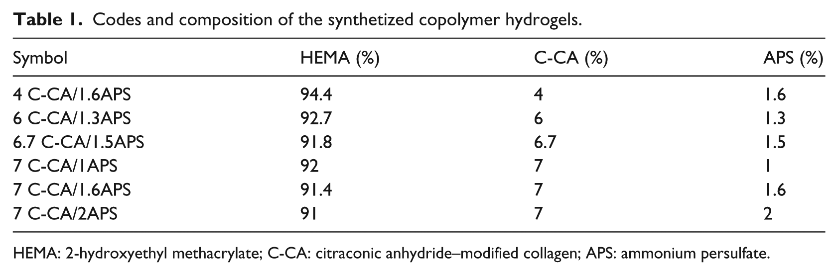
HEMA: 2-hydroxyethyl methacrylate; C-CA: citraconic anhydride–modified collagen; APS: ammonium persulfate.
Fourier transform infrared spectroscopy
The Fourier transform infrared (FT-IR) spectra were recorded in potassium bromide (KBr) pellets using a Bruker Vertex 70 spectrophotometer (Karsruhe, Germany) in the range of 400–4000 cm−1 (resolution of 4 cm−1, 100 scans), at ambient temperature. To prepare the pellets, 500 mg KBr was mixed with 5 mg of the sample in powder form. The processing of spectra was achieved using OPUS software version 5, developed by Bruker, Germany.
Scanning electron microscopy
The morphological aspects of investigated samples were visualized with a QUANTA 200 scanning electronic microscope with integrated EDS system, GENESIS XM 2i EDAX with SUTW detector (FEI Company, USA), the magnification being indicated on the figures. Before test, the drug-loaded hydrogels were freeze-dried and the degraded ones were washed with bidistilled water for 3 days and then dried by lyophilization. The fracture surface of the freeze-dried hydrogels was registered and examined. ImageJ software was used to investigate the porous aspect and to calculate the pore size distribution.
Determination of swelling degree
The hydrogel samples (30 mg) were immersed in buffer solutions of different pH values ranging between 3.0 and 12.0 at 37 °C and periodically weighed after removing excess water from the surface of the gel with a filter paper. The experiments were performed in triplicate. The swelling degree (SD) was calculated using the following equation (1)
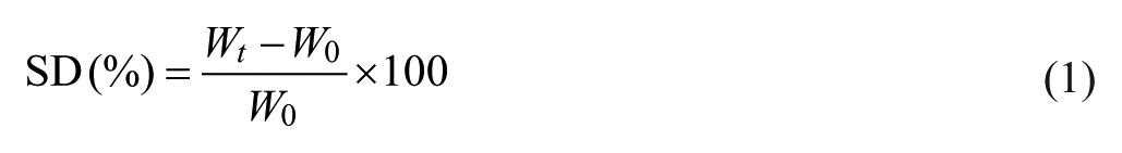
where Wt is the weight of the swollen gel sample at time t and W0 is the initial weight of the dry gel samples submitted to swelling.
To establish the swelling kinetics of the cryogels in the buffer solutions of different pH values, equation (2) was used
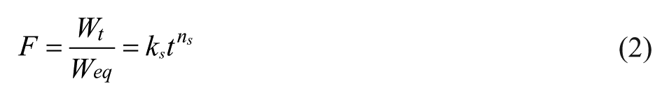
where Wt is the mass of the swollen sample at time t, Weq is the mass at equilibrium taken from swelling profiles as maximum mass of swelled hydrogels, ks (min−n) is the swelling rate constant describing the rate of swelling, and ns is the power diffusion law exponent which takes into account the type of solvent transport. Equation (2) applies to the initial states of swelling (with SD less than 60 %) where the linearity of ln(Ft) as a function of ln(t) is obtained.
The in vitro testing of enzymatic degradation on hydrogels
The in vitro degradation was analyzed by photometric ninhydrin method. The capability of hydrogels to degrade on exposure to enzymes was studied using collagenase. The ninhydrin reagent was prepared by dissolving 0.8 g SnCl2·2H2O in 500 mL 0.2 M citrate buffer solution (pH 5) to give a 0.007 M SnCl2 solution, followed by addition of 500 mL 2-methoxyethanol containing 20 g (4 % (w/v)) of crystalline ninhydrin. The mixture was purged with nitrogen and stored in dark bottles for up to 1 week at 4°C.
Hydrogel scaffolds and native collagen of 0.045 g were measured and incubated at 37°C in 30 mL of 0.1 M phosphate buffer solution (pH 7.4) containing 0.005 % collagenase (C. histolyticum). At predetermined time intervals, 0.5 mL of sample solution was withdrawn and replenished with fresh phosphate-buffered saline (PBS) solutions containing the same amount of collagenase. The sample solution was mixed with 2 mL ninhydrin reactive and heated in a boiling water bath for 20 min at 100°C. After cooling down, 2.5 mL of distilled water–2-propanol (1:1 v/v) was added, and the colorimetric determination was performed measuring the absorption at 570 nm (Cary 60 UV–VIS spectrophotometer; Agilent Technologies, Santa Clara, CA, United States) within 1 h. Three samples of each group of hydrogels were evaluated, and the average value was recorded. The degradation degree (DD) was estimated using the calibration curve of gelatin (concentration range 0.015 %–0.115 %).
CF hydrochloride loading
The applicability of HEMA/C-CA hydrogels as carriers for controlled delivery of active principles in pharmaceutical and medical fields was tested using CF hydrochloride. Drug-loaded hydrogels were prepared by mixing the drug (5 wt % of the hydrogels weight) with the dried matrices in powder form, followed by swelling at room temperature for 1 h in a phosphate buffer solution (pH = 5.5) while the drug penetrates and/or attaches to the matrices. The quantity of added buffer solution for each formulation corresponds to the maximum quantity of liquid uptake measured during swelling experiments. The obtained bioactive compounds were freeze-dried for 24 h using a Labconco FreeZone device (Kansas City, USA) and then were used for the dissolution experiments.
In vitro controlled release of CF from hydrogels
The in vitro dissolution profiles of the drug-loaded hydrogels were carried out using an Agilent 708-DS Dissolution Apparatus coupled with a Cary 60 UV–Vis spectrophotometer. Each hydrogel specimen, with a mass of 0.02 g, was placed in a basket support and immersed in a phosphate buffer release medium of 100 mL with pH values of 3.0, 4.3, 5.5, 7.4, or 10.1 or 12.0. The release medium at 37°C was stirred with 100 r/min. Aliquots (10 mL) of the dissolution medium were withdrawn at predetermined time intervals and were assayed spectrophotometrically at λmax; the wavelength of the maximum absorbance characteristic for CF hydrochloride in phosphate buffer of different pH values is as follows: 278 nm for pH = 3.0 and 3.4, 275 nm for pH = 5.5, 270 nm for pH = 7.4, 271 nm for pH = 10.1, and 272 nm for pH = 12.0. The amount of active drug released by swelling and diffusion from the hydrogel matrices was determined according to the calibration curve of CF hydrochloride monohydrate absorbance versus concentration in ultraviolet–visible (UV–Vis) range. The experiments were performed in triplicate.
To find out the mechanism of drug release, first 60 % drug release data were fitted in Korsmeyer–Peppas model27,28 (equation (3))
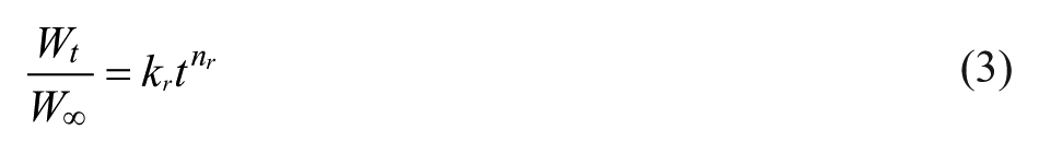
Wt/W∞ represents the fraction of the drug released at time t; Wt and W∞ are the absolute cumulative amount of drug released at time t and the maximum amount released in the experimental conditions used, respectively, at the plateau of the release curves; kr is the release rate constant incorporating the characteristics of the macromolecular drug-loaded system; and nr is the release exponent, which is indicative of the release mechanism. In the equation above, a value of n ≤ 0.5 indicates a Fickian diffusion mechanism of the drug or solvent, while a value 0.5 < n < 1 indicates an anomalous or non-Fickian behavior. When n = 1, a case II transport mechanism is involved with zero-order kinetics, while n > 1 indicates a special case II transport mechanism. 29
Antimicrobial activity
The antimicrobial activity of the samples was tested against standard strain Staphylococcus aureus ATCC 25923 by the disc diffusion method in Mueller–Hinton agar, according to the guidelines recommended by the National Committee for Clinical Laboratory Standards (NCCLS). 30
The cylinder in plate bioassay technique was used. A standard suspension of standard bacteria was prepared from fresh overnight culture and was mixed with 15 mL portions of molten Mueller–Hinton agar in a sterile Petri plate resulting in a final concentration of about 106 cells/mL. When the plates were solid, metal cylinders (6 mm diameter) containing samples (10 mg hydrogel with 5 % CF) were placed on the seeded agar surface. A standard commercially disc of CF (30 mg) was used for comparison.
Antibiotic standard
The antimicrobial agent delivered from the hydrogel samples in the seeded agar was determined by comparing the size of zone diameters of inhibition with those of a series of standard antibiotic concentrations by the test bacteria. The antibiotic diffuses directly against bacteria, and the production of the zone of inhibition around a source of antimicrobial agent is directly proportional to the amount of antibiotic present in that source. Alternatively, from the stack CF solutions, a standard antibiotic concentration was made; the dilution range should be such that it covers the levels found in the samples. The cylinders are then filled with standard dilutions of antibiotic of known potency. After overnight incubation at 37°C, the diameter of each inhibition zone around the wells was measured. The values of diameter of the inhibition zones are expressed as the mean of triplicates.
A dose–response curve of the standard antibiotic is made, and the average diameter of each sample is projected to the standard line. The level of antibiotic in the samples can then be read by reference to the graph.
In vivo biocompatibility studies by subcutaneous implantation
Ethics statement
The experimental research protocol was approved by the local Animal Ethics Committee of the “Grigore T. Popa” University of Medicine and Pharmacy, Iaşi, Romania, in strict observance of the international ethical regulations on laboratory animal work (AVMA Guidelines on Euthanasia, 2007). Biocompatibility tests were performed according to the “Grigore T. Popa” University of Medicine and Pharmacy guidelines for handling and use of experimental animals and in accordance with the recommendations and policies of the International Association for the Study of Pain. 31
The in vivo biocompatibility studies of obtained hydrogels consisted of the determination of the hemodynamic, immune, and biochemical profile of laboratory animals (white Wistar rats weighing between 280 and 300 g uniformly distributed by sex, from Public Health Institute, Iaşi, Romania). The rats were housed in standard conditions of laboratory being maintained at a constant temperature of 21°C ± 2°C on a 12-h dark cycle (light period, 07:00–19:00) and a relative humidity of 50 %–70 %. Food and water were available at all times except during the experiments. To avoid chronobiological influences, the tests were performed in the interval between 8 and 12 a.m. The animals were killed with ether immediately after each experiment. The unloaded and loaded hydrogel samples (60 mg) have been intraperitoneally delivered as pellets to rats which were divided into 14 groups of six animals, for 7 days. The control rat groups received a dose of physiological serum without pellet (M) (0.5 mL/100 g body)—Group I, sterile cotton pellets and physiologic serum (Mp) (0.5 mL/100 g body)—Group II, or sterile cotton pellets and prednisone (PDN) (5 mg/kg body)—positive control Group III. Hydrogels without drug were implanted to the next five groups as follows—Group IV: 4C-CA/1.6APS, Group V: 6C-CA/1.3APS, Group VI: 7C-CA/1APS, Group VII: 7C-CA/1.6APS, and Group VIII: 7C-CA/2APS. Group IX (control) received a dose of sterile cotton pellets and physiologic serum with 3 mg/kg body CF (M-CF). The following five groups (X–XIV) received the same types of hydrogels in the same order but loaded with 3 mg of CF.
For in vivo biocompatibility investigation, granuloma test in rats was performed. On the eighth day, the pellet surrounded by new tissue formed by inflammatory reactions (granuloma) was excised, dried at 57°C, and weighed. The granuloma weight was determined by decreasing the amount of initial pellet before implantation. Blood samples were taken from retro-orbital plexus to assess leukocyte formula and the following immune parameters: phagocytic capacity of peripheral neutrophils (NBT test) and serum complement activity. Also, hepatic transaminases were evaluated: transaminase glutamate pyruvate (TGP), transaminase glutamic oxaloacetic (TGO), and lactate dehydrogenase (LDH).
The histological images of granuloma tissue after contact with cotton and polymeric matrices were collected and analyzed using optical microscopy. The optical images are acquired on a reflected light bright field microscope Leica DM 2500 (Leica Microsystems GmbH, Wetzlar, Germany), at a magnification across the sample surface of 50×, with a 3.3 Mpix Leica DFC320 R2 digital camera (with a resolution of 2088 × 1550 pixels) mounted on the trinocular head.
Statistical analysis
Statistical analysis of the data was performed using the GraphPad Prism software (version 5.0). Statistical significance of differences between data was evaluated by one-way analysis of variance (ANOVA) using the Tukey test. A p-value of <0.05 was considered significant. Data were expressed as mean ± standard error of mean.
Results and discussions
Hydrogels’ obtaining and their characterization
Many possible chemical and physical reactions can occur during hydrogels’ preparation. Among them, the chemical grafting reaction on the modified collagen chains of HEMA and the homopolymerization of HEMA monomers were detailed in a previous study. 24 In Scheme 1, the formation of HEMA–C-CA copolymer and crosslinking by a chemical joint between the active carboxyl groups (COOH) of modified collagen macromolecules and the reactive hydroxyl groups (OH) of HEMA in hydrogels is presented.
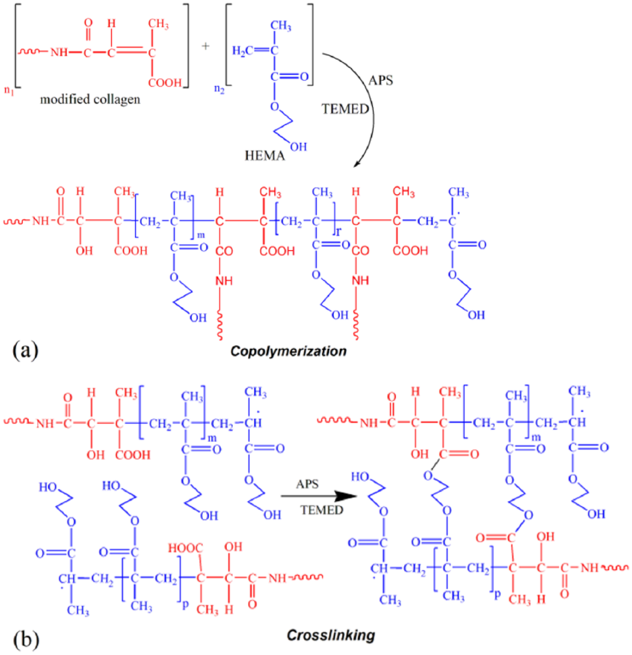
(a) Possible reactions between HEMA and CA-modified collagen and (b) chemical crosslinking of formed copolymers in hydrogels formation.
FT-IR studies revealed the chemical polymerization of modified collagen with HEMA monomer. Figure 1 illustrates the FT-IR spectral details of C-CA, PHEMA, and hydrogels based on HEMA and C-CA.
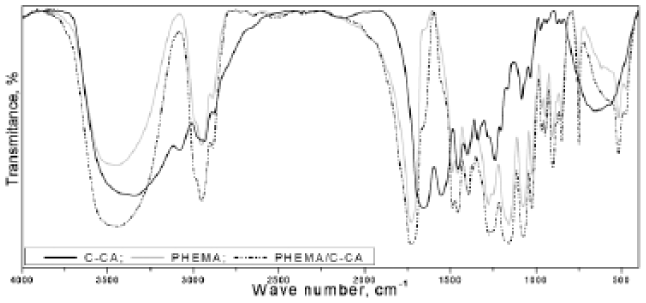
FT-IR spectra of modified collagen (C-CA), PHEMA, and representative HEMA/C-CA hydrogels.
In the hydrogels’ spectra, few significant changes were observed by interference between the bands of PHEMA and collagen. A broad, strong absorption in the region of 3655–3178 cm−1 resulted from superimposed –OH (from pHEMA) and –NH3+ (amide A from collagen) stretching band. In the same way, Amide I band (νC=O) from collagen appearing at 1656 cm−1 interferes with the band from 1650 cm−1 attributed to the C=C vibration of PHEMA. As a result of this, a shoulder at 1666 cm−1 is observed in the hydrogels spectra. Amide II band (δNH) characteristic for collagen is observed at 1550 cm−1. This absorption is found in the hydrogels’ spectrum at 1540 cm−1 as a shoulder, and the absorption band at 619 cm−1 from PHEMA disappeared in hydrogels spectra.
The yield in the hydrogels varied between 74.9 % and 89.3 % meaning that the mass conservation during the chemical reactions is high proving the copolymerization and crosslinking occurrence between the polymeric components. The obtained hydrogels are very stable in aqueous and buffer media of different pH values ranging from acidic to alkaline and consequently present adequate strength for their use as wound dressings.
The porous morphology of hydrogels revealed in the scanning electron microscopy (SEM) images (see Figure 7) was acquired via freeze-drying of the hydrogels swollen in deionized water and via stabilization of the three-dimensional polymeric network gained after the free radical copolymerization process. Their morphology is characterized by interconnected pores of various sizes and distributions (Figure 8) which mainly depend on the degree of crosslinking.
Swelling tests
As the new materials are intended to be used in the tissue engineering field, the capacity to absorb body fluids and transfer nutrients represents a key element. We have studied the swelling capacity of all types of developed hydrogels in PBS 7.4 at 37°C, while monitoring the weight change at pre-established time intervals.
According to their swelling behavior (Figure 2) and SEM images (see Figure 7 and Table 2), the obtained polymeric systems may be classified as super-porous hydrogels with high porosity and interconnected open pore structure. Most of the pores inside the hydrogels are connected to form the open channel system, which acts as a capillary system causing a rapid solvent uptake into the porous structure. 32 The first impact of the hydrogels with the buffer solution produced a very fast swelling in a matter of a minute due to the absorption of solution by capillary force rather than by simple absorption. The solvent uptake degree of the hydrogels reached the equilibrium within 2–3 h.
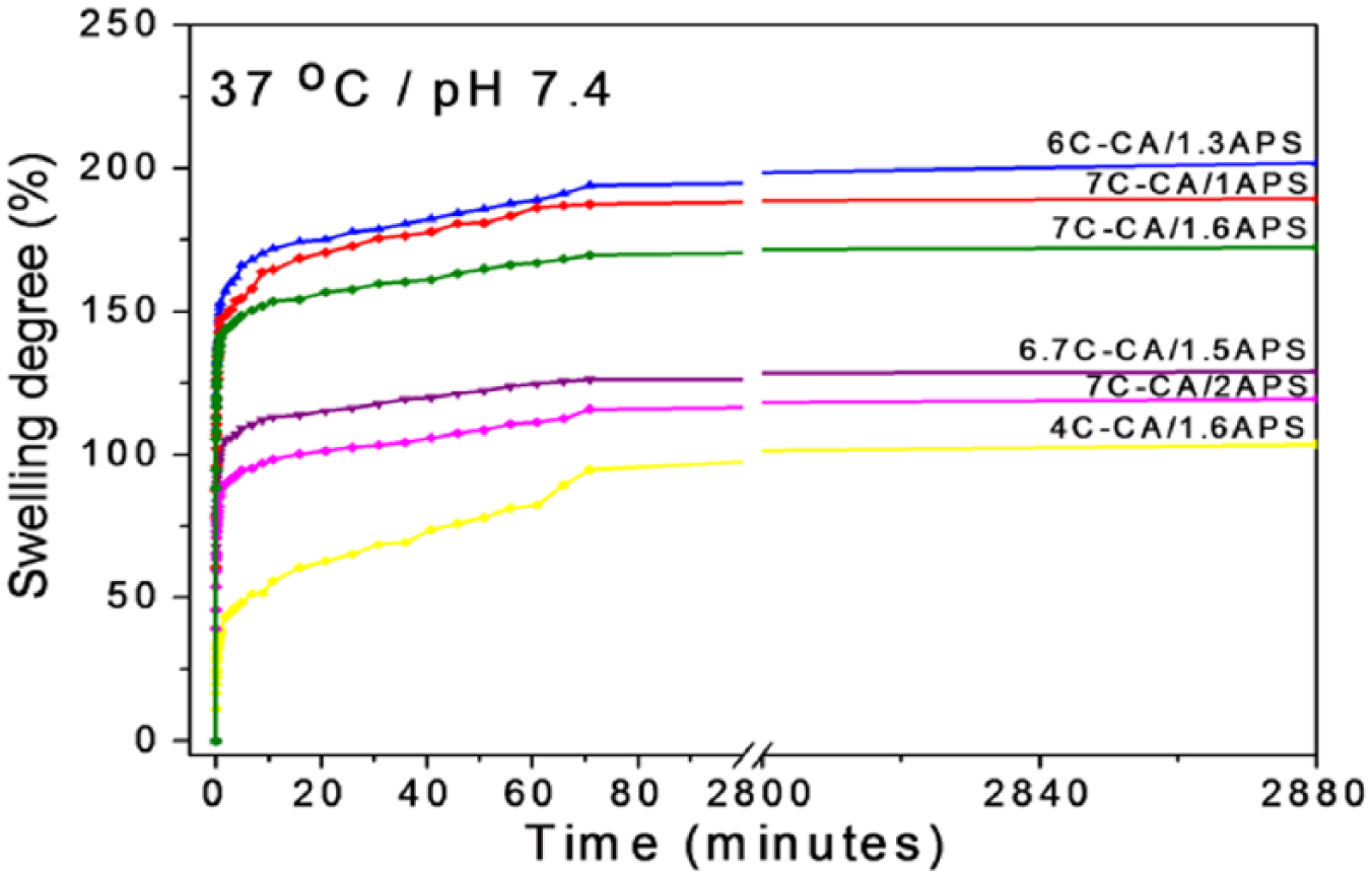
Swelling profiles of HEMA/C-CA hydrogels at pH 7.4.
Effect of reaction conditions on SD and pore size distribution of HEMA/C-CA hydrogels.
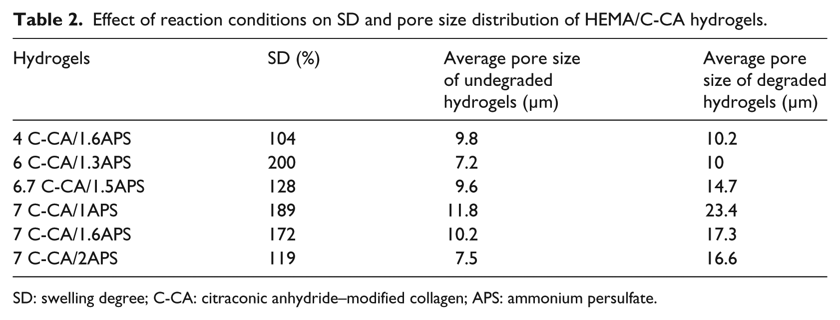
SD: swelling degree; C-CA: citraconic anhydride–modified collagen; APS: ammonium persulfate.
The presence of different amounts of modified collagen and APS in the synthesis of hydrogels will lead to the formation of pores with different dimensions in the hydrogel matrices which will influence the SD. A higher density associated with less flexibility and poor elasticity was observed in the formulations with a decreased percent of C-CA (4C-CA/1.6APS with SD = 104 %). The other copolymer hydrogel samples show higher SD. This behavior can be caused by the hydrophilic functional groups coming from the modified collagen moieties. 33
The swelling behavior of the bicomponent hydrogels was significantly influenced by the concentration of the APS initiator used in the initial recipe. Thus, the presence of an increased initiator content in the polymeric network (up to 2 % APS) will induce a decrease in the average pore size from 11.8 to 7.5 µm and consequently will decrease the absorbent character of the hydrogels (189 % compared to 119 %; Table 2) due to the intensifying of interpolymeric interactions which will cause a higher densification of polymer network and diminishes the solvent penetration inside the network.
The values of the swelling exponent ns, exposed in Table 3, are below 0.5 indicating a special case of Fickian diffusion usually called as “less-Fickian,” “quasi-Fickian,” or “pseudo-Fickian” transport mechanism.34,35 This situation is associated with a solvent penetration rate slower than the polymer chains’ relaxation rate. The 4C-CA/1.6APS and 7C-CA/2APS hydrogels registered the smallest value of ns exponent of 0.073 and 0.064, respectively, meaning that the relaxation of polymeric chains is dominated by rather less flexible structure of polymer matrix because of the presence of a low content of modified collagen (4 % C-CA) or of a high content of initiator (2 % APS). The swelling rate constant ks value corresponding to 4C-CA/1.6APS hydrogel was the smallest and this result is correlated also with the most decreased swelling degree, phenomenon also well observed in Figure 2.
Kinetic parameters (ns and ks) of swelling behavior for collagen-based hydrogels.

ns: swelling exponent describing the mode of the transport mechanism; ks: swelling rate constant; Rns2 and Rks2: correlation coefficients corresponding to ns and ks, respectively; C-CA: citraconic anhydride–modified collagen; APS: ammonium persulfate.
These findings concerning the swelling behavior represent the first proof that the synthesis of the hydrogels was successful and they also demonstrate that the materials present stability at 37°C in phosphate buffer solution. Also, the HEMA/collagen-based hydrogels characterized by fast swelling and high swelling ratio confirmed their potential use as more hydrated and flexible biomaterials for tissue engineering applications.
Dependence of the swelling behavior on pH
Many anionic, cationic, zwitterionic, and non-ionic hydrogels show pH-responsiveness, 36 and these kinds of hydrogels are very useful in drug delivery application. In this study, zwitterionic C-CA was chosen to obtain pH-responsive HEMA-based hydrogels. To investigate the influence of the environmental conditions on the swelling behavior of HEMA/C-CA hydrogels, the following selected hydrogels with different compositions, namely, 4C-CA/1.6APS and 6.7C-CA/1.5APS, were immersed in buffer solutions with different pH values ranging from 3.0 to 12.0 (Figure 3).
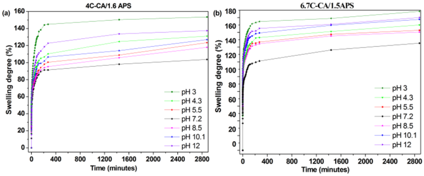
Dependence of hydrogels’ swelling profiles on pH of the medium indicated in figure legend.
Collagen is a well-known natural protein which has pKa values for both amino groups and carboxylic groups. The average pKa for the C-CA functional groups was determined by potentiometric titration method in a previous work 23 as being 4.7 for COOH groups and 10.0 for NH2 groups. According to these findings, the pH responsiveness of the tested porous hydrogels was evaluated. Hence, it was observed that HEMA/C-CA hydrogels showed an “U”-shaped pH-swelling profile (Figure 4), with good swelling properties at pH values below 4.7 and above 10.0, and poor swelling properties close to neutral pH. Below pH 4.7, the increase in SD of hydrogels is caused by the ionization of all the amino groups, and above pH 10, the increase in SD is due to the ionization of all carboxyl groups which will cause the increase in the electrostatic repulsion between negatively charged carboxyl groups. 37 In addition, at the pH value of 3.0, the SD of the 4C-CA/1.6APS and 6.7C-CA/1.5APS hydrogels was higher (153.5 % and 188.4 %) than those at pH 12.0 (137.4 % and 180.2 %), probably because of the presence of a low amount of free carboxyl groups after hydrogels’ synthesis which were reacted with HEMA monomers during copolymerization and crosslinking reactions.
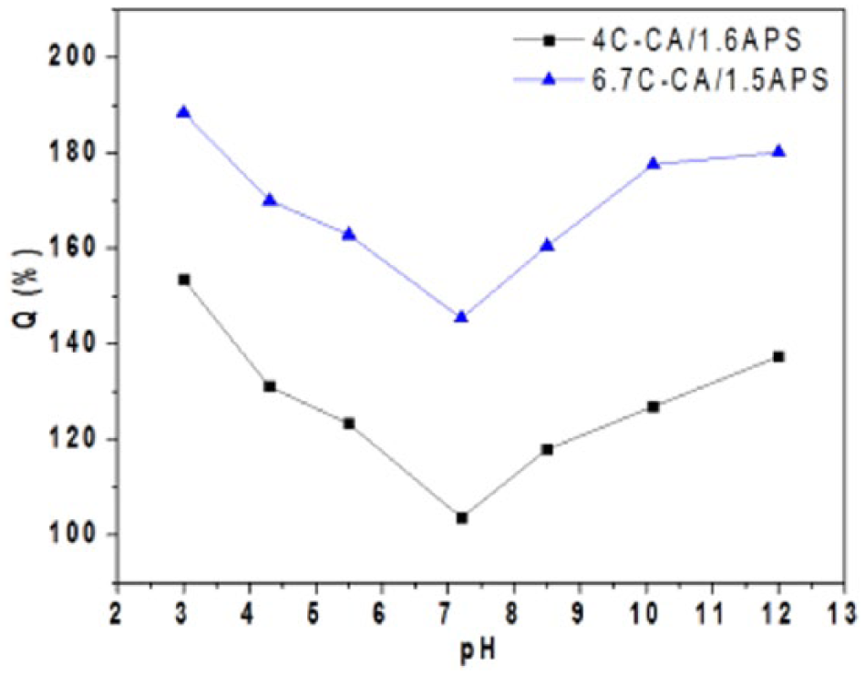
Dependence of the equilibrium swelling degree of hydrogels on the pH of the swelling media.
The lowest SD was recorded by immersing hydrogels in buffer solution of pH 7.2 which is close to the isoelectric point (P.I.) of modified collagen (P.I. = 7.3) where there is a balance between the absorption and release of protons. Thus, at this pH, the 4C-CA/1.6APS and 6.7C-CA/1.5APS investigated samples reached an equilibrium SD of 103.6 % and 145.4 %, respectively.
In Figure 5, the variation in the kinetic parameters (ks: swelling rate constant and ns: swelling exponent) values for 4C-CA/1.6APS and 6.7C-CA/1.5APS copolymer hydrogels at different pH values of the swelling media is given. The ns parameter took values ranging from 0.047 to 0.104, which are lower than 0.5 and this case was still regarded as Fickian diffusion called “less Fickian” behavior as already mentioned above and also in case of the swelling of other copolymer hydrogels.38,39
It was also remarked from the kinetic swelling data (Figure 5) that at extreme pH values, namely, at 3.0 and 10.0, swelling exponent ns registered the highest values of 0.086–0.082 or 0.104–0.083 for 4C-CA/1.6APS and 6.7C-CA/1.5APS, respectively. It can conclude that the sweilling of these hydrogels at pH 3.0 and 10.0 is more diffusion-controlled. The ks values increase at both extreme pH values, e.g. in acidic being of 0.149 or 0.248 min−n and in alkaline 0.171 or 0.283–0.274 min−n for 4C-CA/1.6APS and 6.7C-CA/1.5APS, respectively, and then they decrease at neutral pH the values of 0.137 or 0.237 min−n for 4C-CA/1.6APS and 6.7C-CA/1.5APS, respectively, are evaluated. These changes in ks values at particular pH values which are associated with the change in degree of ionization of the functional groups of C-CA highlight the pH-responsive character of the obtained hydrogels. The variation in the ks values at pH 4.7, 10.0, and 7.3 is associated with the pKa values for carboxylic groups and amino groups of modified collagen and with its isoelectric point. It can be concluded that the swelling mechanism is changed around critical pH values of copolymer hydrogel which shows a pH-responsiveness for swelling, even by incorporation of small amount of C-CA.
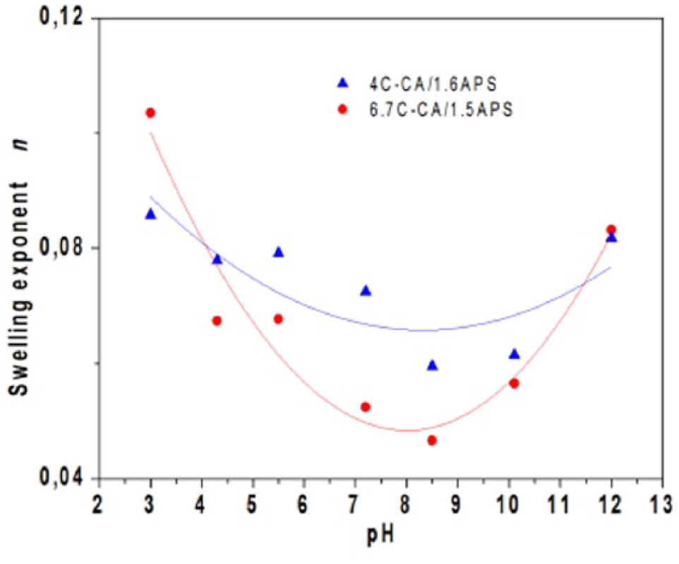
Variation in ns values on pH of the swelling solutions.
Resistance of HEMA/C-CA hydrogels to collagenase degradation
Understanding and control of the degradation of PHEMA is important for its wide range of applications. PHEMA is degradable only in extreme conditions. 40 Control of degradation is achieved by crosslinking 41 and copolymerization 42 with other components. PHEMA–peptide conjugates did not undergo any significant degradation when incubated with papain. 43 To study the in vitro degradation of polymeric hydrogels, samples were exposed to the bacterial collagenase solutions. Therefore, the degradation of hydrogels occurs mainly by collagenase action onto modified collagen which is distributed in bulk of network. The collagenase extracted from the anaerobic bacteria C. histolyticum cleaves the main body helical polypeptide chains of collagen under physiological conditions, resulting in a wide range of degradation end-products. 44 Specifically, it cleaves at alanine and nonpolar amino acids adjacent to glycine within the triple helix of collagen. The degradation of hydrogels was evidenced in FT-IR spectra by disappearance of absorption band corresponding to amide II from modified collagen structure (Figure 6).
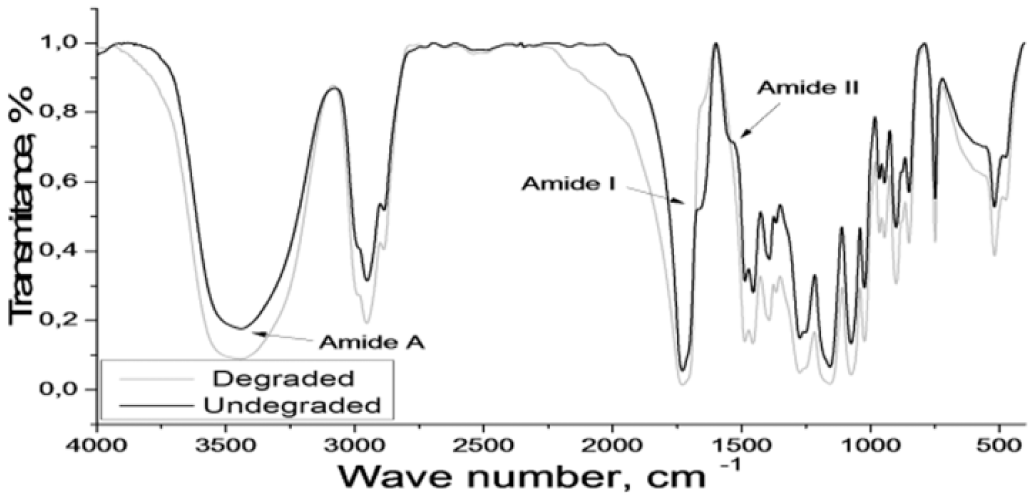
FT-IR spectra for 4C-CA/1.6APS before and after degradation process.
From SEM images (Figure 7), it can be observed that the enzymatic degradation of solid polymer matrices took place in the entire mass of sample and it affected the morphology by changing the pore sizes and their distribution (Figure 8). The diameters were determined on eight different representative images.
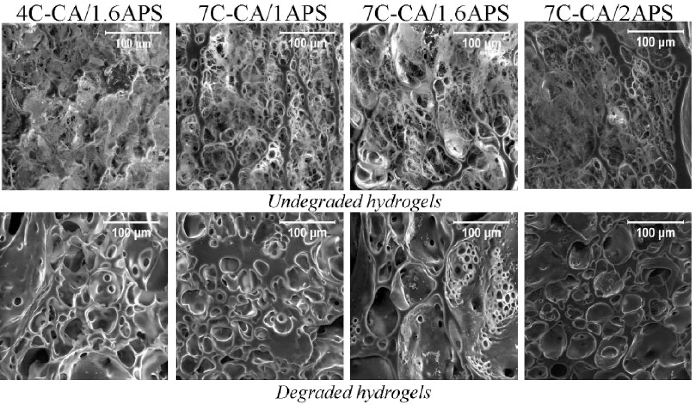
SEM images of same undegraded (top) and degraded (bottom) freeze-dried hydrogels; the scale bars from SEM images represent 100 µm.
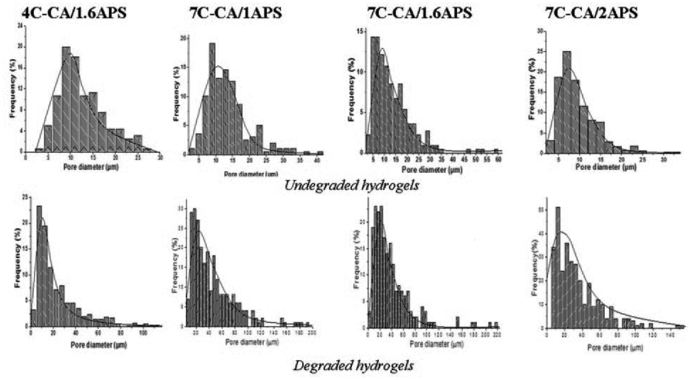
Pore size distributions of same undegraded (top) and degraded (bottom) freeze-dried hydrogels.
By comparing the aspect of the undegraded and degraded hydrogels with various compositions, it can be observed that the pores become larger after degradation and they are not more interconnected probably because the collagenous component was eliminated by enzymatic degradation. A wider pore size distribution (Table 2) with an average diameter between 10.2 and 23.4 µm after hydrogels’ degradation process than before when the average diameter ranged from 7.5 to 11.8 µm was found. In view of the different contents ranging between 1 % and 2 % of APS present in the initial recipe, it was possible to achieve different pore size distributions where a smaller average diameter of pores before and after degradation was observed with the increase in APS amount in the initial recipe which suggests an increase in the resistance to collagenase degradation.
After degradation process, the porosity of hydrogels changed. The enzymatic disintegration of collagen matrix generated the formation of smaller pores in the walls that surround the large pores. The pore size and pore size distribution of polymeric hydrogels influence their behavior. 45 This complex morphological architecture gained by degradation can be useful in tissue engineering. Lesný et al. 46 and Přádný et al. 47 observed that cell growth in the gel scaffold was reduced or even stopped after a period of time, due to an increasingly limited nutrient supply when the large pores became blocked with cells, suggesting that pore wall permeability to nutrient molecules was a key issue.
In general, the degradation rate of the scaffold should match the rate of tissue formation, or at least be comprised within an optimal range, since a too slow degradation would interfere with remodeling and a too fast degradation would lead to premature scaffold resorption. 48 The hydrogels are not fully degradable because only the collagen portion of the scaffold was digested by collagenase. Accordingly, even though the rate of degradation is considered to be fast (few days), their semi-biodegradability caused by the HEMA component makes them suitable for potential biomedical application in wound healing as partial dressings. This observation can be strengthened by some old studies which demonstrated that a part of the degradation products of collagen types I + III induce chemotaxis of human fibroblasts, 49 and such degradation is thought to promote restoration of tissue structure and functionality. 50 The variation in the DD during time for some HEMA/C-CA hydrogels is presented in Figure 9. The values of the maximum DD increased with C-CA content from 9.71 % to 12.05 % and decreased with increased crosslinking density (increased APS content) from 15.31 % to 7.27 %.
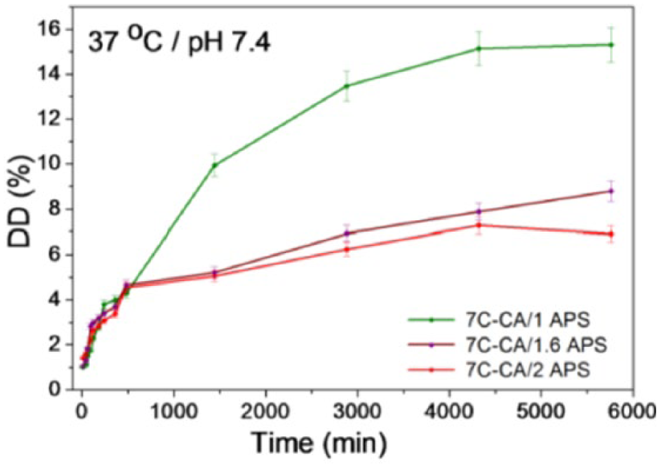
Effect of collagenase on degradation of some HEMA–C-CA copolymer hydrogels.
A dependence of DD on hydrogels’ composition (initiator content, collagen content) was observed. While the modified collagen amount controls the maximum DD, the APS influences both the rate of degradation and maximum DD which decrease with increasing APS amount in the initial recipe. Increasing concentration of the initiator agent (APS) along with the constant maintaining of collagen content leads to a higher density of network, consecutively decreasing water uptake and restricting the enzymatic degradation of the hydrogels. The 7C-CA/1APS hydrogel, with lowest initiator content (1 %), manifested the most accented DD (15.31 %). Increasing APS content to 2 %, the DD decreased at 7.27 %. According to the data of degradation results, it can be noted that the enzymatic DD is influenced by a competition between the contents of the initiator and the amount of modified collagen. As a conclusion, the most great resistance to collagenase degradation was accomplished by the hydrogels with 2 % APS, namely, for 7C-CA/2APS.
Drug release study
Characterization of CF-loaded hydrogels by FT-IR spectroscopy and SEM
The FT-IR spectra for CF-unloaded hydrogel and lyophilized CF-loaded hydrogel (Figure 10) confirm the presence of CF inside hydrogels. In FT-IR spectra of pure CF, a band at 3532 cm−1 attributed to the stretching vibrations of O–H belonging to carboxyl group was found. At 1706 and 1624 cm−1, the bands are associated with the stretching vibrations of the carbonyl group of carboxylic acid and ketone, respectively. The bands centered at 1449, 1271, and 1045 cm−1 are assigned to C–H bending or C–O, C–CO–C of ketone, and C–F bond vibration, respectively. 51 No significant differences were observed in the registered spectra of drug-loaded hydrogel, although a shift of O–H stretching to 3554 from 3532 cm−1 has been observed for antibiotic-loaded hydrogel indicating the interaction between the antibiotic and modified collagen–based hydrogel most likely through hydrogen bonding. 52 Also, this kind of interaction is the same reason for the shift of C=O stretching to 1632 from 1624 cm−1.
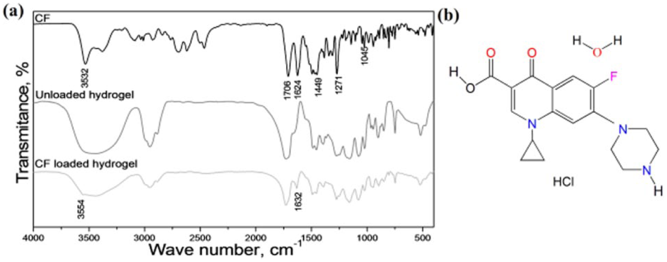
(a) FT-IR spectra of pure ciprofloxacin hydrochloride monohydrate, unloaded and loaded hydrogel and (b) chemical structure of ciprofloxacin hydrochloride monohydrate (CF).
Both FT-IR spectra (Figure 10) and SEM images (Figure 11) can conclude that the CF drug is present in the polymeric matrix of hydrogels. In SEM images of CF-loaded hydrogels (Figure 11), various crystal forms of the drug powder are present entrapped or not in the polymer matrix.
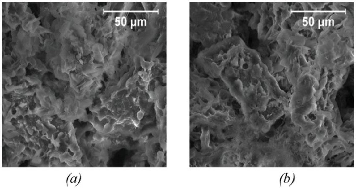
SEM images of (a) 4C-CA/1.6APS and (b) 7C-CA/1.6APS loaded with CF.
In vitro controlled release of CF from polymeric hydrogels
CF, a zwitterionic molecule containing two proton-binding sites, exhibits pH sensitivity. At 37°C, its pKa1 and pKa2 are 6.2 and 8.59, 53 respectively, and it shows a U-shaped pH-solubility profile, with good solubility at pH values below 5 and above 10, and poor solubility close to neutral. 54 It is semi-synthetic quinoline carboxylic acid. It belongs to DNA gyrase inhibitor pharmacological group on the basis of mechanism of action and also classified in antibiotics pharmacological group used in the treatment of various bacterial infections caused by Gram-positive and Gram-negative microorganisms.
The release behavior of CF from the hydrogels at 37°C was investigated in a PBS with pH of 7.4. Drug release profiles’ shapes vary with hydrogel compositions, especially with APS amount from the initial recipe (Figure 12).
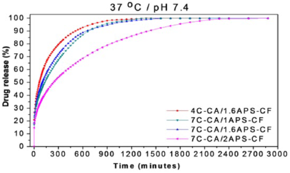
Drug release profile of ciprofloxacin from polymeric hydrogels at pH value of 7.4.
In the initial stage, the release rate of CF from hydrogels was high (few minutes) and is associated with the “burst” effect which is due to the fast swelling behavior of hydrogels or to the fraction of drug on or near the surface of the polymeric network. 55 Then a gradual release process that extends over several hours (up to 50 h) was recorded which could be attributed with the slow CF diffusion through the polymeric matrix. When the release process was complete, no drug was detected in the hydrogel and the total released amount (DR %) of antibiotic from the hydrogels ranged between 96 % and 100 %. Both the time to reach the maximum amount released (tmax) and the time for the drug concentration to decrease to one half of its original value (t1/2) increased with C-CA and APS amount in the initial recipe (Table 4) because of the entrapment of CF by hydrogen bonds through polar groups of all components of the loaded hydrogels. The presence of a higher amount of APS initiator used for hydrogels’ preparation allows the formation of more physical and chemical interactions which cause the maintaining of drug molecules as in a cage into the network slowing down their release in the simulated physiological environment. For example, samples 7C-CA/1APS-CF, 7C-CA/1.6APS-CF, and 7C-CA/2APS-CF have registered increasing t1/2 values in the following order: 102 min < 130 min < 241 min.
In vitro release characteristics of drug from investigated hydrogel materials at pH = 7.4 and their kinetic parameters.
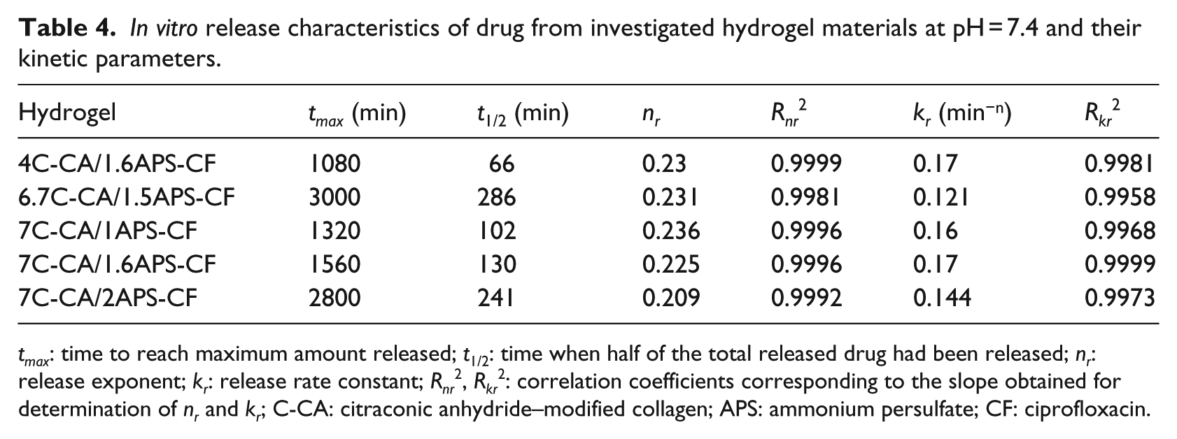
tmax: time to reach maximum amount released; t1/2: time when half of the total released drug had been released; nr: release exponent; kr: release rate constant; Rnr2, Rkr2: correlation coefficients corresponding to the slope obtained for determination of nr and kr; C-CA: citraconic anhydride–modified collagen; APS: ammonium persulfate; CF: ciprofloxacin.
The data of the in vitro drug release of CF were analyzed to establish the mechanism and rate of drug release by the kinetic parameters’ (nr and kr) evaluation (Table 4). The nr values ranging between 0.21 and 0.27 indicate that the release mechanism of CF from polymeric hydrogels follows a “less-Fickian” diffusion. The small values recorded by nr parameter in the basic medium (pH 7.4) are correlated with a slow drug release profile and are caused by the rate of polymer relaxation which is much greater than the rate of drug diffusion. The release rate constant kr values are approximately constant.
Dependence of drug release on pH
The dependence of CF release on pH from investigated hydrogels was evaluated at different pH values as follows: 3.0, 4.3, 5.5, 7.4, 10.1, and 12.0, and their release profiles are presented in Figure 13.
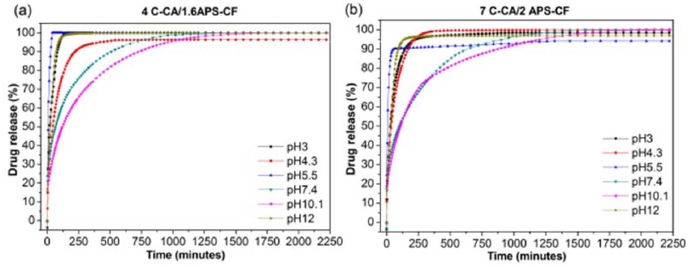
Drug release profile of ciprofloxacin from (a) 4C-CA/1.6APS-CF and (b) 7C-CA/2APS-CF hydrogels in media with different pH values ranging from 3.0 to 12.0.
A slower release of CF with a more sustained release behavior was registered for all hydrogels when the dissolution medium of pH 7.4 or 10.1 was used. In case of pH 7.4, this effect is caused by the decreased swelling properties of hydrogels close to neutral pH medium which will slow down the drug diffusion through the polymeric networks and also because of the poor solubility of CF. In case of pH 10.1, the reason for which the rate release is very slow is only due to the poor solubility of CF, because at this pH the SD of the hydrogels is more greater. The time to reach the maximum CF released amount (tmax) significantly increased up to 1080–2220 min in the pH interval 7–10 with respect to those at both low and high pH values, and therefore, the hydrogels exhibit a good pH sensitivity due to the C-CA presence (Figure 14).
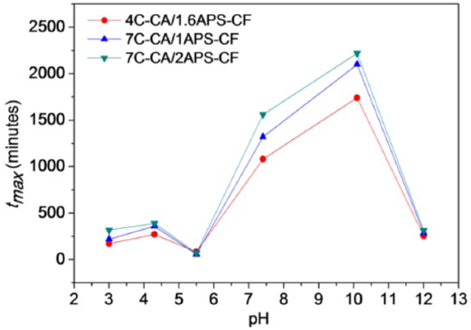
Influence of the pH values on the time to reach maximum released amount (tmax).
Instead, at the lowest and highest selected pH values of 3.0 and 12.0, the release equilibrium state was reached very fast after only 170–345 and 255–390 min, respectively (Table 5). Moreover, at pH 5.5, the CF release rate from the hydrogels was even faster than in conditions of pH 3.0 or 4.5 probably caused by the highest solubility of CF at this value of pH. 26
In vitro release characteristics of CF from investigated hydrogels at different pH media.
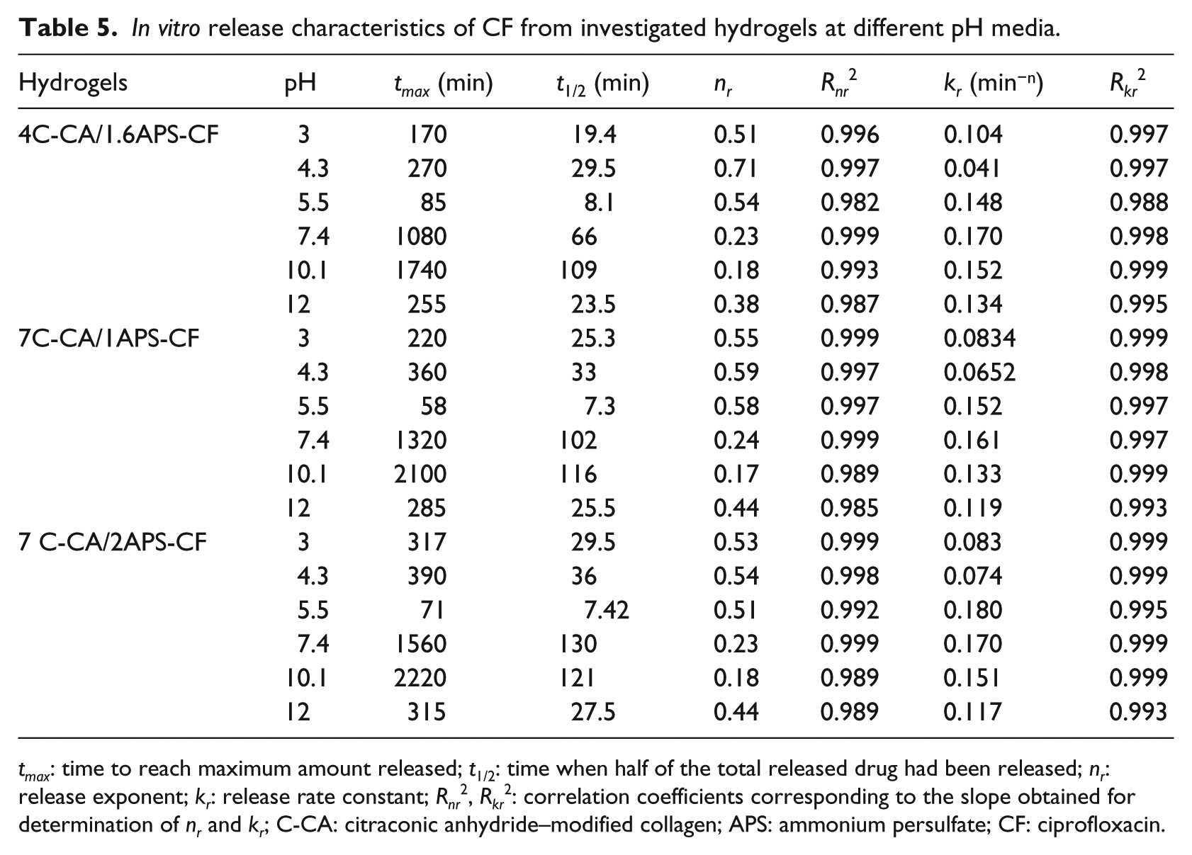
tmax: time to reach maximum amount released; t1/2: time when half of the total released drug had been released; nr: release exponent; kr: release rate constant; Rnr2, Rkr2: correlation coefficients corresponding to the slope obtained for determination of nr and kr; C-CA: citraconic anhydride–modified collagen; APS: ammonium persulfate; CF: ciprofloxacin.
Based on the kinetic parameter values (Table 5), the CF hydrochloride release behavior of the HEMA/C-CA hydrogels might be explained by a variation in the type of transport mechanism at pH fluctuation. At low pH (3.0, 4.5, and 5.5), the values of the release exponent are approximately 0.5 indicating a Fickian diffusion mechanism. Furthermore, with the pH raising to neutral and alkaline, the values of the release exponent nr ranged between 0.17 and 0.44 suggesting a “less-Fickian” diffusion mechanism. This change of the drug transport mechanism from Fickian to less-Fickian type which practically means a shift from diffusion to relaxation-controlled behavior demonstrates the pH-responsiveness of the CF release better illustrated in Figure 15(a).
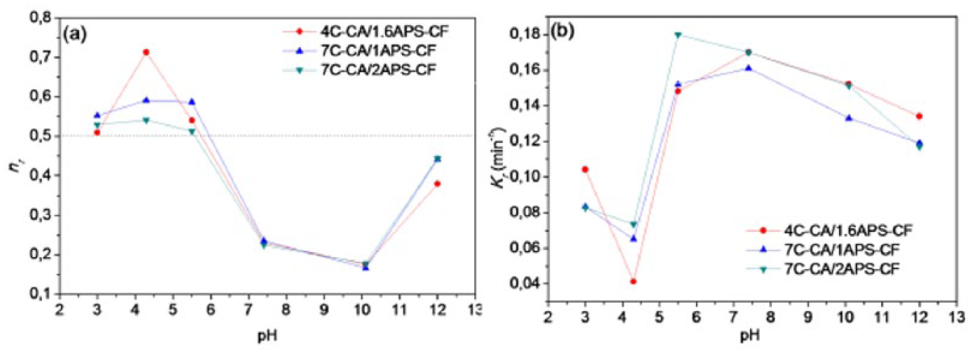
Influence of pH on kinetic parameters ((a) the release exponent: nr and (b) release rate constant: kr) resulting from CF release profiles.
The kinetic constant kr values are a little higher when the antibiotic release takes place in a neutral and low alkaline pH medium (from pH 7 to 10) comparing with the release in solution with acidic or basic pH (Figure 15(b)). The appreciable changes in release rate constant kr between 4.5 and 5.5 pH could be associated with the changes in degree of ionization of the functional groups of polymeric networks and also with their responsiveness to pH.
It is known that the pH of the skin surface of healthy adults and children ranged between 4.2 and 5.6. 56 When the pH is increased, the number of bacteria on the skin surface is increased. The pH environment of chronic and acute wounds has been recorded within the range of 7.15–8.9. 57 The CF release from HEMA/C-CA hydrogels was very fast at the pH level of the intact skin (pH 4.5 and 5.5).At pH 7.4, similar to the physiologic blood pH or to the pH environment of wounds, the release of CF from HEMA/C-CA-CF hydrogels is slower and this may represent a breakthrough in providing effective wound dressing materials for general wound infections that require a controlled drug delivery rate where the release of CF is pH dependent.
Antibacterial activity
The antibacterial activity of CF delivered from hydrogel samples was measured against Staphylococcus aureus ATCC 25923, and the results are presented in Table 6 and Figure 16.
In vitro activity of ciprofloxacin delivered from four samples of hydrogels against Staphylococcus aureus ATCC 25923.

C-CA: citraconic anhydride–modified collagen; APS: ammonium persulfate.
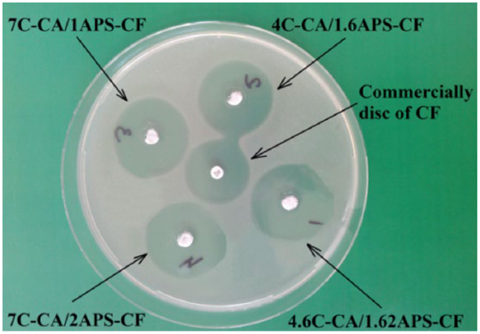
Microbiological assay of antibiotic levels delivered from tested hydrogel samples.
The CF standard of 50 µg/mL yields one value of 36 mm, 25 µg/mL yields one value of 32 mm, 12.5 µg/mL yields one value of 28 mm, and 6 µg/mL yields one value of 21 mm. The effect of CF release from samples 4.6C-CA/1.62APS, 7C-CA/1APS, and 7C-CA/2APS displays a very good efficacy against Staphylococcus aureus ATCC 25923 approximately equal to that of 50 µg CF standard. The sample 4C-CA/1.6APS was slightly less active than the other samples, with a zone diameter of 34.5 mm; this value is equivalent to the antibiotic concentration of about 46 µg. In this case, the release rate is 92 %.
Biocompatibility test results
The HEMA/C-CA hydrogel functions as a semi-biodegradable template that induces organized regeneration of dermal tissue (neodermis) by the body and the infiltration of fibroblasts, macrophages, lymphocytes, and endothelial cells that form a neovascular network. 58 The implantation of hydrogel pellets produced no significant changes in the weight of the animals compared with control pellets (Mp) after 7 days of experiment. The implant of pellet with PDN caused a decrease in the granuloma weight compared to control pellet. At subacute inflammation test by subcutaneous implant of pellets, CF-loaded hydrogels produced a statistically significant weight reduction in granuloma (*p < 0.05) compared with the pellets made of hydrogels without CF (Table 7). Compared to the effects caused by PDN, the effects of the CF-loaded hydrogel pellets on the decrease in the weight of granuloma are in descending order as follows
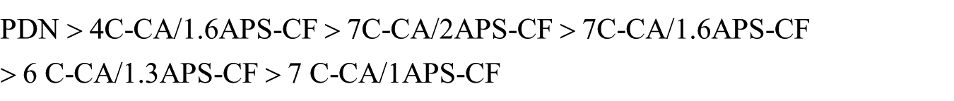
The effect of hydrogel pellets on granuloma weight after 7 days of implantation on rats.
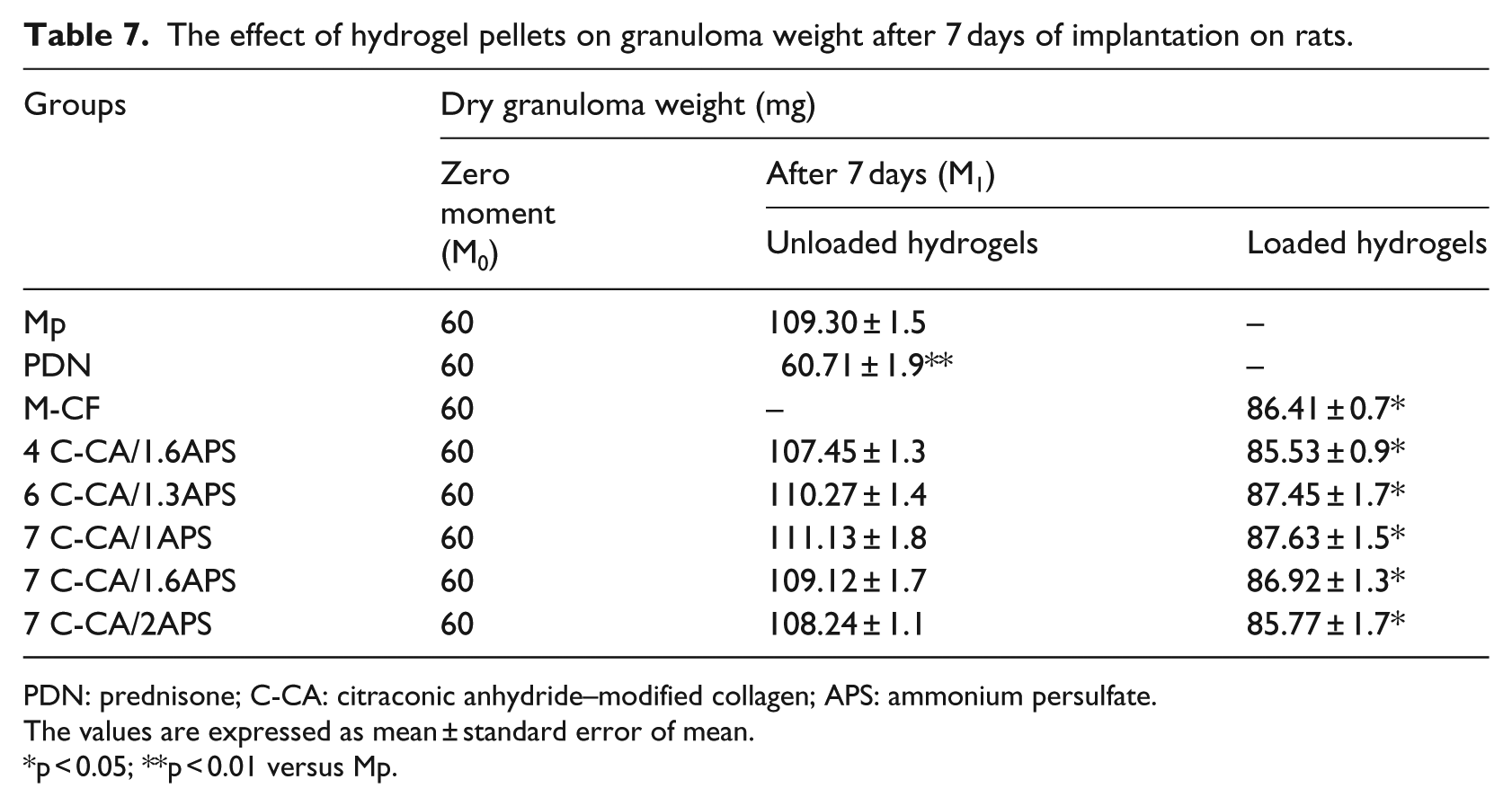
PDN: prednisone; C-CA: citraconic anhydride–modified collagen; APS: ammonium persulfate.
The values are expressed as mean ± standard error of mean.
p < 0.05; **p < 0.01 versus Mp.
The in vivo biocompatibility of investigated hydrogels was evaluated by hematological and immune system parameters and hepatic enzymes’ level comparatively with control groups of rats (Table 8). Comparing to the unloaded hydrogels, the CF-loaded hydrogels significantly inhibited the subacute inflammatory reaction according to the following results: the loaded hydrigels led to more reduced dry granuloma weight, to lower polymorphonuclear neutrophils and lymphocytes’ percentages, and significant decrease in phagocytic capacity of polymorphonuclear neutrophils in peripheral blood.
Leukocyte formula, hepatic enzymes, and immune parameters in control groups and in groups treated with polymeric pellets with and without CF after 7 days.
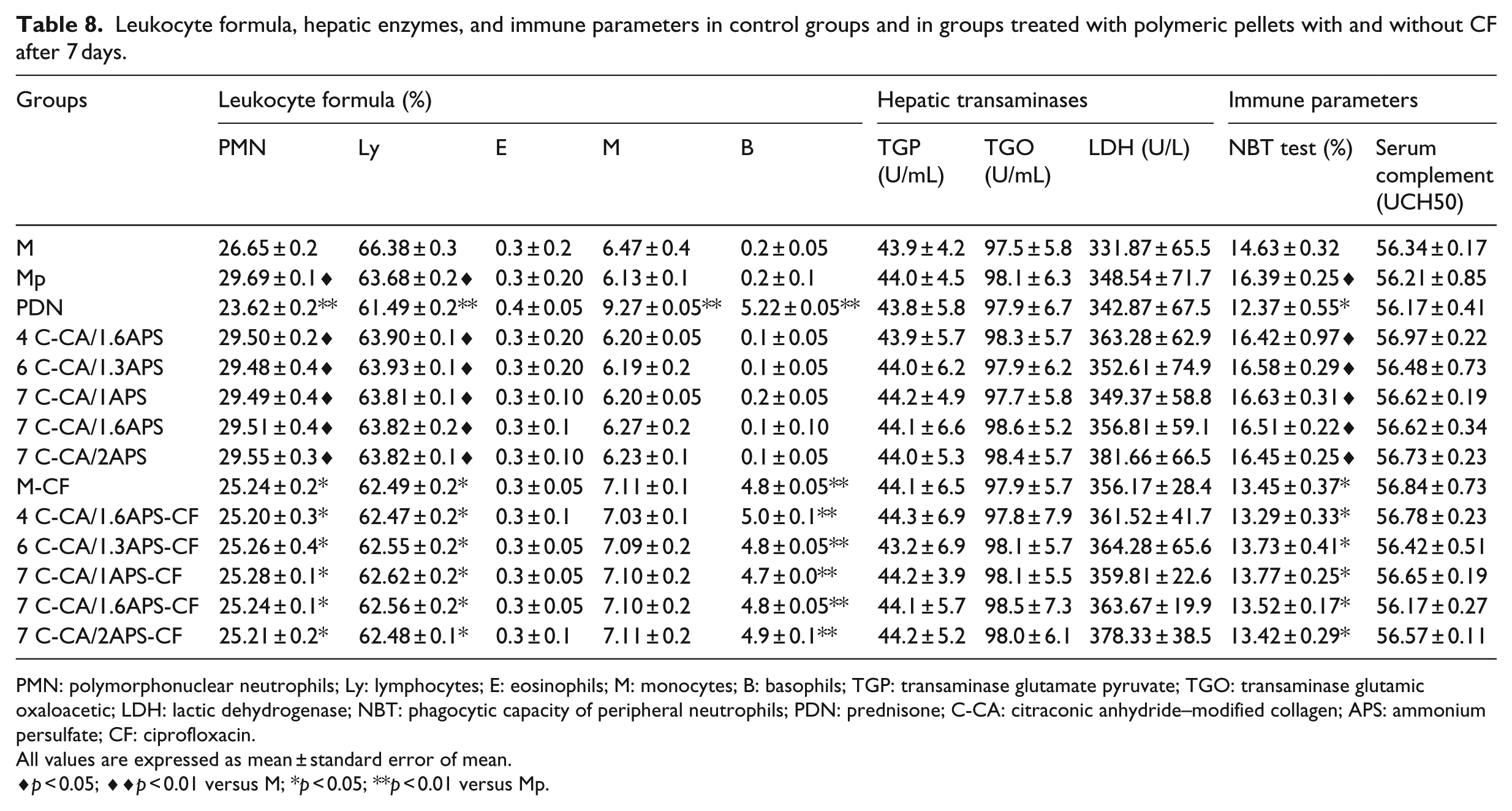
PMN: polymorphonuclear neutrophils; Ly: lymphocytes; E: eosinophils; M: monocytes; B: basophils; TGP: transaminase glutamate pyruvate; TGO: transaminase glutamic oxaloacetic; LDH: lactic dehydrogenase; NBT: phagocytic capacity of peripheral neutrophils; PDN: prednisone; C-CA: citraconic anhydride–modified collagen; APS: ammonium persulfate; CF: ciprofloxacin.
All values are expressed as mean ± standard error of mean.
p < 0.05; ♦♦p < 0.01 versus M; *p < 0.05; **p < 0.01 versus Mp.
The values of the hepatic enzymes (TGP, TGO, LDH) are presented in Table 8. After 7 days from experimental granuloma induced with pellets based on hydrogels with or without CF, no significant modifications of TGP and TGO values were observed, as compared with both control group with subcutaneous sterile cotton pellets and control group without pellets. Following the LDH enzyme, the results have shown a small variation in its value in animals with hydrogel pellets as compared with the control group M, but in comparison with the Mp control no significant variation was observed.
The absence of significant variations in the values of leukocyte formula, hepatic transaminases, lactic dehydrogenase, and immune parameters attests the good biocompatibility of the HEMA/C-CA hydrogels and can be used as biomaterials in tissue engineering.
The sterile cotton pellets’ (Figure 17(a)) implantation determined an acute local inflammation with giant cells, macrophages, and lymphocytes with exudate and necrosis. The histopathological examination of granulomas taken from animals treated with PDN (Figure 17(b)), or with M-CF implants (Figure 17(c)), shows the presence of collagen in the tissue formation and a low number of small macrophages.
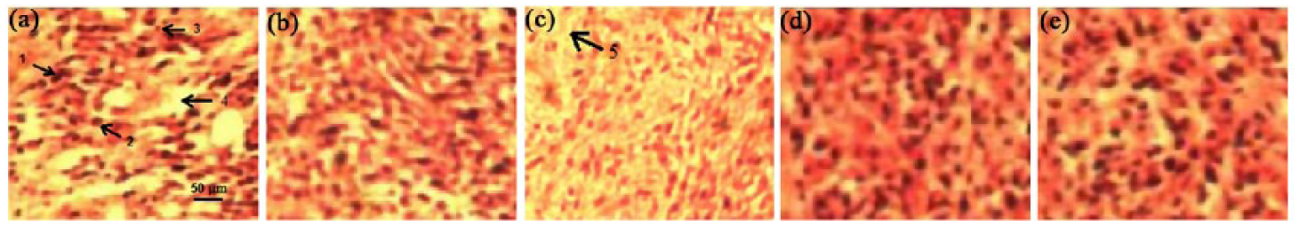
Histopathological images (HE coloration 10×) of the inflammatory granuloma tissue after implantation with (a) sterile cotton pellets (Mp), (b) cotton pellets after prednisone treatment (PDN), (c) M-CF pellets, (d) 4C-CA/1.6APS-CF pellets, and (e) 7C-CA/2APS-CF pellets.
In the granulation tissues taken from animals with implant of 4C-CA/1.6APS-CF and 7C-CA/2APS-CF pellets (Figure 17(d) and (e)), the presence of collagenase and a small number of macrophages is observed. The presence of collagenase and a small number of macrophages could be explained by healthy tissues surrounding copolymer hydrogels because of the antimicrobial activity of CF.59,60
Conclusion
HEMA/C-CA hydrogels with three-dimensional open-cell scaffold and interconnected porous structure which may allow facile communication between the biological cells and scaffold were obtained. The hydrogel networks retain a high amount of solvent being superabsorbent and their swelling degree depends on the hydrogel structure and the environmental conditions, more precisely on the pH of the solutions in contact with the tested hydrogels. Degradation by collagenase increases with collagen content in hydrogels and decreases with APS amount in the initial recipe because of more compact material obtained. The microbiological test shows that the 4.6C-CA/1.62APS, 7C-CA/1APS, and 7C-CA/2APS hydrogel samples have a good efficacy against S. aureus ATCC® 25923. The data reported proved that the release rate of CF from hydrogel samples is very high. Biocompatible and semi-biodegradable polymeric hydrogels were in vitro studied as carriers for CF. All these systems behave like polymer matrices convenient for controlled/sustained/targeting drug delivery systems. It was established that both the profiles of swelling of the HEMA/modified collagen hydrogels and CF release from these hydrogels are pH-responsive. The HEMA/C-CA hydrogel scaffolds described in this study possess key characteristics useful for tissue engineering, especially for wound dressings.
Footnotes
Declaration of conflicting interests
The author(s) declared no potential conflicts of interest with respect to the research, authorship, and/or publication of this article.
Funding
The research leading to these results has received funding from the Romanian—EEA Research Programme operated by the Ministry of National Education under the European Economic Area Financial Mechanism 2009–2014 and Project ACTIBIOSAFE, contract no. 1SEE/30.06.2014, and UEFSCDI Project BIONANOMED 164/2012.