
Abstract
Injectable in situ-forming hydrogels appears to be a promising approach for tissue engineering applications. In this study, the effect of phenol moiety (Ph) addition to gelatin in enzymatically-gellable modified pectin hydrogel (Pec-Ph) was studied. Addition of gelatin-Ph to Pec-Ph (Pec-Ph/Gel-Ph) altered the physical properties of Pec-Ph-based hydrogels as compared to unmodified gelatin (Pec-Ph/Gel) addition. Swelling ratio and degradation rates of the Pec-Ph/Gel-Ph hydrogel decreased 35% and 50%, respectively, and the elasticity of Pec-Ph/Gel-Ph hydrogel was higher than the Pec-Ph/Gel hydrogels. Scanning electron microscopy images showed that the existence of phenolic groups in gelatin decreased the pore size of Pec-Ph/Gel-Ph hydrogels. Culture of chondrocyte cells in the Pec-Ph/Gel-Ph hydrogels showed more metabolic activity (4×) during a 14-day culture period. Hydrogels subcutaneously implanted in rats could also be identified readily without complete absorption and signs of toxicity or any untoward reactions after 1 month. The work showed the potential of Pec-Ph/Gel-Ph hydrogels as a promising in situ injectable hydrogel for soft tissue engineering applications.
Keywords
Introduction
Among the scaffolds for tissue-engineering applications, the in situ injectable hydrogels are highly attractive in their interest to researchers in clinical applications especially for soft tissue engineering, since they provide a minimally invasive approach for implant surgery and can easily be formed to a desired shape to match irregular complex defects.1–4
Biomaterials and fabrication methods of scaffolds play crucial roles in developing injectable hydrogels. 5 There are various physical and chemical approaches for the fabrication of injectable hydrogels. Recently, the enzymatic cross-linking method as a chemical approach has been highlighted for development of novel injectable hydrogels, owing to the fast gelation, ability to work in mild conditions, and low cytotoxicity.6–9 In recent years, a variety of hydrogels have been developed taking advantage of the enzymatic method for the cross-linking of phenol-conjugated polymers, such as dextran, hyaluronic acid, alginate, gelatin, chitosan, glycol polypeptide, polyethylene glycol, and poly-propylene oxide as injectable biomaterials for various biomedical applications.10,11 Among them, gelatin has been widely used in cartilage tissue engineering due to its good adhesion, similarity to the native bone tissue, biodegradability, cost-effectiveness, commercial availability, and facile chemical modifications with other biomaterials or biomolecules.12–14
It has been demonstrated that the design of scaffolds based on blends of proteins and polysaccharides has emerged as a valuable strategy for soft tissue engineering.15–17 Chen et al. 11 developed successfully an injectable hydrogel system based on combination of hyaluronic acid and pectin derivatives. Their results showed, by varying their weight ratios, the physicochemical properties of hydrogel such as gelation time, mechanical properties and degradation rate were easily adjustable. Additionally, chondrocyte behavior was strongly dependent on the hydrogel composition and the presence of integrin binding moieties. Recently, Wang et al. 18 studied the applicability of cross-linked hyaluronic acid and gelatin derivatives as injectable hydrogels in cartilage tissue engineering. They reported the hyaluronic acid and thiolated-gelatin had uniform pore structure and low degradation rate, and RT-PCR analyses demonstrated that this hydrogel greatly improved the expression level of the associated cartilage matrix. Moreover, Morshedloo et al. 4 prepared injectable alginate hydrogel by introducing phenolic groups into alginate. Gelatin, a well-known protein having RGD sequence, could improve the enzymatically-gellable injectable alginate hydrogel for soft tissue engineering applications. The chondrocyte cells maintained their original phenotype and revealed statistically more metabolic activities in the alginate-based hydrogel.
Pectin, a natural polysaccharide extracted from the plant cell walls, is a cost-effective, non-toxic and easily functionalized component. Thus, it has been widely explored recently for various biomedical applications, such as drug and gene delivery, wound dressing and tissue engineering.19,20
In our previous study, we introduced phenolic moiety into pectin and evaluated the potential of enzymatically-gellable injectable pectin/collagen hydrogel for skin tissue engineering applications. 21 In this work, peroxidase-mediated pectin hydrogel was improved by addition of gelatin (Gel) and gelatin modified with the phenolic group (Gel-Ph) to evaluate the enzymatically-gellable injectable pectin-based (Pec-Ph) hydrogel for chondrocyte bioactivity. Physical properties of Pec-Ph/Gel and Pec-Ph/Gel-Ph hydrogels, behavior of chondrocyte cells cultured in pectin-based hydrogels as well as subcutaneous implantation of the hydrogels in a rat model was performed to examine the natural-based injectable hydrogel potential for use in soft tissue engineering applications.
Materials and methods
Materials
Pectin citrus peel (LM, 30,000–100,000), gelatin (type A, 300 Bloom), tyramin hydrochloride, n-hydroxysuccinimide (NHS), horsradish peroxidase (HRP 150 units/mg), phosphate buffer saline (PBS) and methylthiazolium bromide (MTT) were purchased from Sigma. Pencillin streptomycin, fetal bovine serum (FBS) and DMEM (low glucose) were obtained from Gibco (France). N-(3-dimethylaminopropyl)-n-ethylcarbodimide hydrochloride (EDC) was obtained from Merck (Germany). C28 chondrocyte (human cells) were prepared from Pasteur Institute (Tehran, Iran).
Modification of gelatin and pectin with phenolic hydroxyl groups (Ph)
Gel-Ph and Pec-Ph were synthesized by conjugating amino groups of tyramine with carboxyl groups in gelatin and pectin via the carbodiimide as described elsewhere.22,23 Briefly, 1 g pectin was dissolved in 100 mL of distilled water for 24 h. Then, 0.3 g tyramine hydrochloride, 0.1 g NHS, and 0.1 g EDC were added to the pectin solution (pH = 6.0) and were stirred for 24 h at room temperature. For modification of gelatin, 0.5 g gelatin was dissolved in 50 mL distillated water. After dissolving gelatin, 0.2 g tyramine hydrochloride, 0.05 g NHS, and 0.05 g EDC was added to the solution, respectively. The solution was stirred at room temperature for 12 h and after addition of 50 mM sodium phosphate a further 30 min of stirring was carried out.
Gel-Ph and Pec-Ph were precipitated by 90% ethanol and washed several times with alcohol until the absorbance peak at 275 nm (using UV-VIS spectroscopy) was undetectable in the remaining alcohol solutions and lyophilized. To test the introduction of Ph groups, the synthesized Gel-Ph and Pec-Ph were dissolved in distilled water at 0.1% (w/v) and the absorbance of the solution were evaluated at 275 nm.
Gelation time
The gelation time of the Pec-Ph/Gel-Ph and Pec-Ph/Gel (2%/1% w/v) were measured by dissolving the conjugates in CF-KRH, based on a previously reported method. 23 Briefly, the solutions containing of the pectin and gelatin derivatives and HRP were poured into a 48-well at 500 µL/well. Then H2O2 was added into each well and stirred using a magnetic stirrer until the magnetic stirring was hindered. The test was carried out at room temperature.
Swelling and degradation tests
To assess swelling behavior of hydrogels, cylindrical molds were used to prepare hydrogels with approximately 10 mm in diameter and 5 mm in thickness. The samples were then placed in an incubator in PBS with pH 7.4 at 37°C. During the period of incubation, the hydrogels weight was recorded at 1, 3, 6, 18, 24, 48, and 72 h. According to equation (1), the weight change was used to calculate the swelling ratio of the hydrogel:

in which, WI and WF are, respectively, the initial and final hydrogels weights. The experiment was carried out in triplicate for every sample. For in vitro biodegradation assessment, hydrogels from each sample were immersed into 10 mL of PBS solution (pH = 7.4) and then incubated at 37°C for 15 days. At the predetermined time intervals, the samples were removed from the PBS solution and then washed with distilled water and lyophilized for 48 h. Remained mass of sample to its initial mass was considered as degradation rate (equation (2)):

where, W0 and WF were initial mass and the mass of each sample at predetermined time intervals, respectively.
Mechanical property
The mechanical behavior of the cylindrical hydrogel samples with 1 cm in diameter and 10 mm in height were investigated by using a compressive strength test, as described in detail elsewhere. 22 The prepared hydrogels were placed in a material testing machine (Zwick/roell Z010, Germany) with a crosshead speed at 2.0 mm/min to determine compression–resistance stress profiles. The test was done in triplicate for hydrogel samples.
Scanning electron microscopy
Porosity and microstructure of hydrogel samples were investigated through scanning electron microscopy (SEM) (MV2300, CamScan, Japan). For SEM imaging, hydrogel samples were rapidly frozen and lyophilized. The cross section of freeze-dried samples was then coated with gold for 2 min at 20 mA. To evaluate pore size, at least 200 pores were selected from each sample and their areas were calculated by BEL View image analysis software (Version 1.49). Each pore was assumed to be a spherical shape, and pore size was reported as equivalent diameter of spheres. Distribution of pore sizes in the hydrogel samples was assumed to be Gaussian.
Cell culture
For cell culture in hydrogels, 1 × 10⁶ cells/mL of chondrocyte cells were mixed with Pec-Ph/Gel-Ph (2%/1% w/v) and Pec-Ph/Gel (2%/1% w/v) hydrogels and were used for cell culture test after peroxidase-mediated gelation. The HRP and H2O2 concentration in the final hydrogel were 3 units/mL and 7.7 mM, respectively. After 10 min of standing, the hydrogels containing cells cultured in DEME low glucose supplemented with 10% fetal bovine serum and 1% penicillin streptomycin under a humidified atmosphere at 37°C, 5% CO2 incubator. The MTT assay was applied to study the viability of chondrocyte cells at 7 and 14 days.
In vivo animal test
For implementation of in vivo animal test, two adult 8-week-old male Wistar rats weighing 220 and 240 g were used after 7 days of acclimatization period. The rats were kept in individual cages and fed with pelleted food and water ad libitum. In order to anesthetize the rats, a combination of xylazine (10 mg/kg) and ketamine (40 mg/kg) was intraperitoneally injected. Then, under general anesthesia, the dorsal region of each rat was prepared for aseptic surgery by shaving the hair and rinsing the skin with povidone iodine solution and alcohol. A skin incision (about 2–3 cm) was made on both sides of the spinal column of each rat and a subcutaneous pocket was prepared. Afterward, the skin incisions were closed using 3/0 nylon sutures and the rats were allowed to recover from anesthesia inside their cages. After 4 weeks of hydrogel injection, the rats were weighed prior to being euthanized with an overdose of the aforementioned anesthetic drugs. Routine histological sections with 5 μm thickness were provided from the specimens and stained with hematoxylin and eosin. Sections were examined under the light microscope.
Results and discussion
Characteristics of synthesized gelatin-Ph and pectin-Ph
Phenolic groups (Ph) were introduced into pectin and gelatin to improve their properties to form pectin-based injectable hydrogel. As shown in (Figure 1(a)), Pec-Ph (0.1% w/v) had an absorbance peak at around 275 nm attributed to the presence of Ph groups as compared to pectin (0.1% w/v). Similar profiles were observed in comparison of Gel-Ph and gelatin (0.1% w/v) as shown in (Figure 1(b)). The presence of the peak in this wavelength range, indicates the presence of phenolic ring and the proof of successful synthesis.23,24
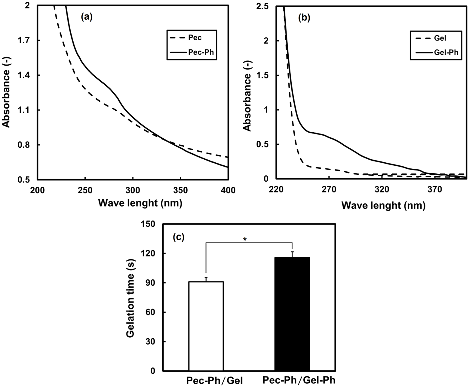
UV-vis absorbance spectra of Pec-Ph (a) and Gel-Ph (b) showing the absorption peak at 275 nm respectively, and gelation times of Gel-Ph/Pec-Ph and Gel/Pec-Ph at the presence of 3 unit/mL of HRP and 7.7 mM H2O2 (c), The statistical significance among the data sets was evaluated by the Student’s t-test (*p < 0.05, **p < 0.01).
To demonstrate phenolic groups introduced in the Pec-Ph and Gel-Ph derivatives modifies the gelation time of Pec-Ph in the presence of Gel-Ph and gelatin was also examined at a defined HRP and H2O2 concentration. Gelation time is one of key factors in designing in situ hydrogels as a scaffold for tissue engineering and other biomedical uses. Based on results (Figure 1(c)), gelation time in the Pec-Ph/Gel-Ph hydrogel was longer than Pec-Ph/Gel hydrogel (p < 0.05). Gelation time directly depends on the HRP and H2O2 concentrations and can be adjusted by changing the concentration of the HRP and H2O2.4,23 At the presence of 3 units/mL of HRP and 7.7 mM H2O2, the gelation time of Pec-Ph/Gel and Pec-Ph/Gel-Ph hydrogel was 1.5 and 2 min, respectively, which were desirable for in situ gel formation due to fast gelation at low concentrations of H2O2. 23 The results indicate successful introduction of phenolic moieties to pectin and gelatin to form enzymatically-gellable pectin-based injectable hydrogels.
Influence of gelatin and Gel-Ph addition on physical properties of Pec-Ph-based hydrogels
Microstructure of hydrogels
The pore size and structure are of crucial factors in designing an appropriate extra cellular matrix (ECM) in tissue engineering. The scaffold pore size as well as pore interconnectivity have been shown to influence osteochondral repair. 25 The microstructure of the hydrogels was observed by SEM and the results are shown in Figure 2(a). The appearance of pores revealed the presence of phenolic group on the gelatin in the Pec-Ph/Gel-Ph hydrogel reduced pore size as compared to Pec-Ph/Gel hydrogel due to higher crosslinking density in the hydrogel. The quantitative analysis of pore size confirmed that the pore size distribution for the Pec-Ph/Gel-Ph was in the range of 50–100 µm, while for the Pec-Ph/Gel was observed 100–200 µm (Figure 2(b)). The average diameter of the pores was observed to be 92 and 212 µm for the Pec-Ph/Gel-Ph and Pec-Ph/Gel hydrogels, respectively.
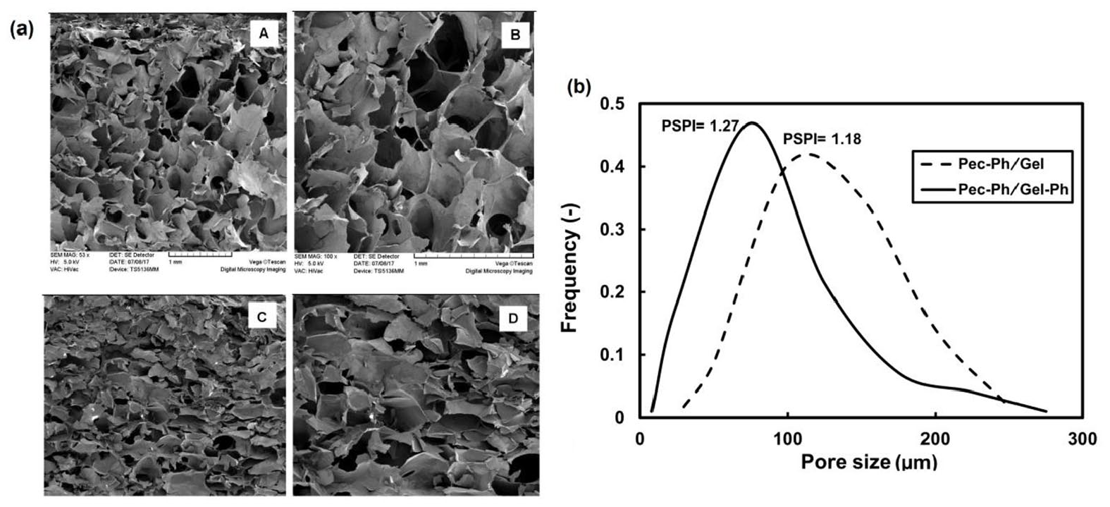
SEM images showing morphology of Pec-Ph/Gel (A, B) and Pec-Ph/Gel-Ph hydrogels (C, D) (a), pore size distribution (b).
Various cell types have different dimensions and geometries; thus, depending on the application, the pore size of the scaffold used should be no smaller than the dimensions of the cell in suspension. Han et al. 26 reported that the silk scaffolds with smaller pore sizes (90–180 µm) which were fabricated through salt extraction method represented better chondrocyte cell proliferation and higher levels of sulfated glycosaminoglycan, collagen and chondrocytes marker aggrecan. The microstructure results showed the Pec-Ph-based hydrogels with the concentrations used in the present study can be used for soft tissue cell proliferation such as chondrocytes.
Swelling and degradation
Hydrogels act as dynamic environments which mimic cellular ECM. However, only hydrogels with an appropriate swelling and degradation rate may be used as successful tissue engineering scaffolds. 27 In this study, the ability of water absorption by hydrogels was measured for 72 h by immersion in PBS solution in a humidified incubator at 37°C. As shown in Figure 3(a), the Pec-Ph/Gel-Ph hydrogels swelled during the incubation period, reaching to 7.7% and 10.6% after 24 and 72 h, respectively. In the case of the Pec-Ph/Gel hydrogel, the swelling rate showed a similar trend and was higher than the Gel-Ph/Pec-Ph hydrogel, reaching to 14.4% at the end of the incubation period. The swelling degree of hydrogels depends on the extent of cross-links. 28 The microstructure of the scaffolds could be considered as an effective parameter on the swelling rate of hydrogels. Since the Pec-Ph/Gel hydrogel has higher pore size, so it can accommodate more water molecules, thereby results in more swelling of the hydrogel. Indeed, the substitution of Ph group in gelatin hydrogel reduces the number of carboxyl groups in gelatin that increases swellings through the formation of hydrogen bonds between water and gelatin.
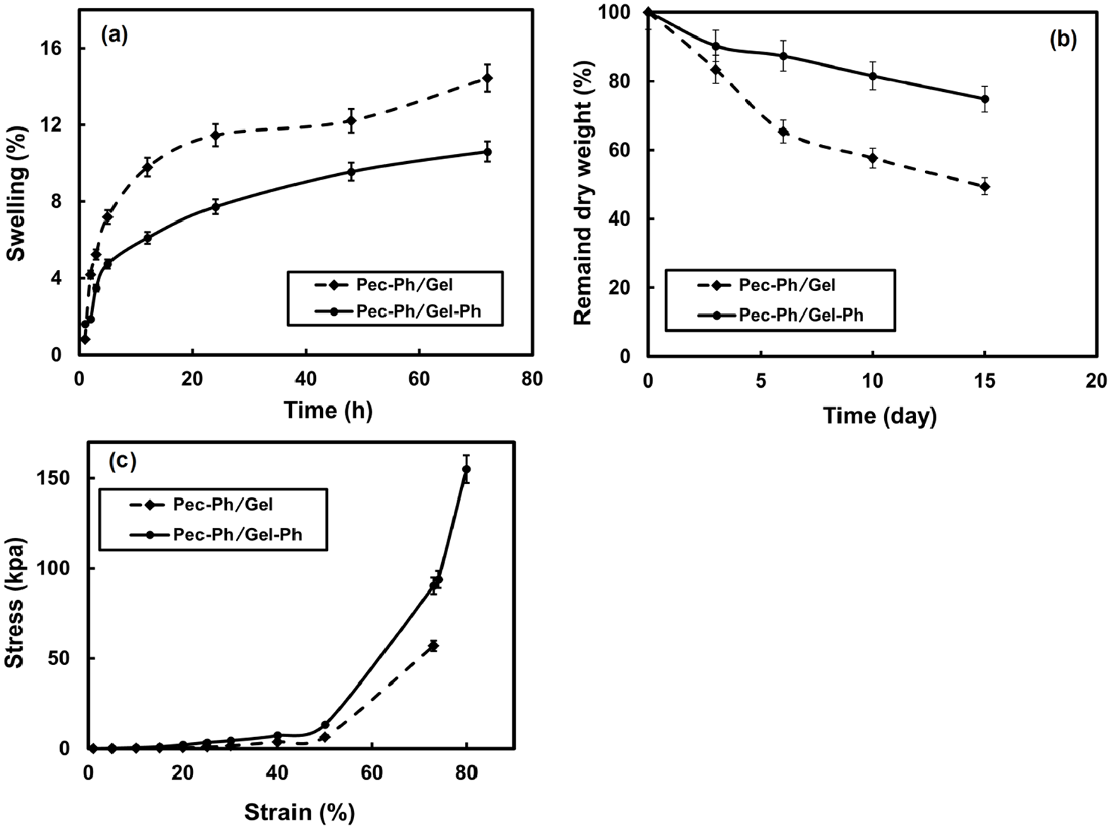
Physical properties of swelling ratio (a) and degradation rate (b) during 3 days and 15 days incubation periods, respectively and mechanical strength of the hydrogels (c).
The hydrogel scaffolds should have an appropriate microstructure and possess adequate mechanical strength and biodegradation rate. Figure 3(b) compares the biodegradation behavior of the Pec-Ph-based hydrogels in terms of the residual weight of hydrogels in PBS during 2-week incubation. Based on the results obtained, the degradation rate of the Pec-Ph-based hydrogels in the presence of Gel-Ph decreased, and as shown, the Pec-Ph/Gel-Ph hydrogel maintained approximately 74.8% of its initial weight, while the Pec-Ph/Gel hydrogel retained 49.4% of its initial weight after 2 weeks of the degradation period (p < 0.05). The degradation rate is in relation with mechanical strength and pore size. In brief, scaffolds with lower porosity or larger pore size are degraded rapidly compared to those with higher porosity or smaller pore size. The reason can be that the scaffolds with smaller pore size possess thinner pore walls and therefore a larger surface area to volume ratio, which accelerates the diffusion of small acidic by-products, resulting in a weaker acid-catalyzed hydrolysis. The carboxylic end groups of gelatin may also be disassociated and produce H+, which acts as a catalyst, accelerating the hydrolysis reaction. 29 In agreement with this expression, it can be seen that the biodegradation rate of the Pec-Ph/Gel hydrogel which had larger pore size and higher carboxylic end groups was approximately two times than of Pec-Ph/Gel-Ph hydrogel.
Mechanical properties
The mechanical properties of an injectable hydrogel are very important for successful regenerative application. The hydrogel must possess suitable mechanical stability to withstand stress incurred during culturing and provide temporary support for the cells in the defect tissue. 30 Figure 3(c) shows the mechanical properties of the hydrogels assessed using a compression test. As shown, the strain at the rupture point of Pec-Ph/Gel hydrogel was 72%, whilst this point for Pec-Ph/Gel-Ph hydrogel occurred at 80% strain. Adding Gel-Ph to Pec-Ph hydrogel led to higher mechanical strength, in which the mechanical strength for the Pec-Ph/Gel-Ph hydrogel at 72% strain was 2.9 times higher than that of the Pec-Ph/Gel hydrogel. The higher mechanical strength seems to be due to the presence of more phenolic bonds in the Pec-Ph/Gel-Ph hydrogel which causes more elasticity. 31 Studies show porosity and pore sizes of the hydrogels also have an effect on the mechanical properties. Large pores can reduce the structural strength of the scaffold.32,33 The images of SEM showed that the pore size of the Pec-Ph/Gel hydrogel was higher as compared to the Pec-Ph/Gel-Ph hydrogel, leading to the reduction of mechanical strength.
Influence of gelatin and Gel-Ph addition on model cell proliferation in Pec-Ph-based hydrogels
Figure 4 shows the proliferation behavior of chondrocyte cells cultured in the Pec-Ph/Gel-Ph and Pec-Ph/Gel hydrogels as model for 2 weeks culture period. As shown in Figure 4(a), the chondrocyte cells could proliferate inside the both Pec-Ph-based hydrogels. The proliferation results showed an approximately fourfold increase in cell expansion rate for the cells cultured in the Pec-Ph/Gel-Ph hydrogels, the value of which was nearly two times higher than that for the cell-laden Pec-Ph/Gel hydrogel samples at day 14 (p < 0.01). The chondrocyte cells could also form more globular aggregates during the culture period in the Pec-Ph/Gel-Ph hydrogels (Figure 4(b) and (c)).
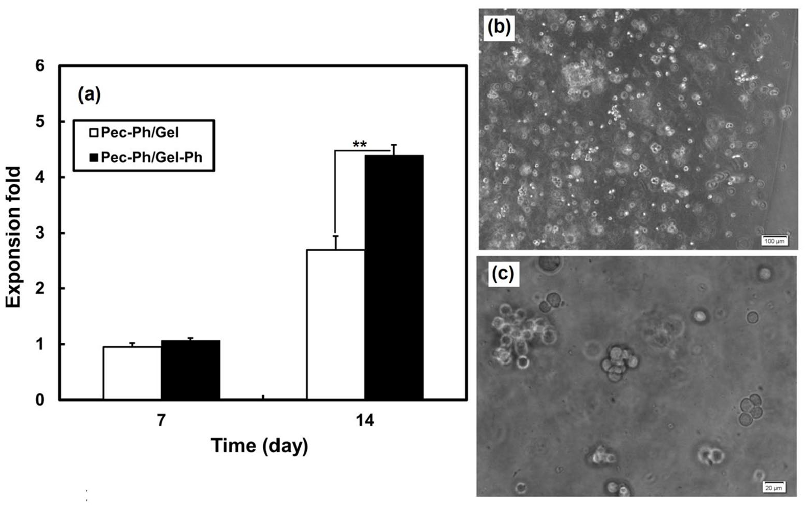
Metabolic activities of the cells cultured in Pec-Ph/Gel-Ph and Pec-Ph/Gel hydrogels at seeding density of 1 × 10⁶ cells/mL gel and culture days of 14 days (a), and microscopic images of chondrocyte cells and aggregates in Pec-Ph/Gel-Ph hydrogels after 14 days (b and c).
The higher cell proliferation in the Pec-Ph/Gel-Ph hydrogels can be due to the increase in the ligand density as specific surface area increases in the scaffold with smaller pores, allowing for more binding sites for the cells. 34 Indeed, unbound gelatin in the Pec-Ph/Gel hydrogels can be released to medium during the culture period, modulating adhesive sites for cell attachment and growth. Moreover, the higher degradation of the Pec-Ph/Gel with a significant change in the microstructure and mechanical properties may have an adverse effect on the behavior of the cells that populate the scaffold. The results of chondrocyte behavior inside the Pec-Ph-based hydrogels demonstrated that the Pec-Ph/Gel-Ph hydrogel has potential to use for soft tissue engineering applications.
In vivo test
Figure 5 shows subcutaneous implantation of injectable in situ Pec-Ph/Gel-Ph hydrogel with and without chondrocyte cells in rat. Animal test results indicated that after 1 month, the scaffolds could be identified easily at the site of implantation without complete absorption. There was no sign of toxicity or any untoward reactions due to the hydrogel implantation during this time. The body weight of rats at the time of sampling increased, indicating their overall health. Macroscopically, inflammatory reaction around implantation sit of the hydrogel is visible. Complete cartilaginous tissue was not formed at implantation sites, but single or multiple chondrocyte like cells were observed indicating the survival of originally placed cells inside the scaffolds. The results revealed the Pec-Ph-based hydrogels have potential for clinical soft tissue engineering applications.
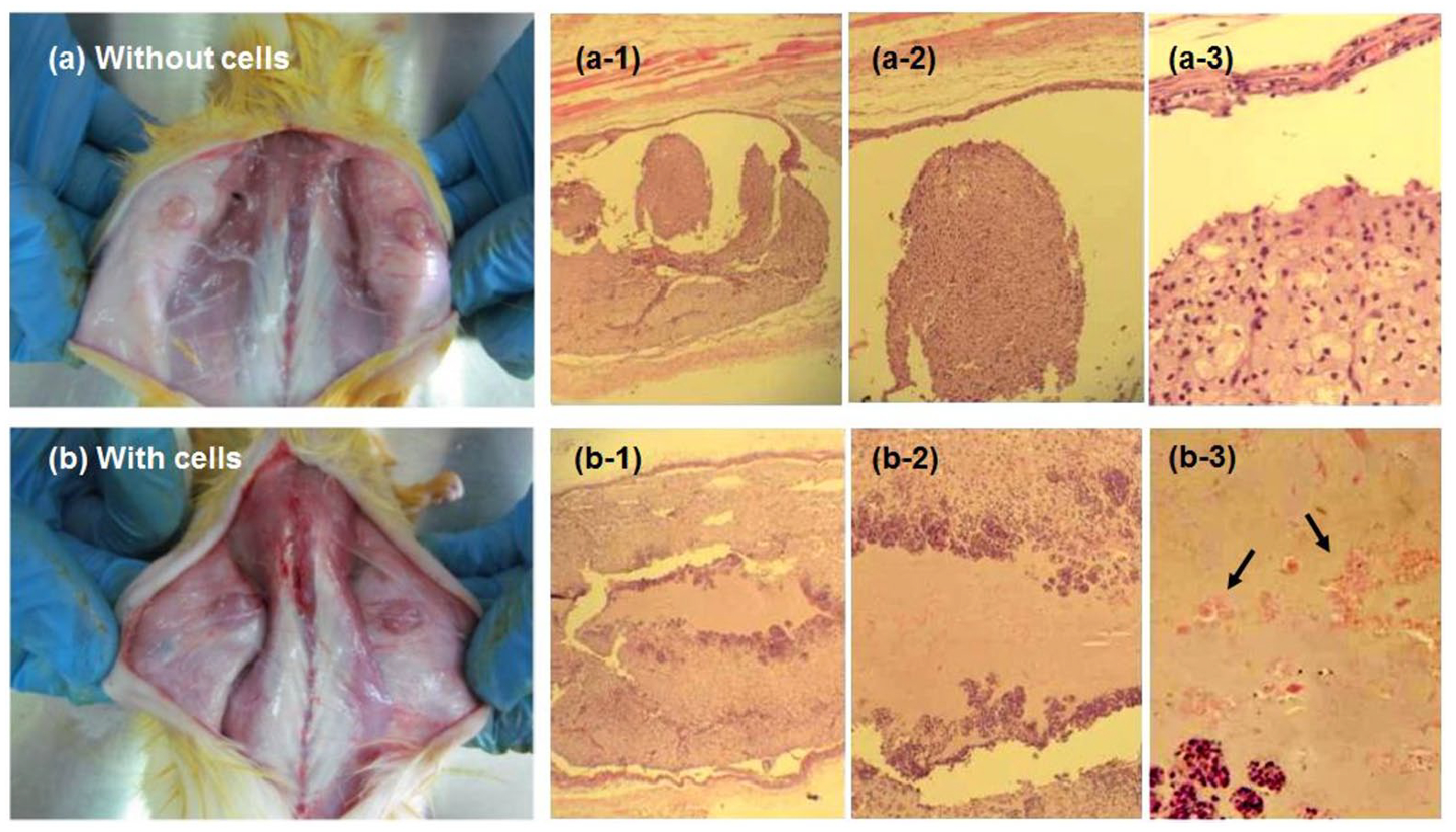
Subcutaneous implantation of the Pec-Ph/Gel-Ph hydrogel without cells (a) and with chondrocyte cells (b) in rats after 1 month. a-1 to a-3 and b-1 to b-3 show microscopic images of the hydrogels without and with chondrocyte cells with different magnifications. Block arrows show the chondrocyte cells after 1 month implantation.
Conclusion
This study investigates the impact of Gel-Ph on the properties of peroxidase-mediated injectable Pec-Ph/Gel-Ph hydrogel and evaluated the behavior of cultured chondrocyte cells as a model for soft tissue engineering applications. Pec-Ph/Gel-Ph hydrogels revealed higher gelation time, lower degradation rate as well as swelling properties. Pore size of the Pec-Ph/Gel-Ph hydrogels in the presence of Gel-Ph decreased and become more uniform in comparison with the Pec-Ph/Gel hydrogels. Gel-Ph use in the Pec-Ph-based hydrogels increased considerably the mitochondrial activities of the chondrocyte cells as compared to the Pec-Ph/Gel hydrogels during culture period. In animal test carried out for the Gel-Ph/Pec-Ph hydrogel subcutaneous injection in rat, the viable chondrocyte cells were clearly observed as red colored areas in the central regions of cell-laden subcutaneous hydrogels after 1 month. The results arising from our investigation showed the potential of Pec-Ph/Gel-Ph hydrogels for producing in situ formed injectable hydrogel with improved overall properties for soft tissue engineering application.
Footnotes
Declaration of conflicting interests
The author(s) declared no potential conflicts of interest with respect to the research, authorship, and/or publication of this article.
Funding
The author(s) received no financial support for the research, authorship, and/or publication of this article.