
Abstract
Pectin has recently attracted increasing attention for biomedical and pharmaceutical applications. Due to the lack of adhesion molecules in polysaccharides, phenolic hydroxyl conjugated gelatin was added to enzymatically-gellable peroxidase-modified pectin derivative and compared with phenolic hydroxyl -pectin/collagen. Both pectin and gelatin were modified by tyramine hydrochloride in the presence of EDC/NHS. The phenolic hydroxyl -pectin/phenolic hydroxyl -gelatin, phenolic hydroxyl-pectin/collagen, and phenolic hydroxyl -pectin hydrogels were prepared using horseradish peroxidase and hydrogen peroxide. The hydrogels were characterized by gelation time analysis. Morphology, enzymatic biodegradation, mechanical and swelling properties as well as water vapor transmission rate were also evaluated. Fibroblasts were cultured for 7 days, and the survival rate was evaluated using conventional MTT assay. Hydrogels composed of Ph-pectin/Ph-gelatin showed decreased biodegradation rate, and WVTR and further improved mechanical performance in comparison with other groups. Both phenolic hydroxyl -pectin/collagen and phenolic hydroxyl -pectin/phenolic hydroxyl -gelatin hydrogels exhibited porous structures. The hydrogels composed of collagen promoted cell survival rate 1.4 and 3.5 times compared to phenolic hydroxyl -gelatin and phenolic hydroxyl -pectin based hydrogels at the end of 7 days, respectively (p < 0.001). The study demonstrated the potential of enzymatically-gellable pectin-based hydrogels as cost-effective frameworks for use in tissue engineering applications.
Introduction
Tissue engineering serves as a significant and efficient platform for healing tissue damages and deficiencies. It includes an evolution of biological substitutes as remedies for injured organs. There are three substantial elements for the regeneration of damaged tissues as follows; cells, scaffolds, and growth factors. 1 In the tissue engineering technique, scaffolds have an inevitable and crucial role in regulating the cell bioactivity, morphological adaptation, cell-to-cell connection, and providing a favorable tissue microstructure. 2 Materials such as pectin, collagen, and gelatin have been extensively exploited to design biodegradable and biocompatible hydrogels while promoting cell attachment, proliferation, and differentiation. 3 Hydrogels as hydrophilic and cross-linked porous polymers have a high capacity of water absorption. 4,5 Having an analogous structure to natural tissues, hydrogels have had far-reaching applications in biomedical fields. 6 Owing to non-toxicity, cost-effectiveness, and their superior characteristics, pectin-based hydrogels were selected for developing effective structures in this study. 7 Pectin is a hetero-polysaccharide that exists abundantly in the plants and fruits walls with a molecular structure of 1, 4-linked α-d-galactosyluronic acid residues.8,9 Two forms of pectin are high and low methyl ester proteins with more than 50% of esterified acid units and less than 50% methyl ester groups. Pectin has excellent properties such as biocompatibility, biodegradability, and the ability to support various bioactive substrates. 10 Takei et al. previously developed sugar beet pectin hydrogels by enzymatic cross-linking without detrimental effects on encapsulated cells during the gelation process. 11 Neves and co-workers modified pectin hydrogels by using RGD containing peptide. They showed stimulatory effects of RGD-enriched pectin hydrogels on cell adhesion and substrate degradation capacity. 7 Gelatin is a natural water-soluble biopolymer derived from partial hydrolysis of insoluble collagen. Therefore, all of the basic factors of collagen dominate the attributes of the gelatin. This biopolymer has various pharmaceutical or medical applications. 12 Gelatin contains amino and carboxylic groups, which enable chemical cross-linking with diverse polysaccharides. This trait enables us in order to develop different suitable matrices with a special ultrastructural feature in favor of cell adhesion. 13 Previously, it has been reported that pectin-gelatin hydrogels are cytocompatible while having suitable mechanical properties. 14 Gelatin-based hydrogels can be created by the application of the physical crosslinking method. 15 However, at temperatures higher than 30–35°C, non-covalent bonds are easily destroyed, resulting in the collapse of the physical network. 16 Of note, gelatin-based hydrogels have poor mechanical strength and low elasticity. The synthesis of scaffolds solely based on gelatin leads to quick biodegradation in aqueous environments or its easy elimination from the in vivo systems 16 To improve the properties of gelatin, it can be covalently cross-linked by chemicals such as glutaraldehyde, formaldehyde, or carbodiimide.18,19 Collagen is another natural biopolymer that commonly exists in skin, bone, cartilages, and tendons. Collagen can be obtained through animal sources like bovine skin, rat tail, or cow skin. Collagen is a non-toxic substrate and can support excellent cell adhesion and cell-to-cell interaction.20,21 However, unmixed and unbounded collagen has weak mechanical strength. 22 Cells easily interact with unmixed collagen resulting in the shape deformation of the collagen matrix if it is not stabilized or cross-linked with another material. Therefore, the development of compound hydrogels based on bounded collagen is under the attention of numerous authorities.20,23 Enzymatically gellable hydrogels have attracted great interest in filling any shape of the defect. Cells are evenly distributed in gellable hydrogels, and they can appropriately fill irregular-shaped defects. 24 The introduction of horseradish peroxidase (HRP) and hydrogen peroxide (H2O2) into hydrogels and their reaction with hydroxyl phenolic (Ph) groups can lead to producing cross-linked biopolymers which possess an improved regenerative behavior. The advantage of Ph conjugated polymer hydrogels, as a matrix of bioactive agents, has been widely studied before. 25 It was found that these hydrogels have suitable mechanical integrity and can control cell behavior in a controlled manner. 26 The potential capability of combining phenolic groups with polymers such as gelatin has been demonstrated in the earlier literature and the work done by our group.24,27 In this study, we aimed to evaluate the physical traits and regenerative potential of Ph-modified pectin hydrogels in combination with modified gelatin and unmodified collagen, which were manufactured using an enzymatic reaction. There are few reports about regenerative potential of hydrogels and substrates enriched with pectin in the cutaneous tissue.
Here, we tried to show a unique concept of cross-linking of pectin to collagen and gelatin and studied their application in the regeneration of cutaneous tissue. Due to public acceptance and attention of the scientific community, the application of plant-based natural components in natural hydrogels with usability in the cutaneous pathologies is at the center of attention. In this paper, we prepared Ph-pectin, Ph-pectin/Ph-gelatin, and Ph-pectin/collagen hydrogels and analyzed how collagen and gelatin make a difference in the attributes of Ph-pectin hydrogels and modulate the bioactivity of fibroblasts. To achieve this goal, mechanical strength, swelling, biodegradability, and scanning electron microscopy (SEM) analyses were conducted. We hypothesized that data from this study could help us to find an appropriate hydrogel for the regeneration of injured skin.
Materials and methods
Materials
Pectin (citrus peel), Type A porcine gelatin (Cat no: G2500), tyramine hydrochloride, HRP (700 units/mg; Cat no: T2879), Phosphate buffer saline (PBS), and 3- [4,5 dimethylthiazol-2-yl]-2,5 diphenyl tetrazolium bromide (MTT; Cat no: M5655) powder were purchased from Sigma-Aldrich. Collagen type II was extracted from bovine skin using porcine pepsin enzyme (Cat no: P0609; Sigma Aldrich). 1-Ethyl-3–(3-dimethylamino propyl) carbodiimide hydrochloride (EDC; Cas no: 25952–53-8), N-Hydroxysuccinimide (NHS; Cas so: 6066–82-6), and dimethyl sulfoxide (DMSO; Cas no: 67–68-5; Merck; Germany) were obtained from Merck. Fibroblasts were purchased from the Iranian Cell Bank (Pasteur Institute, Iran). Culture medium RPMI-1640 (Cat no: 11875101) containing 10% FBS (fetal bovine serum, Cat no: 10100139) and 1% Penicillin-Streptomycin (Cat no: 10378016) were purchased from Gibco. Cells were incubated in a humidified atmosphere at 37°C with 5% CO2. 0.25% Trypsin-EDTA (Cat no: LM-T1721) solution was purchased from Biosera.
Methods
Modification of pectin and pectin/gelatin
Ph-Pectin, Ph-pectin/Ph-gelatin, and Ph-pectin/collagen groups were fabricated using tyramine hydrochloride through the reaction of carboxylic groups of gelatin and pectin and amino acid groups of tyramine as previously described (Figure 1). 20 In short, 10 g/L pectin and 7 g/L pectin/3 g/L gelatin powders were dissolved in distilled water for 1 hour at 40°C to yield pectin and pectin/gelatin solutions. 0.3 g tyramine, 0.1 g EDC, and 0.1 g NHS were overlaid to each solution. After 24 hours, the hydrogels were washed with 90% ethanol (EtOH) (v/v) solution for 10 minutes. Then, EtOH was withdrawn, and the remaining precipitates were respectively washed with 80% and 100% EtOH for 45 min and air-dried to produce Ph- pectin and Ph- pectin/Ph-gelatin powders.
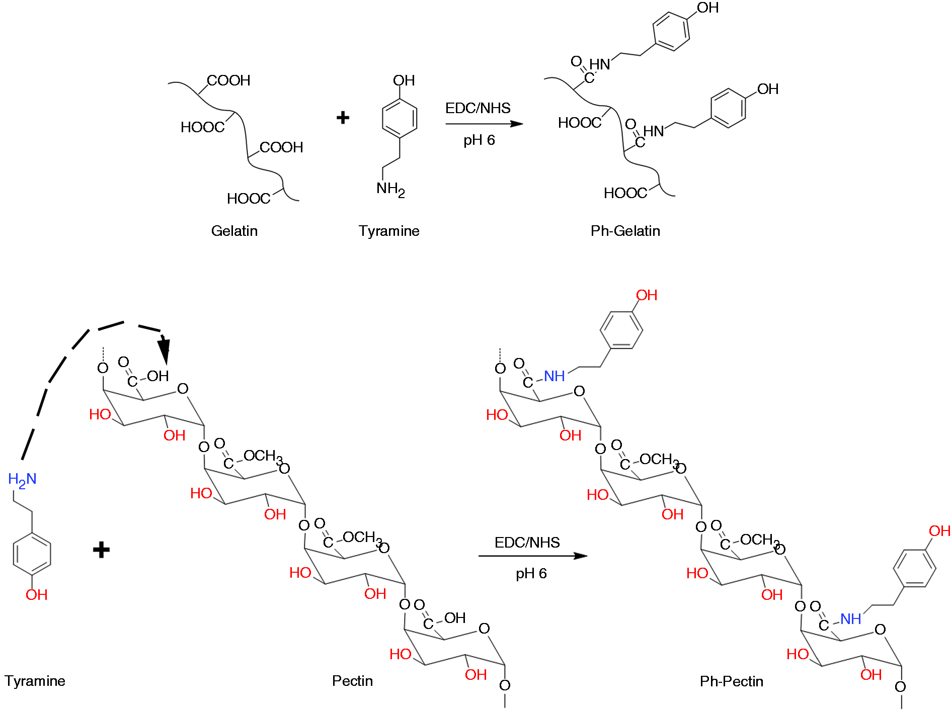
Synthetic scheme for the formation of Ph-Gelatin and Ph-Pectin using tyramine.
Preparation of Ph-pectin, Ph-pectin/Ph-gelatin, and Ph-pectin/collagen hydrogels
3% (w/v) Ph-pectin and 2% (w/v) Ph-pectin/1% (w/v) Ph-gelatin solutions were prepared by dissolving the powders in PBS. Also, 1% (w/v) collagen powder was dissolved in 0.01 M acetic acid solution to yield a collagen solution. Then, solutions of Ph-pectin and collagen were mixed together to form Ph-pectin 2% (w/v)/collagen 1% (w/v) mixture. Hydrogels of Ph-pectin, Ph-pectin/Ph-gelatin, and Ph-pectin/collagen were formed by mixing 0.35 U/mL HRP and 0.1 mL H2O2 (0.1 mM) per ml of gel. The gelation time of the prepared hydrogels was also determined when the movement of the magnet was obstructed by the gellified hydrogels, and the surface of the gel swelled.
Ph content of the modified pectin and gelatin
Ph-pectin and Ph-pectin/Ph-gelatin powders were dissolved in distilled water with a concentration of 0.1% (w/v). The qualitative content of Ph groups was assessed using an ultraviolet-visible spectrometer (Cecil BioQuest CE 2501 Life Science), and the absorbance of the solutions analyzed at 275 nm.
Hydrogels characterization
Cylindrical hydrogels were produced with a diameter of 14.6 mm and a height of 1 cm in a syringe to use for the characterization tests.
SEM analysis
For effective regeneration, the scaffolds should possess high porosity, even pores, and excellent interconnectivity. The impact of collagen and Ph-gelatin on the pore morphology of Ph-pectin hydrogel was analyzed by SEM imaging. The cross-section of samples was covered with a thin layer of gold for 2 min at 20 mA. The samples were studied with an SEM (Model: TescanMV2300) functioning at 5 kV. About 150 pores from each SEM image of hydrogels were picked out by BEL View image analysis software to calculate the pore size polydispersity index (PSPI). For this purpose, the diameter of the selected areas was determined by considering the pores as a sphere. The Gaussian distribution for the pores of the hydrogels was plotted, and the value of PSPI was employed to investigate its deviation from 1. This value was calculated by the following equation.
28
Where “n” and “d” are the number and diameter of calculated pores. If all the pores of a sample were in the same size, the amount of PSPI would be 1.
Swelling ratio
The swelling analysis was conducted to assess the capacity of the hydrogels to absorb water. Each group was weighed in triplicate, immersed in 20 mL PBS (Ph = 6.8), and incubated at 37°C. At specific periods (1, 3, 6, 18, 24, 48, and 72 hours) scaffolds were pulled out, and excess PBS was removed by filter papers. The weight of the swelled hydrogels was recorded using the A&D GR-300 lab balance. The swelling ratio was evaluated as follows:
Where Ws and Wd attribute to the swollen and dry weights of the hydrogels.
Mechanical strength
Cylindrical hydrogels with a diameter of 14.6 mm and a height of 1 cm were prepared as mentioned above. The maximum sustainable stress of the hydrogels was tested with a Zwick/Roell Z010 tensile machine with a 5 N load cell at the speed of 2 mm/min and a strain of 80%. This test was repeated three times.
Biodegradation
Degradation of the hydrogel is a key factor in tissue engineering since it has a profound effect on cell viability, growth, and host cell response against the transplant cells. The hydrogel should degrade as new tissue formation occurs. After the regeneration of the injured tissue, the hydrogel should degrade thoroughly or be absorbed by the body. 29 Hydrogels of equal weights (W1), in triplicate, were submerged in 20 mL PBS and (1.5 μg/mL) Lysozyme enzyme to mimic the in vivo degradation operation at 37°C. The control hydrogels of the same samples with equal weights were incubated in 20 mL PBS without Lysozyme at 37°C. After 3, 9, and 15 days, the hydrogels were sampled, washed with distilled water, and dried by a freeze drier. After lyophilization, hydrogels were weighed (W2). The degradation ratio was calculated by the following equation: 30
WVTR
The moisture permeability of the hydrogel was calculated by the WVTR test in the simulated body physiological environment (37°C). Prepared cylindrical hydrogels with equal weights were initially weighed. The hydrogels were placed on the mouth of cylindrical tubes (30 mm diameter) containing 10 mL water with insignificant water vapor transfer. The hydrogels were fixed with a Teflon tape to avert any water vapor loss and maintained at 37°C and 35% relative humidity in an incubator. At predetermined periods, samples were weighed, and their water loss was evaluated by the slope of the plotted graph based on time. WVTR was assessed using the following formula; 31
where A is the surface area of the sample (m2). This test was performed in triplicate.
Cell culture study
To evaluate the biocompatibility of the hydrogels, cellular toxicity was analyzed by MTT assay. To this end, fibroblasts were cultured in RPMI-1640 medium containing 10% FBS and 1% Pen-Strep solutions. Cells were incubated at 37°C with 5% CO2. After 7 days, cells were trypsinized and cultured in hydrogels with a density of 106 cells per mL of gel in 12-well plates. Two ml culture medium was added to each sample. On days 1, 3, 5, and 7, the culture medium was removed, and 1 mL MTT solution (5 mg/ml) was added to each well, and plates incubated at 37°C for 4 hours in dark. Afterward, the MTT solution was discarded, and 1 mL DMSO solution was added to dissolve the formazan crystals. The absorbance was measured at 570 nm. This analysis was repeated in triplicate.
Statistical analysis
For analyzing the statistical difference between groups, we performed a One‐Way analysis of variance (ANOVA) with the Tukey post hoc test. The differences were considered statistically significant if p < 0.05. All the results were obtained from three sets of experiments.
Results and discussion
Ph content of the modified pectin and gelatin
In this study, tyramine was used to introduce Ph groups into gelatin and pectin via carbodiimide-mediated condensation (Figure 2(a)). We monitored UV–Vis absorbance spectrum of unmodified pectin, gelatin, and their conjugates with a concentration of 0.1% (w/v) dissolved in distilled water. The unmodified gelatin and pectin did not show any characteristic absorbance between 250 and 300 nm. As shown in Figure 2(a), the analysis of Ph-pectin/Ph-gelatin and Ph-pectin samples demonstrated an increase in the absorbance peak at 275 nm in comparison with unmodified pectin/gelatin and pectin groups, confirming the presence of Ph groups in Ph-pectin/Ph-gelatin and Ph-pectin. Therefore, the current protocol contributed to a successful linkage of tyramine with carboxyl groups in the amino acid residues of pectin and gelatin substrates. We also found that the Ph-pectin/Ph-gelatin solution showed an increase in the absorbance at 275 nm compared to Ph-pectin. This can be related to the existence of a higher number of Ph moieties in Ph-gelatin containing conjugates in the mixture of pectin, gelatin incubated with tyramine.
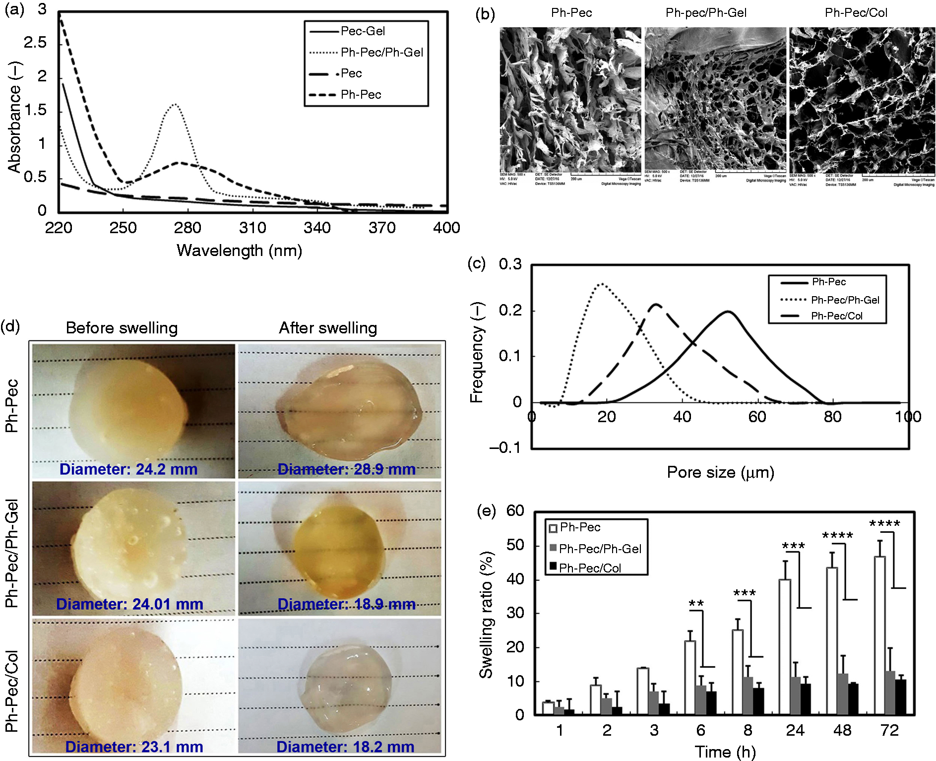
(a) UV absorbance spectrum of unmodified and modified Pec and Pec/Gel, (b) SEM images of Ph-Pec, Ph-Pec/Ph-Gel, and Ph-Pec/Col matrices, (c) Gaussian distribution of the pore size in Ph-Pec, Ph-Pec/Ph-Gel, and Ph-Pec/Col, (d) photographs of cylindrical Ph-Pec, Ph-Pec/Ph-Gel, and Ph-Pec/Col hydrogels before and after swelling in PBS at the end of 72 h (n = 3) (e) In vitro swelling studies of Ph-Pec hydrogels and Ph-Pec hydrogels containing Ph-Gel and Col, **P < 0.01; and ***P < 0.001.
Gelation time
In this study, chemically cross-linked hydrogels were prepared by the reaction of -COOH carboxyl groups with the phenolic groups, and this reaction was catalyzed by the combination of H2O2 and HRP. H2O2 molecules bind to the iron atom of HRP and initiate the chemical reaction. The produced complex oxidizes Ph groups of the polymer and causes gelation of the network. Following other oxidations of Ph moieties, HRP goes back to its original state and enters the crosslinking process again.32,33 Gelation time can be controlled by changing HRP concentration at a fixed concentration of H2O2 or vice versa. In the current experiment, we found that the gelation time for Ph-pectin, Ph-pectin/collagen, and Ph-pectin/Ph-gelatin was 180, 130, and 120 seconds, respectively. Fast gelation of Ph-pectin/Ph-gelatin could be due to the existence of higher phenolic content indicated by UV irradiation. Previous experiments demonstrated that gelation of the hydrogels with higher phenolic content occurs rapidly, suggesting that a larger number of electron acceptors could enhance the production of oxidized donors in the enzymatic reaction. 34 Compared to Ph-pectin, Ph-pectin/collagen hydrogels exhibited a shorter gelation time. The reason may lie in the fact that the introduction of HRP and H2O2 into collagen protein led to the oxidation of tyrosine residues and crosslinking of protein molecules between tyrosine residues and between tyrosine and other amino acid residues. 35 Furthermore, Ph-pectin per se has a large number of free hydroxyl groups that can be cross-linked with collagen fibrils leading to an augmented crosslinking and a decreased gelation time. 36 A short gelation time (1–2 min) is appropriate for encapsulating cells and drugs inside hydrogels. On the other hand, a long gelation time is favorable for injectable hydrogels and filling defect sites before gel formation. 37 Based on this explanation, Ph-pectin/collagen and Ph-pectin/Ph-gelatin hydrogels are suitable for cell or drug loading, while Ph-pectin hydrogel can be used for functional injectable tissue engineering.
SEM analysis
Hydrogels should have appropriate porosity and connection between pores to increase the migration, proliferation, and differentiation of cells. 38 To achieve this aim, the freeze-drying method was used. The freezing process formed ice crystals, and the lyophilization process removed these crystals to form porous materials. 39 The optimum range of pore size reported in the literature for fibroblast ingrowth is 20–125 µm. 40 SEM images of hydrogels are indicated in Figure 2(b). Results revealed that the addition of Ph-gelatin and collagen into the Ph-pectin network contributed to the formation of porous-like structures. As can be seen from the pictures and distribution chart, the average pore size reached 36.3 ± 24 μm in Ph-pectin/Ph-gelatin hydrogel while these values were 63 ± 26 and 71 ± 32 μm in Ph-pectin and Ph-pectin/collagen hydrogels, respectively. These results indicated that the pore diameter of the Ph-pectin/Ph-gelatin group is larger to the optimum range compared to other groups. The existence of large-size pores could facilitate the in situ migration of plated cells and increase the penetration of cell to deeper layers. PSPI analysis showed more regularity and proximity of pore sizes for Ph-pectin/Ph-gelatin and Ph-pectin/collagen indicated by their homogenous network (Figure 2(c)). Therefore, it seems that the resultant pore size of Ph-pectin/Ph-gelatin and Ph-pectin/collagen scaffolds is proper for tissue engineering applications.
Swelling ratio
Provided that the hydrogels are being exploited as medical materials, they should have a suitable capacity for water absorption. The swelling ability is in charge of the enhanced pore size, promoted cell attachment, and cellular interactions of the hydrogels. All the above-mentioned factors are indispensable for enhancing tissue regeneration as well as improved bidirectional diffusion of nutrients and water through the matrix structure. 5 The structures of the hydrogels before and after swelling are indicated in Figure 2(d). As shown in Figure 2(e), the swelling pattern for all three hydrogels was incremental during the time of the study; however, the swelling ratio decreased drastically when collagen and ph-gelatin were combined with Ph-pectin hydrogels. The swelling ratio of Ph-pectin hydrogel increased from 3.5 to 47% at the end of 72 hours. The ability to absorb water in Ph-pectin hydrogels was decreased by the introduction of Ph-gelatin and collagen compartments. The swelling ratio changed from 2.5 to 13% for Ph-pectin/Ph-gelatin and from 1.8 to 10.5% for Ph-pectin/collagen hydrogels during the experiment. As a result, the amount of swelling ratio for Ph-pectin hydrogels was approximately 3.6- and 4.5-folds more than Ph-pectin/Ph-gelatin and Ph-pectin/collagen conjugates, respectively. The swelling capacity of the hydrogel is due to the presence of hydrophilic groups such as primary amidic, hydroxyl, and carboxylic groups. However, crosslinking between the polymer chains makes the hydrogel insoluble in water. 41 High cross-linked hydrogels are less acidic since amino groups are covered, which reduces the ionization process. 42 In tissue engineering, excess swelling should be avoided to prevent matrix disintegration. A useful method for diminishing the swelling is to create crosslinks which are not influenced under physiological situations. In this work, both pectin and gelatin were conjugated with tyramine to include phenol content into the structure of the hydrogels. This can be achieved by oxidative coupling of phenolic groups by using a reaction catalyzed by peroxidase in the aqueous stage. 43 A reduction in the swelling ratio of Ph-gelatin containing hydrogels may be due to its enhanced crosslinking degree and a reduced number of available hydrophilic groups. Although the swelling ratio of Ph-pectin/collagen hydrogels increased during the time of the experiment, they entered the degradation phase in the aqueous solution after 24 hours. As a result, the final weight of hydrogels decreased, coincided with the reduced swelling ratio. It is worth noting that the decreased swelling ratio in the collagen-containing structure is not due to its limited water absorption capacity, but its degradation and weight loss during the experiment. Chemically cross-linked Ph-pectin/collagen hydrogels can be achieved through the oxidation of tyrosine residues, the interaction of pectin and collagen, and the additional reaction of the –COOH carboxyl groups and phenolic groups (amine groups) . 44 However, amine groups are not active in Ph-pectin hydrogels, and chemical bonds did not form between the functional groups of collagen and phenolic groups of Ph-pectin. 45 This led to the reduction of crosslinking density and the rapid degradation of the hydrogel. The group that contains solely Ph-pectin showed a higher swelling ratio compared to other groups. Earlier works suggested that a higher swelling ratio of pectin hydrogels could be due to the presence of a high number of carboxyl groups, which makes it more hydrophilic. 46
Mechanical strength
The favorable mechanical properties of hydrogels, particularly, compressive strength is vital for the manufacturing of hydrogels intended to be used in tissue engineering. 47 From the stress-strain profile of hydrogels (Figure 3(a)), Young’s moduli were measured as the slope of the initial sections of the curves. Results demonstrated that Young’s moduli for Ph-pectin, Ph-pectin/collagen, and Ph-pectin/Ph-gelatin hydrogels were 0.1, 0.13, and 0.27, respectively. It should be noted that the compressive force tolerated up to 45% strain by Ph-pectin/Ph-gelatin that was about 2- and 2.9-fold higher than Ph-pectin/collagen and Ph-pectin counterparts, respectively. The mechanical strength of Ph-pectin/Ph-gelatin was high because of the existence of phenolic moieties and suitable crosslinking density. On the other hand, the Ph-pectin hydrogel was more flexible, which was a result of higher strain and later breaking. Pectin is an anionic substrate with carboxyl groups which can interact with the negatively charged gelatin chains (anionic interaction). However, these interactions could not provide desirable mechanical strength through the hydrogel network since they are physical, reversible, and weak. 48 It was found that phenols crosslink through C-C and C-O linkages. 49 Here, the Ph-pectin/Ph-gelatin hydrogel was formed using the oxidative coupling of both Ph-pectin and Ph-gelatin moieties catalyzed by H2O2 and HRP. This further crosslinking improved the integrity and strength of Ph-pectin/Ph-gelatin hydrogels, which considerably enhanced their resistance when loading a strain compared with two other hydrogels. The mechanical property of hydrogel is tuned by changing the number of cross-linked moieties. 50 The increase of the crosslinking rate enhances mechanical strength. 51 Ph-pectin/collagen conjugates exhibited reduced stiffness due to the presence of only physical crosslinking and a smaller number of phenolic contents in their network (just pectin was conjugated with phenol moieties).
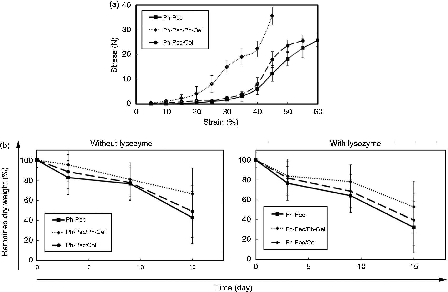
(a) Stress-strain profile of gels prepared from Ph-Pec, Ph-Pec/Ph-Gel, Ph-Pec/Col, (b) In vitro biodegradation rate of Ph-Pec, Ph-Pec/Ph-Gel, Ph-Pec/Col structures in PBS with and without lysozyme.
Biodegradation
Hydrogels should be resorbed by time inside the body to allow endogenous tissue generation. 5 As mentioned before, to simulate body condition, the lysozyme enzyme was added to the PBS solution. The data of remaining dry weight for Ph-pectin, Ph-pectin/Ph-gelatin, and Ph-pectin/collagen hydrogels demonstrated that scaffolds were degraded when they submerged in the PBS solution and the rate of degradation can be accelerated with the addition of lysozyme. The results revealed that after 15 days, approximately 32% and 50% of Ph-pectin hydrogel remained in PBS solution with or without lysozyme, respectively (Figure 3(b)). The percentage of the remaining dry weight in PBS and PBS/lysozyme media for Ph-pectin/collagen was 40% and 49% for 15 days. These values for Ph-pectin/Ph-gelatin were 52% and 66%, respectively at the end of the experiment (Figure 3(b)). These results confirm that the fabricated hydrogels are biodegradable and appropriate for tissue engineering aims. At low pH values, carboxyl groups of Ph-pectin remain protonated, and their repulsive force subsides. In this work, the pH value of the media was more than 4, which caused an increased swelling ratio, and as a result, faster degradation of the Ph-pectin hydrogel matrix due to the ionization of carboxyl groups and electrostatic repulsion of COO-. 42 Inconsistent with results obtained from the mechanical test, the mechanically weakest hydrogels degraded rapidly while the mechanically most robust hydrogels degraded later in both PBS and enzyme-containing solution. In Ph-pectin/Ph-gelatin, there are a higher number of Ph groups, which makes a larger number of crosslinking sites available. Based on the higher Ph content, this hydrogel is more robust and forms mechanically stronger crosslinks, which enable the matrix to tolerate degradation for a longer time compared to Ph-pectin/collagen and Ph-pectin.
WVTR
The desirable wound dressing hydrogels should have proper environmental relative humidity, temperature, and oxygen/gas permeable properties. 52 Thus, these conditions were determined by calculating the slope of the weight loss versus time plot for hydrogels in Figure 4(a) as WVTR. A dressing with a suitable WVTR is required because an unusually high WVTR may cause the dehydration of a wound, whilst an unsatisfied low WVTR may lead to the aggregation of wound moisture content. WVTR values can be explained depending on various situations where a wound dressing may be required. For normal or low injured skin, the optimal value of WVTR is around 150–200 g/m2/day, for first-degree burns, WVTR should be between 250–300 g/m2/day, and for granulating wounds, this value should be between 5000–5200 g/m2/day. Moreover, other external conditions such as environmental relative humidity and temperature can affect WVTR which should be considered. 53 Herein, the amounts of WVTR for Ph-pectin/Ph-gelatin, Ph-pectin, and Ph-pectin/collagen hydrogels were found 5493 gr/m2/day, 5737 gr/m2/day, and 5866 gr/m2/day, respectively that were close to the range of optimal WVTR to keep the sufficient value of water for wound dressing in the granulating wound site. SEM images and pore size results confirm that Ph-pectin/collagen hydrogels have a larger pore size compared to two other networks resulting in a rapid water release. As can be seen, there are no significant differences between the WVTR values, which have proved this fact all the hydrogels are similar and suitable for use in body tissue re-epithelialization.
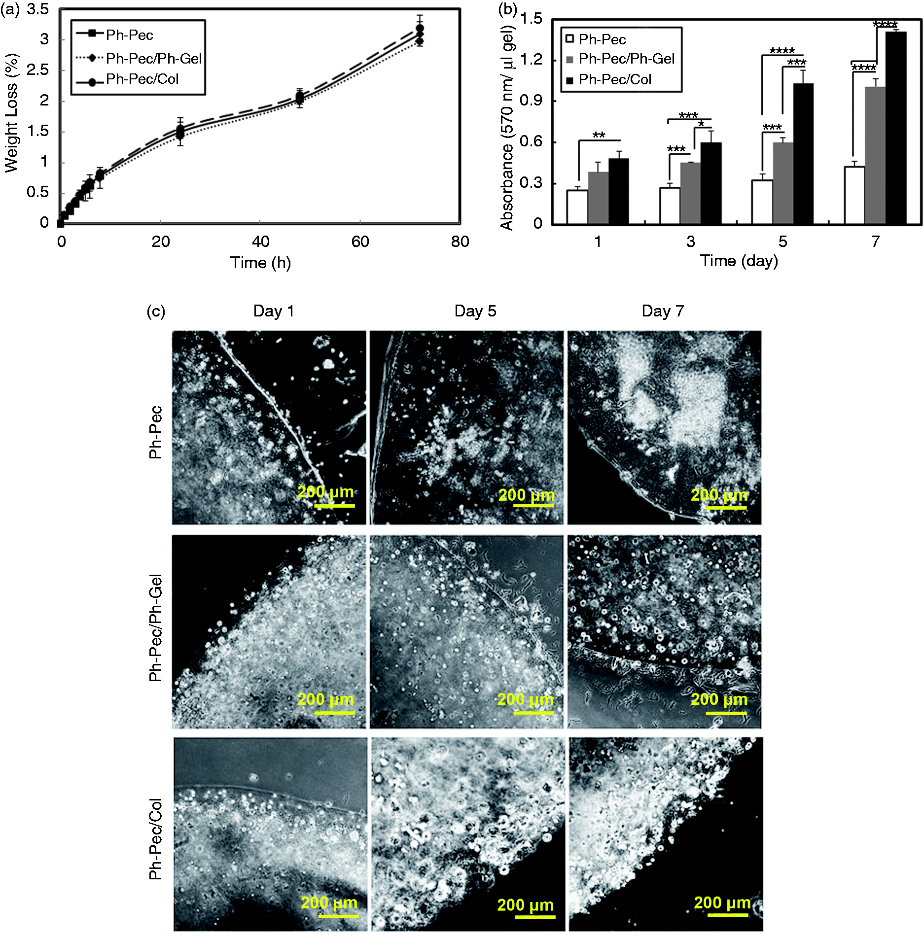
(a) Water vapor transmission rate of Ph-Pec, Ph-Pec/Ph-Gel, Ph-Pec/Col hydrogels, (b) Fibroblast viability in Ph-Pec, Ph-Pec/Ph-Gel, Ph-Pec/Col shown by MTT assay, (c) Light microscopic images of cell proliferation in 3 types of hydrogels at the end of 7 days, **P < 0.01; and ***P < 0.001, ****p < 0.0001.
Cytotoxicity
In this study, we aimed to assess the possible biocompatibility of prepared structures for tissue engineering applications. Thus, we used fibroblasts to address these issues. Since a perfect injectable scaffold should be biocompatible and nontoxic, microscopic bright-field imaging and MTT assay were used to validate the survival rate and cytocompatibility of fibroblasts inside the Ph-pectin, Ph-pectin/Ph-gelatin, and Ph-pectin/collagen hydrogels after 7- days in vitro (Figure 4(b) and (c)). According to data outlined in Figure 4(c), on day 7, the fibroblasts formed cellular aggregates, and colonies, which were in accordance with the results of MTT absorbance (Figure 4(b)). MTT results showed a 3.5-fold and 2.5-fold elevation in the amount of absorbance for Ph-pectin/collagen and Ph-pectin/Ph-gelatin compared to Ph-pectin hydrogels, respectively. Cell proliferation in a 3 D environment depends on various factors such as the type of hydrogels and type of cells in addition to biochemical signaling molecules and mechanical cues. 54 The presence of bioactive molecules such as RGD (Arginine, Glycine, and Aspartate residues - generally found in the natural ECM) in the network of the hydrogels can improve the bioactivity. The existence of RGD sequences in collagen and gelatin can induce cell adhesion because of their capability of providing cell-binding sites and their biological influence on cell function and survival. The porous morphology also facilitates the distribution of metabolites, nutrients, oxygen, and signaling agents. 55 SEM images revealed that Ph-pectin/Ph-gelatin and Ph-pectin/collagen hydrogels possess a desirable porous network. Owing to their structures that resemble the native ECM, the existence of RGD motifs in their backbone, and their porous framework, which allows for cell growth and transplantation, collagen and Ph-gelatin containing hydrogels increased cell proliferation compared to Ph-pectin. However, cell proliferation in Ph-pectin/collagen was even 1.4 fold higher than Ph-pectin/Ph-gelatin. Considering the role of hydrogel stiffness and pore size in cell growth and adhesion, there may be two justifications. 1) In our previous work, we reported that the stiffness of the hydrogels can decrease fibroblast proliferation rate since they lack sufficient proteolytic activity to overcome the physical barrier of compact frameworks. 31 Here, the mechanical properties experiment depicted that Ph-pectin/Ph-gelatin possess higher stiffness compared to Ph-pectin/collagen, which can account for promoted cell growth in collagen incorporated groups. 2) Cell culturing onto 3 D structures depends on large pore size, enabling cell penetration inside the 3 D scaffold and high porosity, which paves the way for nutrients and wastes exchange and cell growth. 56 The presence of pores provided a favorable environment for cell penetration in all three hydrogels. However, cells were only able to attach to the top surface of Ph-pectin and Ph-pectin/Ph-gelatin due to their smaller pore size compared to Ph-pectin/collagen. Promoted cell growth and attachment in collagen-containing conjugates can be explained by the fact that enhancing pore diameter has a positive influence on improving cell penetration and attachment. 11
Conclusion
Ph-modified enzymatically gellable hydrogels composed of Ph-pectin, Ph-pectin/Ph-gelatin, and Ph-pectin/collagen were prepared and evaluated for tissue engineering applications. Ph-pectin/Ph-gelatin hydrogels exhibited a homogenous structure, improved mechanical properties, and decreased degradation rate and WVTR. Despite the excellent physical properties of Ph-pectin/Ph-gelatin, Ph-pectin/collagen hydrogels induced an increased cell proliferation compared to other networks. This study highlights that not a single trait, but the suitable combination of mechanical characteristics, surface morphology, the presence of cell-binding sites, as well as an ECM-like network can dictate the fibroblast proliferation. All these findings together demonstrate the important role of collagen and Ph-gelatin in promoting the physical characteristics and cell function in pectin-based hydrogels.
Footnotes
Declaration of conflicting interests
The author(s) declared no potential conflicts of interest with respect to the research, authorship, and/or publication of this article.
Funding
The author(s) disclosed receipt of the following financial support for the research, authorship, and/or publication of this article: This study was supported by a grant from the Sahand University of Technology, Tabriz, Iran.