
Abstract
Hyperthermia has been suggested as a novel, minimally invasive cancer treatment method. After implantation of magnetic nano- or microparticles around a tumour through blood vessels, irradiation with alternating magnetic fields facilitates the efficient in situ hyperthermia even for deep-seated tumours. On the basis of this idea, if the microspheres are capable of delivering drugs, they could be promising multifunctional biomaterials effective for chemotherapy as well as hyperthermia. In the present study, magnetite microspheres were prepared by aggregation of the iron oxide colloid in water-in-oil (W/O) emulsion. The release behaviour of alendronate, a typical bisphosphonate, from the microspheres was examined in vitro as a model of the bone tumour prevention and treatment system. The alendronate was successfully incorporated onto the porous magnetite microspheres in vacuum conditions. The drug-loaded microspheres maintained their original spherical shapes even after shaking in ultrapure water for 3 days, suggesting that they have sufficient mechanical integrity for clinical use. It was attributed to high aggregation capability of the magnetite nanoparticles through van der Waals and weak magnetic attractions. The microspheres showed slow release of the alendronate in vitro, resulting from tight covalent or ionic interaction between the magnetite and the alendronate. The release rate was diffusion-controlled type and well controlled by the alendronate concentration in drug incorporation to the microspheres.
Introduction
Hyperthermia is a minimally invasive cancer treatment that is based on the finding that cancer cells have lower heat resistivity than normal cells. 1 However, development of hyperthermia effective even for deep-seated cancer cells remains as a problem to be solved. Novel cancer treatments using various ferromagnetic ceramic particles such as magnetite (Fe3O4) and γ-hematite (Fe2O3) may solve it.2,3 Implantation of ferromagnetic microspheres of 20–30 µm diameter into blood vessels around tumours facilitates the induction of more efficient hyperthermia combined with embolization to cut off the nutrient supply to the tumours.4–7 This type of embolization therapy is especially effective for liver and kidney cancers. 8
If these microspheres are also capable of delivering drugs, they could be promising multifunctional biomaterials effective for both hyperthermia and chemotherapy. Alendronate, a typical bisphosphonate, was chosen as a drug in the present study. It is clinically used for bone tumour treatment and inhibition of osteoporosis.9,10 Although oral administration is widely used to deliver alendronate, there are several drawbacks to this method such as low biological availability (less than 1%) and side effects due to excess dosages. 11 Therefore, the development of a local and sustainable drug delivery system is needed. The preparation of bisphosphonate-modified magnetite nanoparticles has been previously reported. 12 However, the release behaviour of bisphosphonate from magnetite-based nano- or microparticles has not yet been reported.
In this study, magnetite microspheres were prepared using emulsion methods. Alendronate was incorporated into microspheres and its release was quantitatively assessed as a model of bone tumour prevention and treatment.
Materials and methods
Preparation of microspheres
All the chemical reagents were purchased from Wako Pure Chemical Industries, Ltd., Japan. An aqueous volume of 37.5 mL of 37 mM FeCl2 and 49 mM FeCl3 was mixed with 8 mL of 26 wt% NH3 solution. The precipitate that formed was collected by centrifugation, washed with ultrapure water, and dispersed in 22.5 mL of 0.2 M HCl. The solution was then heated at 80℃ for 3 h to form a sol. A water-in-oil (W/O) emulsion was then obtained by mixing 2-ethyl-1-hexanol containing 3 wt% Span80 and the sol at weight ratio of 3:1 at a rotation speed of 3400 r/min. A precipitate was immediately formed by addition of the emulsion to 1-butanol. Further details of this preparation technique and mechanism of the microsphere formation were described previously. 6
The microstructure of the microspheres was evaluated by scanning electron microscopy (SEM; S-3500N, Hitachi Co., Tokyo, Japan), X-ray diffraction (XRD; MXP3V, Mac Science Ltd., Yokohama, Japan) and Brunauer, Emmett, and Teller (BET) surface area and pore size analyser (Autosorb-1 C/MS, Quantachrome Instruments, Boynton Beach, FL). A thin film of Au-Pd was deposited on the surfaces of the specimens using an ion sputter coater (E-101, Hitachi Co.) for SEM observations. To evaluate mechanical properties, 10 mg of the microspheres was soaked in 30 mL of ultrapure water and shaken in a water bath (H-10, Taitec Co., Koshigaya, Japan) at a speed of 100 strokes/min for 3 days. Morphological changes of the microspheres were then observed by SEM.
In vitro drug release profiles
A mass of 30 mg of the prepared microspheres was soaked in 20 mL of sodium alendronate trihydrate (NH2(CH2)3COH(PO3H2)(PO3HNa)·3H2O) solution at various concentrations for 1 h under vacuum at 0.03 MPa, gently washed with ultrapure water and dried at 60℃. Next, 10 mg of the drug-loaded microspheres was soaked in 20 mL of ultrapure water at room temperature for various periods. The release test was performed in static conditions. The alendronate concentration was quantitatively determined by inductively coupled plasma atomic emission spectroscopy (Optima 4300DV Cyclon, Perkin-Elmer Co., Cambridge, UK).
Results
Figure 1 shows an SEM image of the prepared microspheres. Porous microspheres with a diameter of approximately 30–40 µm were obtained. The pore size on the surfaces ranged from 1 to 5 µm. The XRD pattern of the microspheres is shown in Figure 2. Broad diffraction peaks assigned to the magnetite (JCPDS#19-0629) were detected at 30°, 35°, 43°, 54°, 58° and 63° in 2θ. The BET surface area was 82.3 m2/g and the average pore diameter was 17.6 nm.
Scanning electron microscopy (SEM) image of the prepared microspheres. X-ray diffraction (XRD) pattern of the prepared microspheres.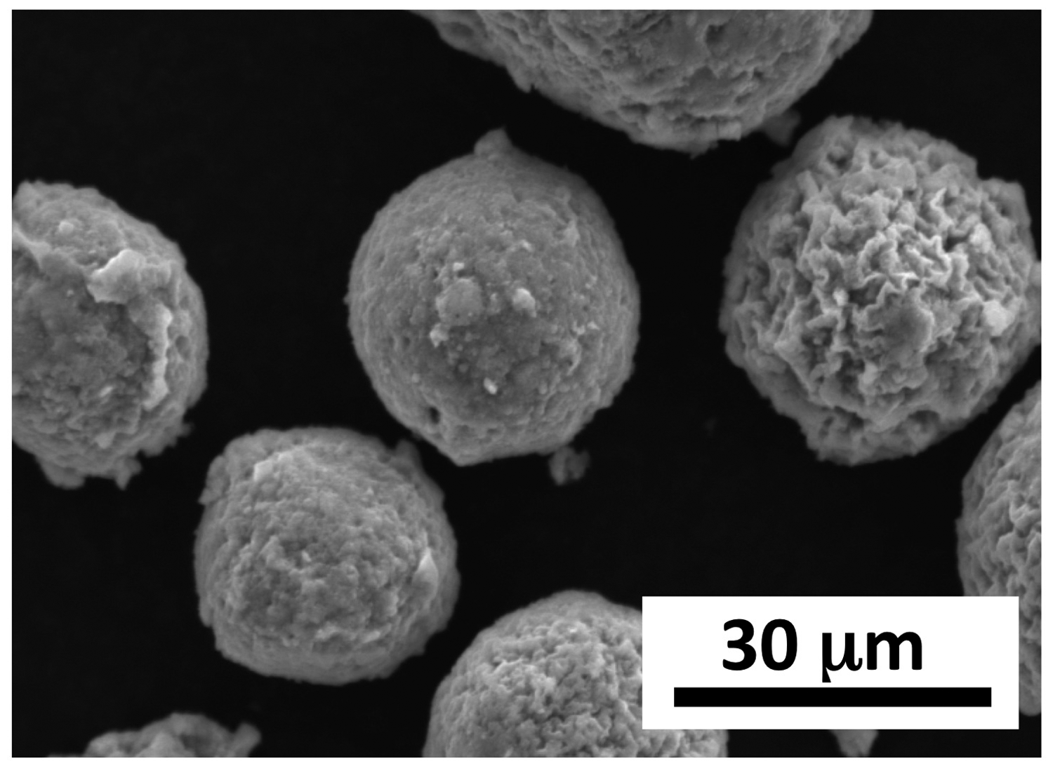
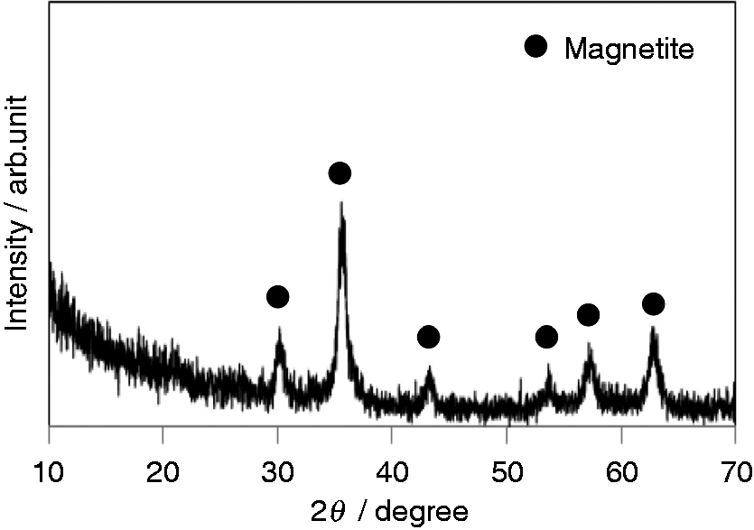
Figure 3 shows an SEM photograph of the prepared microspheres after being shaken in ultrapure water for 3 days. Their original spherical shapes were well maintained and significant deformation was not observed even after the shaking. In addition, the pore size on the surfaces was hardly changed.
Scanning electron microscopy (SEM) image of the prepared microspheres after being shaken in ultrapure water for 3 days.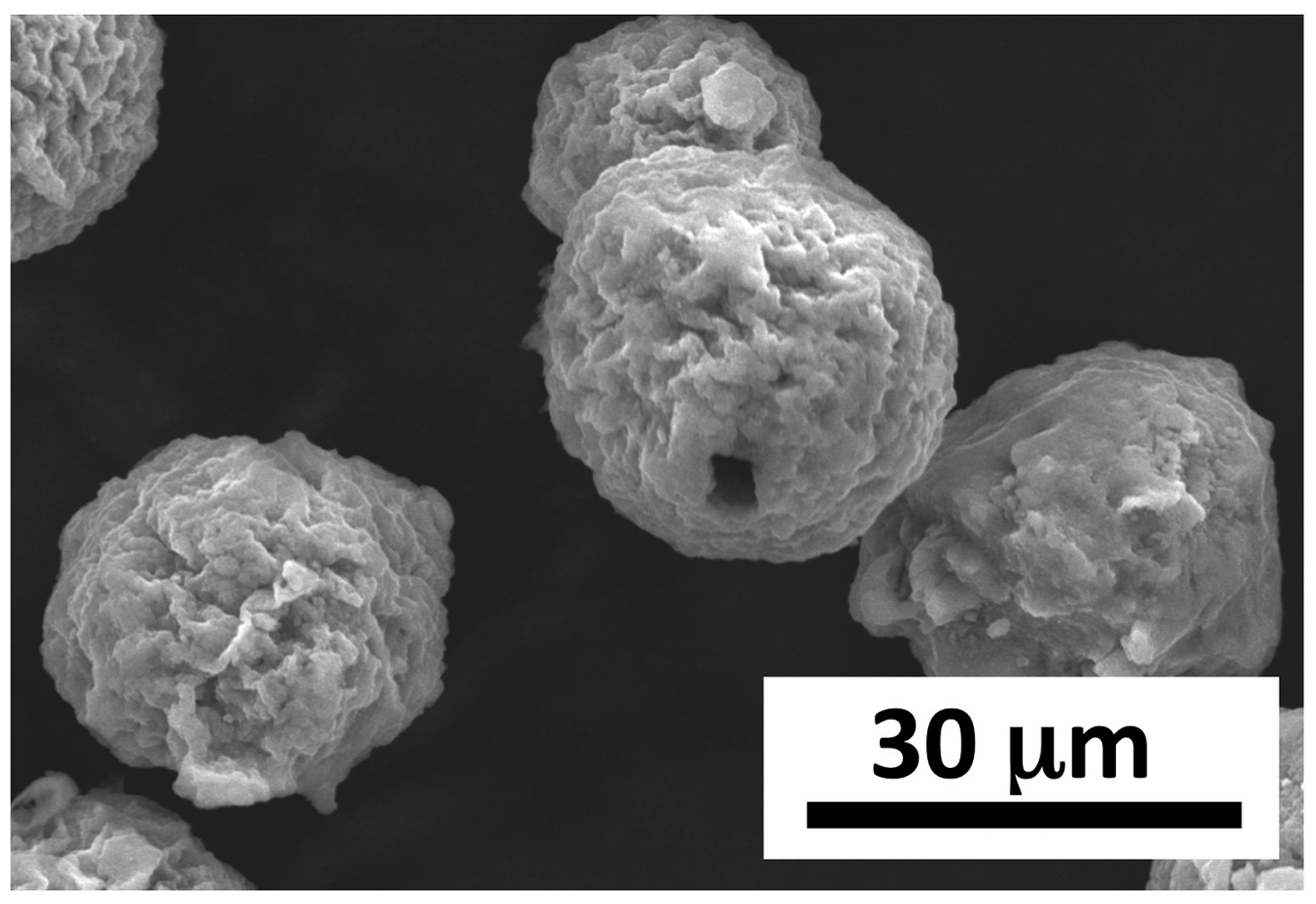
Figure 4 shows the content of alendronate incorporated into the microspheres as a function of the alendronate concentration. The incorporated mass increased with the increasing alendronate concentration in solution.
Mass of alendronate incorporated into the microspheres as a function of the alendronate concentration.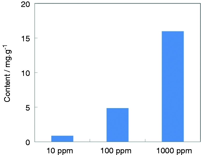
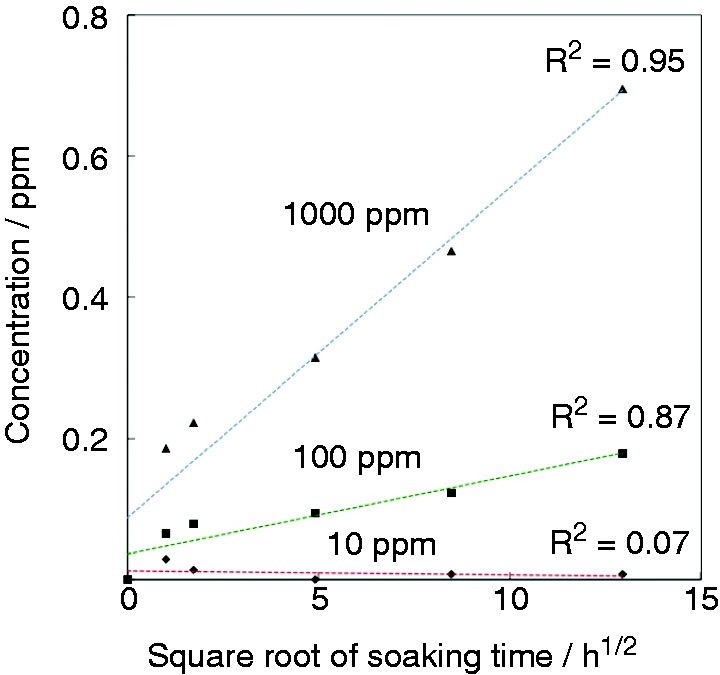
Figure 5 shows the alendronate released from drug-loaded microspheres into ultrapure water after various periods as a function of the initial alendronate concentration. The microspheres with initial concentrations of 100 and 1000 ppm gradually released alendronate. The degree of alendronate release increased with increasing initial alendronate concentration. The released concentration was plotted as a function of the square root of soaking time (Figure 6) showing highly linear correlations for the 100 and 1000 ppm concentration samples. These results indicate that the process of alendronate release is diffusion-controlled.
Alendronate concentration after soaking drug-loaded microspheres in ultrapure water after various periods as a function of the initial alendronate concentration. Alendronate concentration after soaking drug-loaded microspheres in ultrapure water plotted as a function of the square root of time for different initial alendronate concentrations.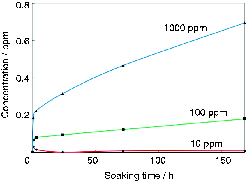
Discussion
Magnetite microspheres were prepared by dehydration of a W/O emulsion containing the magnetite sol derived from Fe2+ and Fe3+. In a prior study by the present authors, non-magnetic microspheres of iron (III) hydroxide were converted into magnetite by hydrothermal treatment in an aqueous solution containing ethyleneglycol and urea. 6 In the present study, magnetite microspheres were obtained without further thermal or hydrothermal treatments. Based on the present results, this streamlined fabrication process is effective. Magnetite nanoparticles can easily aggregate in aqueous conditions through van der Waals and weak magnetic attractions. 13 The present microspheres are assumed to be constructed by dehydration and aggregation of the magnetite nanoparticles in butanol. Because the prepared microspheres were not broken after being shaken in ultrapure water (Figure 3), they are expected to have a low risk of injuring the surrounding blood vessels in vivo caused by broken fragments. The high aggregation potential of the magnetite nanoparticles likely contributes to the high mechanical integrity of the microspheres.
The prepared microspheres were able to incorporate and release alendronate, and that release rate was controlled by the alendronate concentration. Iron oxide and phosphate can construct a complex via formation of an Fe–O–P bond.
14
Therefore, the phosphate group of the alendronate may construct a similar complex with magnetite crystals as shown schematically in Figure 7. The released portion of alendronate from the microspheres after 7 days was estimated to be 8% based on the results in Figures 4 and 5. Tight bonding between the magnetite and the alendronate would contribute to the prevention of an initial bolus loss and increase continuous drug release.
Schematic representation of binding of alendronate to magnetite.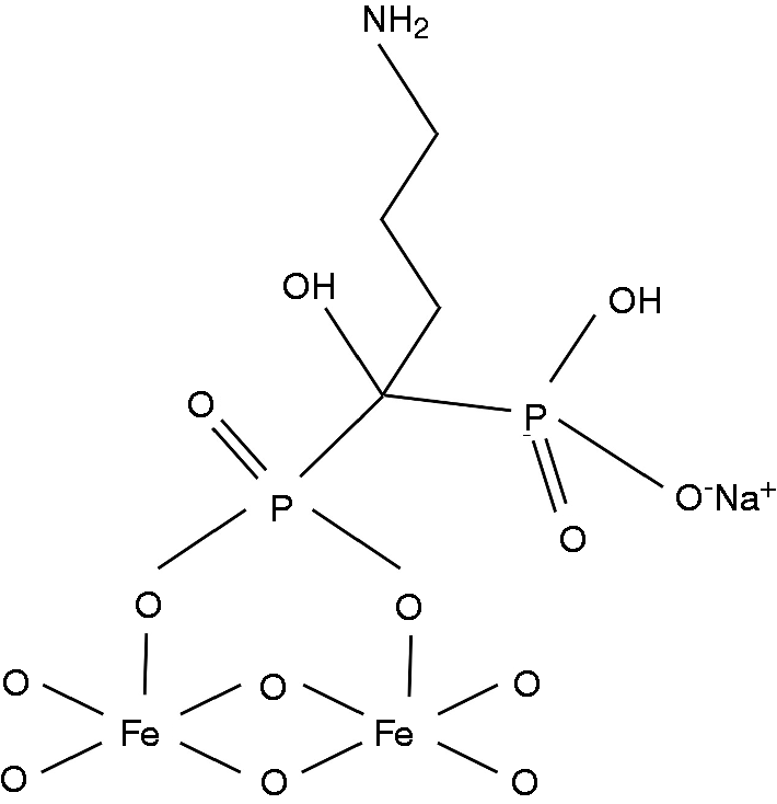
The release of bisphosphonate from hydroxyapatite-based microparticles has been reported by several research groups. Seshima et al. investigated the release of pamidronate, a type of bisphosphonate, from calcined hydroxyapatite granules of 300–500 µm in diameter. 15 After soaking the calcined granules at 400℃ for 3 days, the pamidronate concentration was 0.3 mM and the survival rate of osteoclasts was decreased by up to 40%. This indicates that bone absorption can be suppressed by pamidronate release.
In addition, Shi et al. prepared composite microspheres of poly(
Although the surface area of the magnetite microspheres (82.3 m2/g) prepared in the present study was larger than that of the hydroxyapatite prepared by Seshima et al. (46.2 m2/g), the mass of released alendronate after 3 days (1.5 µM at maximum) was smaller than that reported in the above two papers. This might be attributed to a smaller powder to liquid ratio in the drug release measurement. In addition, magnetite is likely more positively charged than hydroxyapatite because the isoelectric point of the former (6.5) is higher than that of the latter (6.0).17,18 Thus, the tight ionic attraction of the magnetite with the negatively charged alendronate may also contribute to the slower release. However, further increase in alendronate concentration loaded in the microspheres may increase the release up to the level necessary for effective enhancement of bone formation.
Porous magnetite microspheres were prepared using emulsion methods. These microspheres showed slow release of alendronate, a bisphosphonate drug. The release rate could be well controlled by the drug concentration loaded into the microspheres. The microspheres were able to release the drug over a long time and are therefore useful for novel hyperthermia and chemotherapy combined treatments.
Footnotes
Funding
This work was supported by Adaptable & Seamless Technology Transfer Program through Target-driven R&D (A-STEP) from The Japan Science and Technology Agency (JST).
Conflict of interest
The authors declare that there is no conflict of interest.