
Abstract
Dental implants are the most innovative and superior treatment modality for tooth replacement. However, titanium implants still suffer from insufficient antibacterial capability and peri-implant diseases remain one of the most common and intractable complications. To prevent peri-implant diseases, a composite coating containing a new antibacterial agent, (Z-)-4-bromo-5-(bromomethylene)-2(5H)-furanone (BBF) was fabricated on titanium. This study was designed to investigate the antibacterial activity of the composite coating against two common peri-implant pathogens (Porphyromonas gingivalis and Actinobacillus actinomycetemcomitans). The morphology of the composite coating showed that BBF-loaded poly(L-lactic acid) nanospheres were well-distributed in the pores of the microarc oxidation coating, and cross-linked with each other and the wall pores by gelatin. A release study indicated that the antibacterial coating could sustain the release of BBF for 60 d, with a slight initial burst release occurring during the first 4 h. The antibacterial rate of the composite coating for adhering bacteria was the highest (over 97%) after 1 d and over 90% throughout a 30-day incubation period. The total fluorescence intensity of the composite coating was the lowest, and the vast majority of the fluorescence was red (dead bacteria). Moreover, real-time polymerase chain reaction analysis confirmed that the relative gene expression of the adherent bacteria on the composite coating was down-regulated. It was therefore concluded that the composite coating fabricated on titanium, which showed excellent and relatively long-term antibacterial activity against Porphyromonas gingivalis and Actinobacillus actinomycetemcomitans, is a potential and promising strategy to be applied on dental implants for the prevention of peri-implant diseases.
Introduction
Titanium implants have revolutionized dentistry due to their ability for osseointegration and have become one of the most desirable options to repair missing teeth in both fully and partially edentulous patients. The survival rate of dental implants has been frequently reported to be as high as 93.8%–95% in longitudinal studies.1,2 However, it has become clear that a surviving implant is not necessarily a successful implant, and the surviving implants have been found to be affected by a number of risk factors resulting in biological complications. 3 Peri-implant diseases (peri-implant mucositis and peri-implantitis), which have been characterized as a condition of inflammation and infection of the peri-implant supportive tissues associated with pathogenic microorganisms, represent important biological complications in dental implants. 4 A recent systematic review found that peri-implant mucositis occurred in 46.8% of individuals and 29.5% of implants, and peri-implantitis occurred in 19.8% of individuals and 9.2% of implants. 5 Peri-implant mucositis is considered the preceding step in the infection process leading to peri-implantitis. The continued accumulation of inflammatory infiltrate around the implants promotes the disease progression to hard tissues and simultaneous peri-implant bone loss, which may result in implant failure. 6 Because peri-implant diseases do not respond predictably to treatment and no protocol has been shown effective at arresting the condition and/or reversing it, it is highly desirable to prevent peri-implant infection. 7
Understanding the pathogenesis causing peri-implant diseases is required prior to considering any method to prevent these diseases. A well-supported hypothesis suggests that the etiology of peri-implant diseases is bacterial adhesion and biofilm formation on the implant surface. 8 Additionally, recent evidence suggests that peri-implant infection is associated with Porphyromonas gingivalis, Actinobacillus actinomycetemcomitans, Prevotella intermedia, etc.9,10 Antibacterial coatings have been traditionally designed on implants to prevent initial adhesion of bacteria. There have been studies on incorporating antibacterial agents into the surface on titanium.11,12 However, most of these attempts fail to deliver long-term antibacterial effects. In addition, the emerging appearance of bacterial insusceptibility and drug resistance also encourages the search for novel effective antibacterial agents. 13
Halogenated furanones, a group of secondary metabolites originally extracted from the red alga Delisea pulchra, have shown antibacterial properties that inhibit microbial growth, swarming and biofilm formation. 14 Subsequently, a variety of analogs of natural halogenated furanones have been synthesized, and these compounds have been shown to possess potent antibacterial activity against a great number of Gram-positive and Gram-negative bacteria.15,16 Halogenated furanone compounds can inhibit quorum sensing in various microbial species by affecting acyl-homoserine lactones and autoinducer-2, which are used for cell-to-cell communication to control fundamental physiological processes involved in DNA replication, virulence factor expression and biofilm formation. 17 It has been reported that biofilms contribute to drug resistance through several mechanisms. 18 These antibacterial mechanisms would reduce the risk of halogenated furanones developing drug resistance. Moreover, many studies have evaluated the effects of halogenated furanones on eukaryotic cell viability, but few studies have reported on the toxicity of halogenated furanones.19,20 Given their advantages, halogenated furanone compounds have been considered potential and ideal antibacterial agents for preventing infection on medical devices.
In our previous study, a halogenated furanone compound, (Z-)-4-bromo-5-(bromomethylene)-2(5H)-furanone (BBF), was incorporated into poly(L-lactic acid) (PLLA) nanospheres for the purpose of sustained release, and then a composite coating was fabricated by cross-linking these BBF-loaded PLLA nanospheres onto the microarc oxidation coating on titanium (Ti). 21 The composite coating showed excellent osteoblastic compatibility. 22 In this scenario, the current study was specifically designed to further investigate the antibacterial activity of the composite coating against two peri-implant common pathogens (Porphyromonas gingivalis and Actinobacillus actinomycetemcomitans) to provide a laboratory basis for its further dental implant applications.
Materials and methods
Materials
(Z-) - 4 - bromo - 5-(bromomethylene) - 2(5H) - furanone (BBF), poly(L-lactic acid) (PLLA, molecular weight 152,000), poly(vinyl alcohol) (PVA: molecular weight 89,000–98,000) and gelatin powder (type A) were purchased from Sigma-Aldrich Chemicals (USA). Commercial pure titanium (cp-Ti, grade 2) was provided by Xi’an Aerospace New Material Co., Ltd (China). Brain heart infusion (BHI) medium (BHI medium) was purchased from Oxoid (UK). SYTO 9 and propidium iodide dyes (LIVE/DEAD® BacLight™ Bacterial Viability Kits) were supplied by Invitrogen (USA). TRIZOL reagent, PrimeScript™ RT reagent kit, and SYBR® Premix Ex Taq II were provided by TaKaRa (Japan). Other chemicals and solvents (analytical grade) were supplied by Tianjin Fuchen Chemical Factory (China).
Fabrication of the composite coating on titanium
Preparation of the BBF-loaded PLLA nanospheres (BBF/PLLA-NSs)
The BBF/PLLA-NSs were prepared by the oil-in-water (O/W) emulsion solvent-evaporation method. Briefly, 15 mg BBF and 100 mg PLLA were co-dissolved in 2 mL dichloromethane (DCM) to form the oil phase, and 40 mL of a 1% PVA aqueous solution was used as the water phase. In an ice bath, the oil phase was dispersed in the water phase by sonication at 300 W for 200 s using an ultrasonic homogenizer (JY98-IIIN, Xinzhi, China) to form an O/W emulsion. After emulsification, the O/W emulsion was stirred for 6 h under reduced pressure to evaporate the DCM. The formed nanospheres were isolated by centrifugation for 20 min at 12,000 rpm. The collected nanospheres were then washed three times with distilled water in an ultrasonic bath at 37 °C to remove the residual PVA, isolated by centrifugation and freeze-dried. The blank PLLA nanospheres were prepared by the same method, except that no BBF was added to the oil phase.
Characterization of the BBF/PLLA-NSs
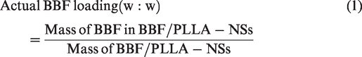
The surface appearance and shape of the BBF/PLLA-NSs were observed with scanning electron microscopy (SEM, S-4800, Hitachi, Japan). The particle size of the nanospheres was assessed by dynamic light scattering using a particle size analyzer (SALD-7101, Shimadzu Corporation, Japan). The amount of BBF incorporated into the BBF/PLLA-NSs was determined by high-performance liquid chromatography (HPLC, Waters, USA). Five milligrams of freeze-dried BBF/PLLA-NSs was dissolved in 1 mL DCM and stirred after the addition of 10 mL methanol to preferentially precipitate the polymer. The precipitate was removed with a membrane filter. Then, the solvent was evaporated until dry with a nitrogen stream, followed by adding 10 mL of a mixture of methanol/water (75/25, v/v). The clear solution obtained was placed into a vial for HPLC to detect the BBF concentration. BBF incorporation was expressed both as actual BBF loading (w/w) and encapsulation efficiency (%) represented by equations (1) and (2), respectively, as
Preparation of the MAO coating (the inner coating) on Ti
The cp-Ti specimens (10 mm × 10 mm × 1 mm) were ground using various grades of SiC paper from 200 to 1200 grit, and ultrasonically cleaned in acetone, ethanol and deionized water. The polished cp-Ti specimens were used as anodes, and stainless steel plates were used as cathodes in an electrolytic bath, in which a mixed aqueous solution containing 0.2 M calcium acetate and 0.04 M β-glycerophosphate sodium was used as the aqueous electrolytic solution. 23 The MAO coating was prepared by MAO equipment (designed and manufactured by Xi’an University of Technology, Xi’an, China) using a pulsed direct current power supply. The applied voltage, frequency, duty cycle and oxidizing time were 300 V, 600 Hz, 8.0% and 5 min, 24 respectively. Then, the MAO-treated titanium (MAO-Ti) specimens were rinsed with distilled water and air-dried immediately. The surface morphology of the specimens before and after MAO treatment was observed with SEM.
Fabrication of the composite coating on Ti
The composite coating was fabricated by cross-linking the BBF/PLLA-NSs onto the MAO coating with gelatin. In brief, 20 mg freeze-dried BBF/PLLA-NSs was dispersed by sonication in 5 mL 0.2% gelatin solution, from which 400 µL suspension was dropped onto each MAO-Ti specimen. Then, the specimens were placed on an oscillator for 1 h to enable the nanospheres to penetrate the pores on the MAO coating. Subsequently, they were dried at 4 °C and immersed in 2.5% glutaraldehyde solution for 30 min to cross-link the gelatin. Finally, the specimens were washed with ethanol three times to remove the remaining glutaraldehyde and freeze-dried. The surface morphology of the composite coating was observed with SEM.
In vitro release of BBF from the composite coating
A release study of BBF from the composite coating was performed by placing one composite coating specimen with 5 mL phosphate-buffered saline (PBS, pH = 7.4) in a dialysis bag (molecular weight cut-off at 3,500 g/mol). This bag was then immersed into 20 mL PBS with continuous gentle magnetic stirring at 37 °C. At predetermined time intervals, 1 mL aliquots were withdrawn and replaced with the same volume of fresh buffer. All samples were first extracted with 1 mL DCM, followed by solvent evaporation with a nitrogen stream, and then 10 mL of the mixture of methanol/water (75/25, v/v) was added. The BBF concentration was also determined by HPLC analysis.
Experimental groups and surface roughness
All the assays were divided into four groups: (1) BBF/PLLA-MAO-Ti (the composite coating): prepared by cross-linking the BBF/PLLA-NSs with gelatin onto the MAO-Ti specimens, (2) PLLA-MAO-Ti: prepared by cross-linking the blank PLLA nanospheres (without BBF) with gelatin onto the MAO-Ti specimens, (3) MAO-Ti: the MAO-treated specimens, and (4) polished cp-Ti. All the specimens were sterilized through exposure to 20 kGy 60cobalt radiation prior to the antibacterial assays.
The surface roughness of the groups was measured by contact profilometry using a surface profilometer (TR240, TIME Group, China). The sampling length was 0.8 mm, and the evaluation length was 4 mm (5 times the sampling length). Four roughness parameters were measured to describe the surface roughness: average roughness (Ra), range of the roughness (Rt), peak-to-valley roughness (Rz), and peak-to-mean height roughness (Rp). Ra is the numerical average of the heights of peaks and depths of valleys observed on a surface profile. Rt is the summation of the height of the highest peak and depth of the deepest valley observed on a surface profile. Rz is the summation of the average peak height of the five highest peaks and the average valley depth of the five deepest valleys observed on a surface profile. Rp is the height from the profile peak line to the profile mean line observed on a surface profile. The surface roughness of the groups would provide an explanation for the results of antibacterial assays.
In vitro antibacterial assays of the composite coating
Bacterial culture
Porphyromonas gingivalis (Pg, ATCC33277) and Actinobacillus actinomycetemcomitans (Aa, ATCC29523) (supplied by School of Stomatology, Fourth Military Medical University) were used in these antibacterial assays. They were cultivated in BHI medium supplemented with blood, and were adjusted to a concentration of 106 CFU/mL in BHI broth. The specimens were placed in separate wells of 24-well culture plates and incubated in 1 mL of the bacterial suspension under standard anaerobic conditions (80% N2, 10% H2, 10% CO2, at 37 °C).
Antibacterial rate (AR) assay
The bacterial suspension was replaced with 1 mL fresh bacterial suspension every day for a total of 30 d. At predetermined time intervals, the specimens were gently rinsed three times with PBS to remove nonadherent bacteria, and the adherent bacteria on the specimens were gently rinsed with 10 mL PBS and ultrasonically detached for 5 min. Then, the bacterial suspensions were recultivated on BHI agar plates for colony counting with ImageJ software. The AR was determined by the following formula: AR (%) = (CFU of control − CFU of experimental groups)/CFU of control × 100%, where polished cp-Ti served as the control group, BBF/PLLA-MAO-Ti, PLLA-MAO-Ti and MAO-Ti constituted the experimental groups.
Viability of the bacteria assay
After incubation for 1 d, the specimens were rinsed three times with PBS, stained with SYTO 9 and propidium iodide dyes for 15 min in the dark and examined by laser scanning confocal microscopy (FV1000, Olympus, Japan). For accurate evaluation of the amounts of colonized bacteria, the fluorescence intensities of green, red and total bacteria were analyzed by ImageJ software.
Real-time polymerase chain reaction (RT-PCR)
After incubation for 1 d, the specimens were rinsed three times with PBS, and the total RNA of the cultured bacteria was extracted by TRIZOL reagent. One microgram of RNA from each specimen was reverse transcribed into complementary DNA by a PrimeScript™ RT reagent kit according to the manufacturer’s protocol. The target genes and primer sequences of the genes are shown in Table 1, and 16S rRNA served as an endogenous housekeeping gene for normalization. Gene expression was quantified using RT-PCR (Bio-Rad iQ5 Multicolor RT-PCR Detection System) with SYBR® Premix Ex Taq II. Data analysis was carried out using iQ5 Optical System Software Version 2.0 (Bio-Rad).
RT-PCR target genes and primer sequences of the bacteria.

RT-PCR: real-time polymerase chain reaction; Pg: porphyromonas gingivalis; Aa: actinobacillus actinomycetemcomitans; 16S rRNA: 16S ribosomal ribonucleic acid; Fim A: fimbrilline A; Cdt B: cytolethal distending toxin B.
Statistical analysis
Each assay in this study was repeated six times, and the data are expressed as the mean ± standard deviation. One-way analysis of variance (ANOVA) combined with a Student–Newman–Keuls (SNK) post hoc test was used to determine the level of significance. Differences were considered statistically significant for values of p < 0.05.
Results
Fabrication of the composite coating on titanium
BBF/PLLA-NSs, MAO coating and composite coating
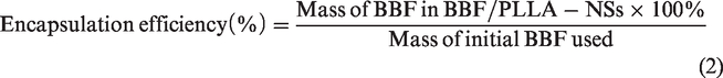
SEM images showed that the BBF/PLLA-NSs were spherical in shape without any apparent aggregation or adhesion, as shown in Figure 1(a). The particle size measured by the particle analyzer revealed that the BBF/PLLA-NSs had a mean particle size of 408 ± 14 nm with a polydispersity index of 0.140 ± 0.008 (Figure 1(b)). The actual BBF loading and encapsulation efficiency of BBF in BBF/PLLA-NSs were 0.102 ± 0.002 (mg/mg) and 72.44 ± 1.27%, respectively. The surface of the polished cp-Ti specimens was smooth (Figure 1(c)). After MAO treatment, a porous microstructure composed of small craters with pores of 1–o µm diameter was observed with SEM (Figure 1(d)). Figure 2(e) and (f) show the surface morphology of the composite coating on Ti. The pores of the MAO coating were covered with cross-linked nanospheres, and only few nanospheres adhered to the flat areas surrounding the pores (Figure 1(e)). At high magnification, the BBF/PLLA-NSs were well-distributed in the pores of the MAO coating, and cross-linked with each other and the wall of pores by gelatin (Figure 1(f)).
Surface morphology and size distribution. (a) SEM image of the BBF/PLLA-NSs (×5,000). (b) Size distribution of the BBF/PLLA-NSs. (c) SEM image of the polished cp-Ti specimens (×10,000). (d) SEM image of the MAO coating on Ti (×10,000). (e) SEM image of the composite coating on Ti at low magnification (×10,000). (F) SEM image of the composite coating on Ti at high magnification (×25,000).

In vitro release profiles of BBF from the composite coating. (a) Initial 24 h of the release study. (b) Experimental values for the complete assay.
In vitro release study of BBF from the composite coating
The in vitro release profile of BBF from the composite coating showed a biphasic release phenomenon (Figure 2). Initially, small amounts of BBF (9.4%) were burst released into PBS during the first 4 h (Figure 2(a)). This rapid release was followed by a slow and sustained release. In the sustained release period, 80.8% of BBF was released from the composite coating during the next 60 d (Figure 2(b)).
Surface roughness of the experimental groups
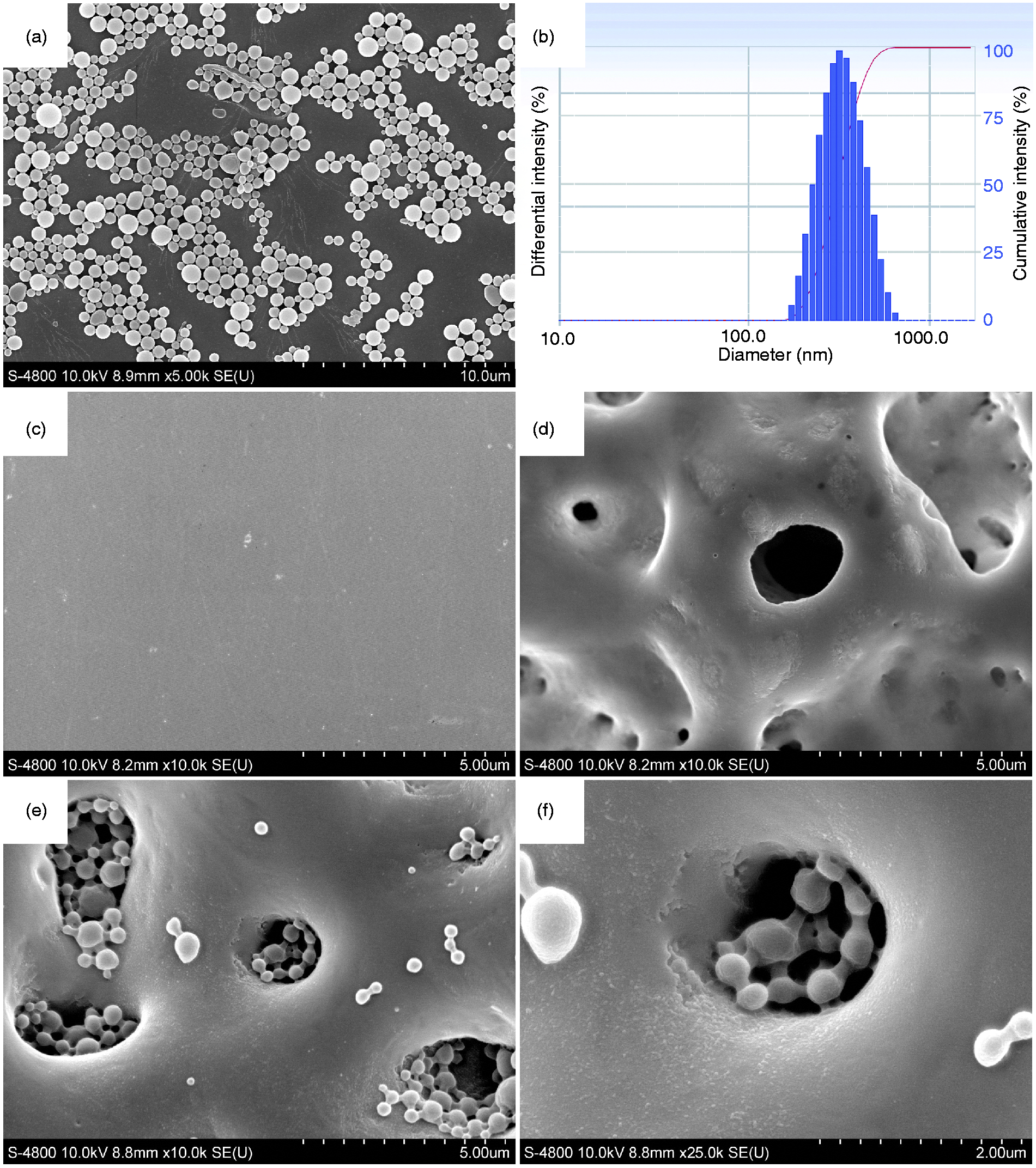
The four roughness parameters of the groups are compared in Figure 3. The roughness of MAO-Ti was the highest, and the roughness of polished cp-Ti was the lowest among the four groups (p < 0.05). There was no statistically significant difference between BBF/PLLA-MAO-Ti and PLLA-MAO-Ti (p > 0.05).
Surface roughness parameters of the groups. *p < 0.05.
In vitro antibacterial assays of the composite coating
Antibacterial rate (AR) assay
At predetermined time intervals, the adherent bacteria (Pg and Aa) were detached from the specimens and recultivated on BHI agar plates to count the colonies with ImageJ software. Figure 4 shows the typical images of the recultivated Pg and Aa colonies from the groups on BHI agar plates after 1 d. The numbers of the recultivated colonies of the two adherent bacteria were similar in the four groups. The number of colonies on BBF/PLLA-MAO-Ti (the composite coating) on BHI agar plates was the lowest (less than 20), and the number of colonies of MAO-Ti was the highest (more than 600) (p < 0.05). The number of colonies on the polished cp-Ti was lower than that on PLLA-MAO-Ti (p < 0.05). The ARs of the experimental groups for the two bacteria for a total of 30 d were also similar, as shown in Figure 5. After incubation for 1 d, BBF/PLLA-MAO-Ti possessed a high AR value of over 97%, and on the second day, the AR decreased slightly to approximately 92% (p < 0.05). There was no significant difference in the AR values throughout the 30-day incubation period (p > 0.05). Compared to BBF/PLLA-MAO-Ti, the ARs of PLLA-MAO-Ti and MAO-Ti were negative, which illustrated that there were more colonies than on polished cp-Ti (as shown in Figure 4). The AR of PLLA-MAO-Ti was higher than that of MAO-Ti at the same incubation time (p < 0.05).
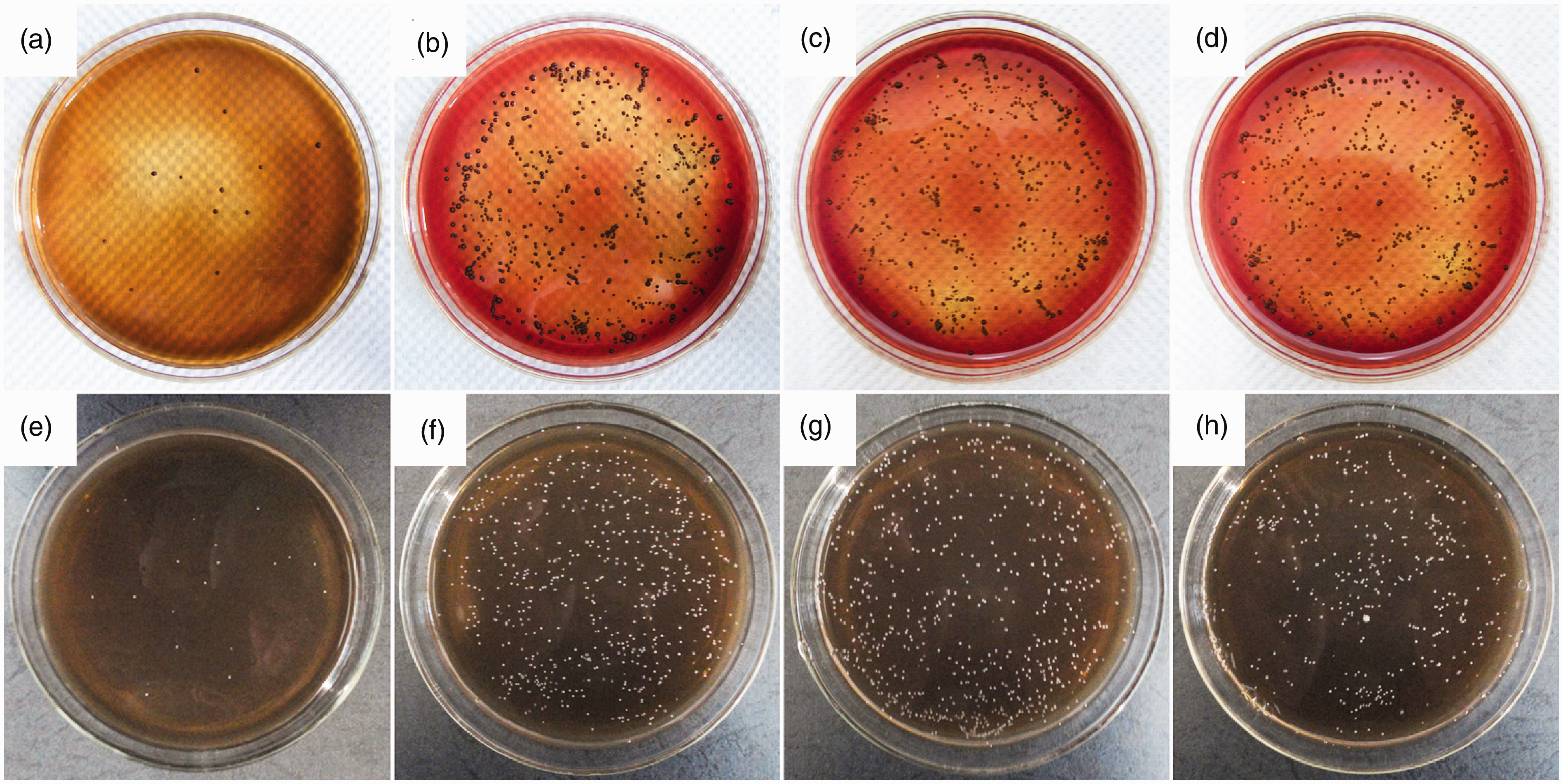
Typical images of recultivated Pg and Aa colonies on BHI agar plates coated with the different groups after 1-day incubation. (a) BBF/PLLA-MAO-Ti, Pg. (b) PLLA-MAO-Ti, Pg. (c) MAO-Ti, Pg. (d) Polished cp-Ti, Pg. (e) BBF/PLLA-MAO-Ti, Aa. (f) PLLA-MAO-Ti, Aa. (g) MAO-Ti, Aa. (h) Polished cp-Ti, Aa.
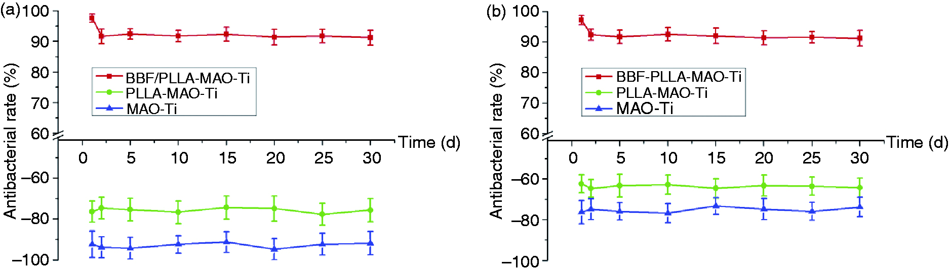
Antibacterial rate of the different groups for adherent bacteria throughout a 30-day incubation. (a) Pg. (b) Aa.
Viability of bacteria assay
The viability of bacteria in the four groups after incubation for 1 d was determined by fluorescence staining, as shown in Figure 6. The red fluorescent propidium iodide and green fluorescent SYTO 9 differ in their abilities to penetrate bacteria. When both dyes are employed, live bacteria with intact membranes are stained by SYTO 9 (green), and dead bacteria with damaged membranes are stained by propidium iodide (red). The fluorescence intensity therefore reflects the amount of adherent bacteria. The fluorescence staining images of the two bacteria adhered to the four groups were similar. Small amounts of red fluorescence (dead bacteria) and almost no green fluorescence (live bacteria) could be observed on BBF/PLLA-MAO-Ti (Figure 6(a) and (e)). In comparison, large amounts of green fluorescence and nearly no red fluorescence were observed on PLLA-MAO-Ti, MAO-Ti and polished cp-Ti. Analyzed by ImageJ software, the green, red and total fluorescence intensities in the images are shown in Figure 7. The total fluorescence intensity of BBF/PLLA-MAO-Ti was low, and the vast majority of the fluorescence was red. In contrast, high total fluorescence intensities were observed on the other three specimens, but the red fluorescence was very low. The total fluorescence intensity on the surface of MAO-Ti was the highest (p < 0.05).
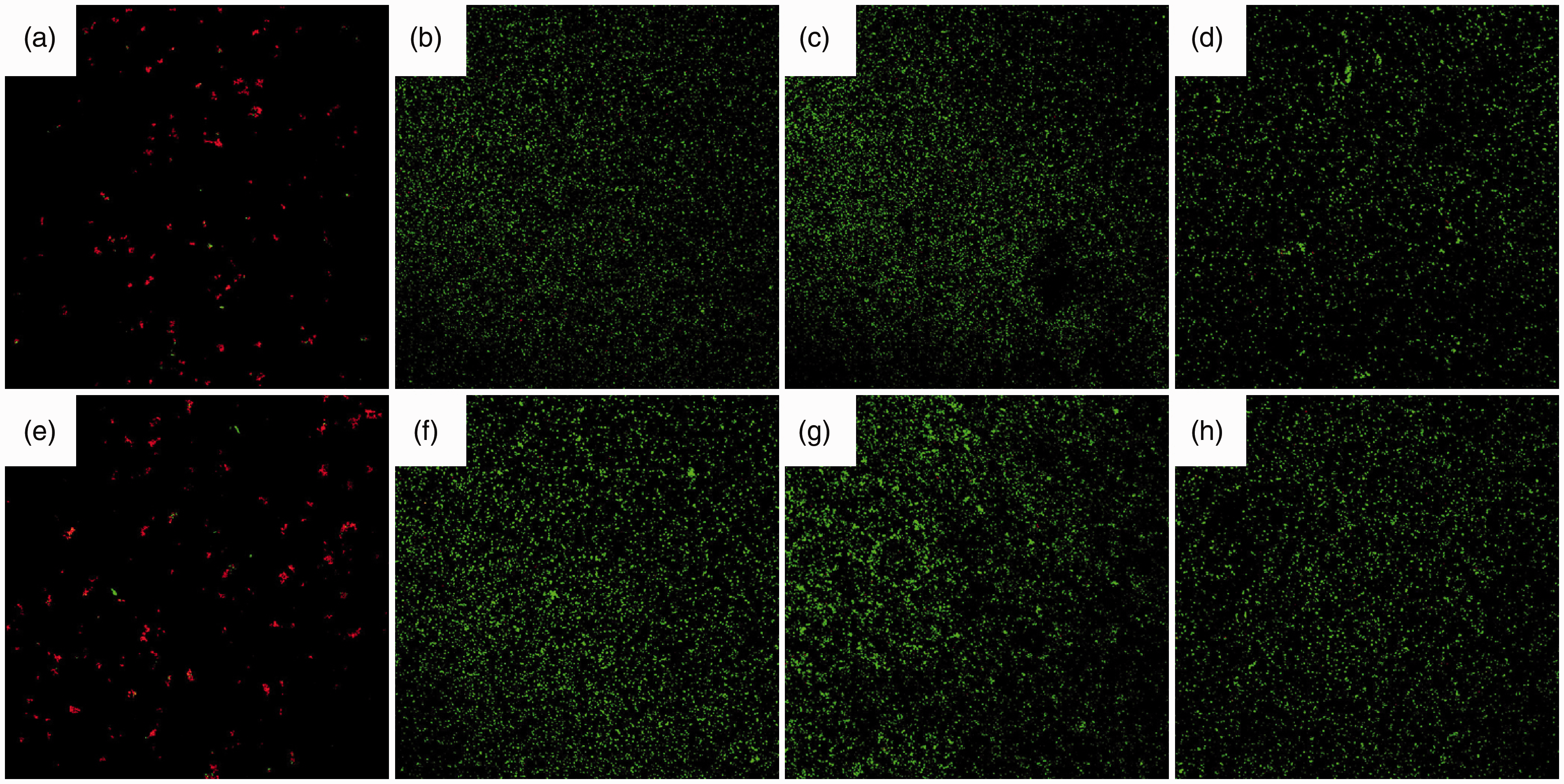
Confocal micrographs of bacteria cultured on the different groups after 1 d with green fluorescence referring to live bacteria and red fluorescence referring to dead bacteria. (a) BBF/PLLA-MAO-Ti, Pg. (b) PLLA-MAO-Ti, Pg. (c) MAO-Ti, Pg. (d) Polished cp-Ti, Pg. (e) BBF/PLLA-MAO-Ti, Aa. (f) PLLA-MAO-Ti, Aa. (g) MAO-Ti, Aa. (h) Polished cp-Ti, Aa.
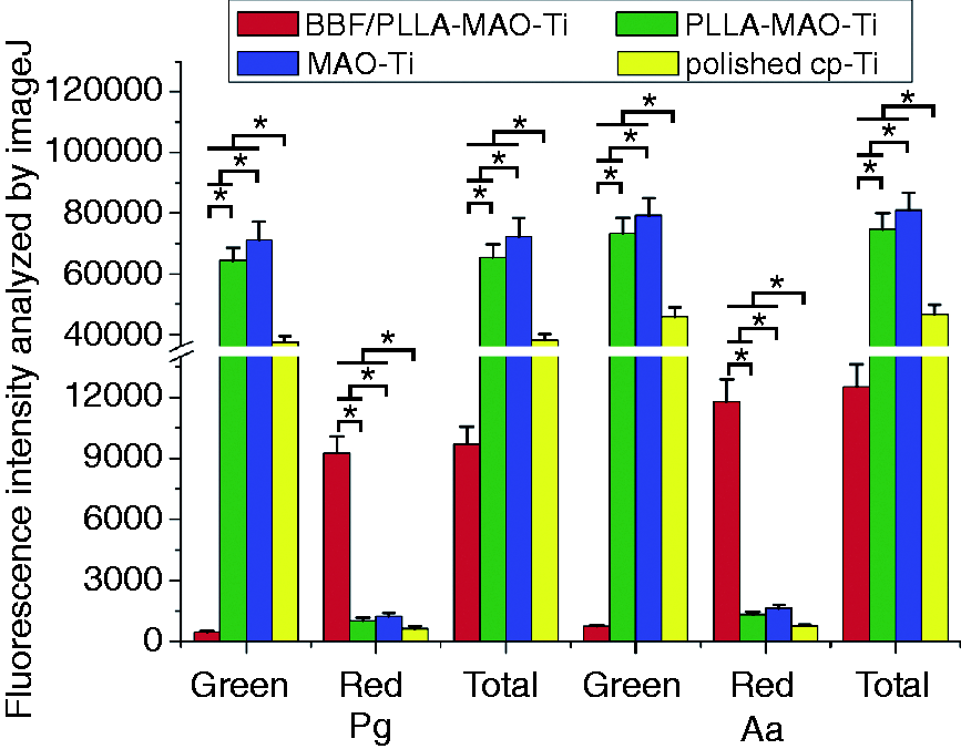
Fluorescence intensities of the bacteria adhering to the different groups after 1-day incubation. *p < 0.05.
RT-PCR
Figure 8 shows the levels of the relative genes of the bacteria grown on the four groups after 1-day incubation by RT-PCR analysis. The levels of mRNA in the two bacteria grown on the four groups were also similar. The fimbrilline A (Fim A) and cytolethal distending toxin B (Cdt B) mRNA expression levels on BBF/PLLA-MAO-Ti were significantly lower than those on the other three groups (p < 0.05). MAO-Ti possessed the highest mRNA expression levels (p < 0.05).
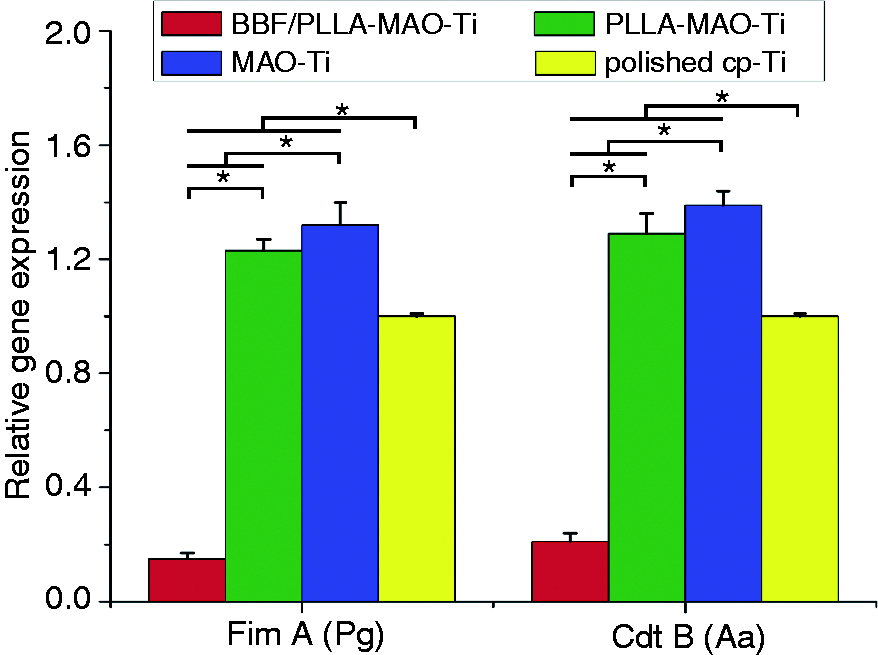
RT-PCR analysis of the levels of relative genes of the bacteria adhering to the different groups after 1-day incubation. *p < 0.05.
Discussion
Dental implants are routinely used to support dental restorations after tooth loss. Although there are data showing the long-term survival rate of dental implants, biological complications still occur. 25 Peri-implant diseases remain one of the most common and intractable biological complications, which may progress and result in implant failure. 26 As a consequence of the increasing number of dental implants, the cases of peri-implant diseases and implant failures would also increase considerably. Prevention of these diseases is therefore a high priority to minimize the occurrence and severity of the problem. 27 Therefore, various antibacterial coatings have been designed on Ti to prevent initial bacterial adhesion. Although some promising results have been achieved, the lack of long-term antibacterial activity and the emergence of drug-resistant bacteria call for novel implant antibacterial coating systems. 28
Halogenated furanone compounds show a stable antimicrobial activity against a great number of bacteria, and are less prone to resistance development. 29 The BBF used in our study is one of the most active halogenated furanone compounds. 30 To achieve the goal of sustained release, BBF was incorporated into PLLA nanospheres. The prepared BBF/PLLA-NSs were spherical in shape with a mean particle size of 408 ± 14 nm (Figure 1(b)). A porous microstructure with 1–3 µm diameter pores (much larger than the particle size) was observed on the MAO coating (Figure 1(d)), which provided space for the adhesion of the nanospheres. Then, a composite coating was fabricated by cross-linking the BBF/PLLA-NSs on the MAO coating on Ti. SEM images showed that the BBF/PLLA-NSs were well-distributed in the pores of the MAO coating, and cross-linked with each other and the wall of the pores by gelatin (Figure 1(f)). This cross-linking would effectively prevent the BBF/PLLA-NSs from detaching during dental implant insertion. The composite coating allowed for a long-term BBF release (approximately 60 d) with a slight initial burst release during the first hours. The burst effect is generally attributed to the drug being adsorbed on or close to the surface of the nanoparticles. 31 During the second stage, the release of BBF was a typical sustained release and mainly depended on polymer matrix degradation and drug diffusion, which is a slower process. 21
In this study, two common peri-implant pathogens, Pg (Gram-negative) and Aa (Gram-negative) were utilized in the antibacterial assays. To monitor the long-term antibacterial effect, the bacterial incubation time was extended to 30 d in the antibacterial rate assay. The AR of the composite coating for the adherent bacteria was over 90% throughout a 30-day period, which illustrated that the composite coating possessed excellent and relatively long-term antibacterial activity against Pg and Aa. Due to the initial burst release of BBF during the first hours, the AR was the highest (over 97%) after 1 d. The antibacterial activity during the first day is critical, because as it has been documented, dental implants are most susceptible to surface bacteria colonization during the initial 6 h after implantation. 32 Therefore, fluorescence staining and RT-PCR analysis were also used in this study to evaluate the antibacterial effect of the composite coating after 1-day incubation. The results showed that the total fluorescence intensity on BBF/PLLA-MAO-Ti was the lowest, and the vast majority of the fluorescence was red (dead bacteria). Moreover, the Fim A gene of Pg and the Cdt B gene of Aa were down-regulated. The Fim A gene encodes the distinct fimbriae of Pg, and plays a key role in periodontal and peri-implant Pg invasion. 33 The Cdt B gene encodes a heat-labile protein cytotoxin by several Gram-negative bacteria, including Aa, and regulates the morphology of the bacteria. 34 These results also indicated the excellent antibacterial activity of the composite coating against Pg and Aa.
As mentioned in the results, the composite coating showed antibacterial activity, but the other three groups did not. Moreover, the results also showed more adherent bacteria on MAO-Ti than on PLLA-MAO-Ti and polished cp-Ti. The number of colonies of MAO-Ti was the highest (more than 600). The total fluorescence intensity on the surface of MAO-Ti was the highest, and the vast majority of the fluorescence was green (live bacteria). In addition, the MAO-Ti specimens possessed the highest mRNA expression levels. Studies have proven that roughened surfaces can encourage bacterial adhesion. 35 In this study, the roughness of MAO-Ti was the highest among the four groups. These results were consistent with the previous findings reported by Mei. 36
Quorum sensing (QS) is the process of cell-to-cell communication in microorganisms. 37 In QS, a single bacterium releases autoinducers into the surroundings. When the concentration of the autoinducers reaches a threshold value, the autoinducer receptors on the surface or in the interior of the bacteria are activated to induce the expression of specific genes, which regulate and control the fundamental physiological processes in bacteria, including DNA replication, virulence factor expression and biofilm formation. 38 Studies have reported that there are three kinds of autoinducers in QS: (1) acyl-homoserine lactones (AHL) existing in Gram-negative bacteria, (2) autoinducing peptides (AIPs) existing in Gram- positive bacteria, and (3) autoinducer-2 (AI-2) existing in both Gram-negative and Gram-positive bacteria. 39 Halogenated furanone compounds are structurally similar to AHL and AI-2, so they combine with the receptors competitively, and finally inhibit the QS activity of various bacterial species. 40 More than 700 species of bacteria have been found in the human oral cavity, which establish mixed-species communities. 41 Studies have suggested that the microbiota in the peri-implant is a polymicrobial anaerobic infection. 42 In addition, the formed biofilm protects adherent bacteria from the host defense system and bactericidal agents via several proposed mechanisms. 43 Therefore, as it has been reported, ideal antibacterial agents, which would be applied on dental implants, should have the following characteristics: (1) a broad spectrum of antibacterial activities, (2) ability to inhibit the formation of bacterial biofilms, and (3) show a low risk to resistance development. 28 Halogenated furanone compounds possess all of these advantages. In our study, the BBF was incorporated into PLLA to prepare sustained-release nanospheres, and then the nanospheres were cross-linked onto the MAO coating to fabricate a composite coating on titanium. The results showed that the composite coating exhibited excellent and relatively long-term antibacterial activity. Therefore, the composite coating is a potential and promising strategy to be applied on dental implants for the prevention of peri-implant diseases.
Conclusion
The composite coating fabricated on titanium exhibited excellent and relatively long-term antibacterial ability against two common peri-implant pathogens (Porphyromonas gingivalis and Actinobacillus actinomycetemcomitans). The antibacterial rate of the composite coating for the adherent bacteria was the highest (over 97%) after 1 d and over 90% throughout a 30-day incubation. The total fluorescence intensity of the composite coating was the lowest, and the vast majority of the fluorescence was red (dead bacteria). Moreover, real-time polymerase chain reaction analysis also confirmed that the relative gene expression of the adherent bacteria on the composite coating was down-regulated. It can be concluded that the composite coating fabricated on titanium is a potential and promising strategy to be applied on dental implants for the prevention of peri-implant diseases.
Footnotes
Declaration of conflicting interests
The author(s) declared no potential conflicts of interest with respect to the research, authorship, and/or publication of this article.
Funding
The author(s) disclosed receipt of the following financial support for the research, authorship, and/or publication of this article: This work was supported by National Natural Science Foundation of China (No. 81901046 and 51371006), Natural Science Foundation of Jiangsu Province (No. BK20170115), Fundamental Research Funds for the Central Universities (No. xzy012019097) and Science Development Funds of Fourth Military Medical University (No. 2019XB060).