
Abstract
Due to the low bioavailability and severe toxic side effects caused by the lack of selectivity of traditional chemotherapy drugs, the targeted delivery of chemotherapy drugs has become the key to tumor treatment. The activity and transmembrane potential of mitochondria in cancer cells were significantly higher than that of normal cells, making them a potential target for chemotherapeutic drug delivery. In this study, triphenylphosphine (TPP) based mitochondria targeting polylactic acid (PLLA) nanoparticles (TPP-PLLA NPs) were synthesized to improve the delivery efficiency of anticancer drugs. The carrier material was characterized by 1H NMR and FT-IR and 7-hydroxyl coumarin (7-HC) was successfully loaded into TPP-PLLA to form 7-HC/TPP-PLLA NPs. Further studies showed that TPP-PLLA NPs were primarily accumulated in the mitochondrial and 7-HC/TPP-PLLA NPs had higher antitumor activity. Taken together, our results indicated that TPP-PLLA NPs could be a promising mitochondria-targeted drug delivery system for cancer therapy.
Introduction
Mitochondria is an energy organelle that is involved in cell life activities.1,2 Since changes in mitochondria always affect the normal activities of cells, and as a part of the cell, the cells reject mitochondria less, many scholars now turn their attention to the inside of cells to study organelles such as mitochondria that can be potential targets.3,4 They look forward to changing the resistance and selectivity of anti-cancer drugs by targeting mitochondria and improving their anti-cancer effects.5–8 Mitochondria have a double-membrane structure, with a membrane potential of 130–150 mV. The outer membrane of the mitochondria is positively charged, while the inner membrane is negatively charged. Compared with other organelles such as the endoplasmic reticulum and ribosomes, mitochondria are rich in electrons and have a higher membrane potential. 9 What’s more, the mitochondrial membrane potential of cancer cells is quite different from that of normal cells. The membrane potential of the former is more negative than that of the latter. 10 Therefore, lipophilic cations can pass through the phospholipid bilayer membrane structure and accumulate in the mitochondria of cancer cells, 3 which will cause less damage to the mitochondria of normal cells.
Studies have shown that triphenylphosphine (TPP) is the most widely used lipophilic cation for mitochondrial delivery.7,11–13 The charges of its three phenyl groups are relatively dispersed and can be dispersed on a larger hydrophilic surface area. 14 TPP can easily cross the mitochondrial double-layer membrane and accumulate in the mitochondrial matrix at concentration hundreds of times higher than that of the cytoplasm, showing a mitochondrial targeting trend. 15 Too much TPP accumulates in the mitochondria, which will destroy the normal potential of the mitochondria, and finally induce apoptosis through the mitochondrial-mediated pathway.6,16–18 In addition, combined with the anti-tumor effect of the introduced anti-cancer drugs, this dual anti-tumor approach will provide a new therapeutic strategy for tumor therapy.
Natural and synthetic derivatives of coumarins have attracted much attention for their potential in the field of anti-tumor therapy. Their antitumor mechanisms are very diverse, including inhibiting carbonic anhydrase (CA), targeting PI3K/Akt/mTOR signaling pathway, inhibiting multiple drug resistance (MDR), and inducing apoptosis, etc.19–21 Biological investigations of coumarin derivatives showed the engrossment of innumerable pathways by which coumarins act as an anticancer agent.
In this study, TPP was conjugated to PLLA through amide bonds to form a new carrier material (TPP-PLLA). The chemical structure of the carrier material was characterized by FT-IR and 1H NMR. Meanwhile, 7-hydroxycoumarin (7-HC) was encapsulated in the carrier material to prepare nanoparticles (7-HC/TPP-PLLA NPs). The targeting of mitochondria and the anti-tumor effect of the nanoparticles were evaluated in vitro.
Materials and methods
Reagents
7-Hydroxycoumarin, stannous Isooctanoate, 3-bromopropionic acid, N-Hydroxysuccinimide (NHS), 1–(3-Dimethylaminopropyl)-3-ethylcarbodiimide (EDCI) were purchased from Shanghai Aladdin Biochemical Technology Co., Ltd. (China). Trichloromethane, Anhydrous ether, Ethanolamine, Di-tert-butyl decarbonate were purchased from Sinopharm Chemical Reagent Co., Ltd. L- lactide was purchased from Jinan Daigangsheng Engineering Co., Ltd. (China). Dichloromethane was purchased from Tianjin Oboke Chemical Co., Ltd. (China). MitoTracker Red, DAPI, Paraformaldehyde were purchased from Service Biotechnology Co., Ltd. (Wuhan, China). Tetrahydrofuran saturated with hydrogen chloride was made in the laboratory.
Cell lines and culture
The rat C6 glioma (C6) cells were obtained from the Shanghai Institute of Life Science Cell Resource Center (Shanghai, China). Cells were cultured in DMEM containing 10% FBS in a humidified atmosphere of 5% CO2 at 37 °C.
Synthesis of 3-carboxypropyl triphenyl phosphonium bromide (TPP-COOH)
TPP and 3-bromopropionic acid were dissolved in 13 mL of acetonitrile with a molar ratio of 1:1. The mixture solution was magnetically stirred at 80 °C under a nitrogen atmosphere and refluxed for 24 h. Then, the product was precipitated with anhydrous ether. The precipitate was recrystallized with acetonitrile. Finally, the solvent was evaporated and dried in a vacuum to obtain a yellowish powder, which was the product of TPP-COOH, 12 with a yield of 78.66%.
Synthesis of N-(t-butoxycarbonyl) ethanolamine
Di-tert-butyl dicarbonate (20 mmol) was dissolved in a certain amount of chloroform, which was added dropwise to chloroform solution containing amino ethanol (20 mmol) at 0 °C. The mixed solution slowly rose to room temperature and the reaction solution continued to be stirred for 24 h. The colorless viscous liquid obtained by rotary evaporation was N-(t-butoxycarbonyl) ethanolamine (EABoc), 22 with a yield of 92.45%.
Synthesis of BOC-NH-PLLA
An appropriate amount of EABoc and stannous octanoate were dissolved in 1 mL of chloroform, then a corresponding proportion of L-lactide (the molar ratios of EABoc and stannous octanoate to L-lactide were 1:100) was added. The chloroform was then removed by vacuuming. The polymerization reaction was conducted at 110 °C for 3 h under the protection of dry nitrogen. After the polymerization, the resulting solid was dissolved in chloroform, and the desired product was deposited in anhydrous ether. Finally, the precipitate was dried under vacuum to produce a white solid, which was Boc-aminoethanol-PLLA (Boc-NH-PLLA), 23 with a yield of 89.41%.
De-protection of t-butoxycarbonyl (BOC)
BOC-aminoethanol-PLLA was dissolved in dichloromethane, and tetrahydrofuran solution (10 mL) saturated with hydrogen chloride was added. The obtained mixed solution was stirred at room temperature for 30 min, and then the solvent was removed by rotary evaporation. The resulting solid was dissolved in dichloromethane and washed with 0.05% sodium bicarbonate solution and deionized water respectively until the water phase was neutral. Finally, the organic layer was dried with anhydrous sodium sulfate and precipitated with anhydrous ether. The product of amino-terminated PLLA (PLLA-NH2) was collected by centrifugation and dried in a vacuum. 23
Synthesis of TPP-PLLA
TPP bromopropionic acid (0.2300 g), N-hydroxysuccinimide (NHS) (0.1636 g) and 1–(3-dimethylaminopropyl)-3-ethylcarbodiimide (EDC) (0.3090 g) were dissolved in acetonitrile. Then triethylamine (20 µL) was added and the mixture was stirred at room temperature for 4 h to activate the carboxyl group. The amino-terminated PLLA was dissolved in acetonitrile (15 mL) and the PLLA solution was slowly dropped into the TPP bromopropionic acid. The obtained mixture was stirred at room temperature overnight. The next day, the reactant was washed with deionized water and precipitated with ethanol. Finally, the product was dried at room temperature to obtain the carrier material TPP-PLLA,24,25 with a yield of 78.76%. The synthesis path of TPP-PLLA was depicted in Figure 1.
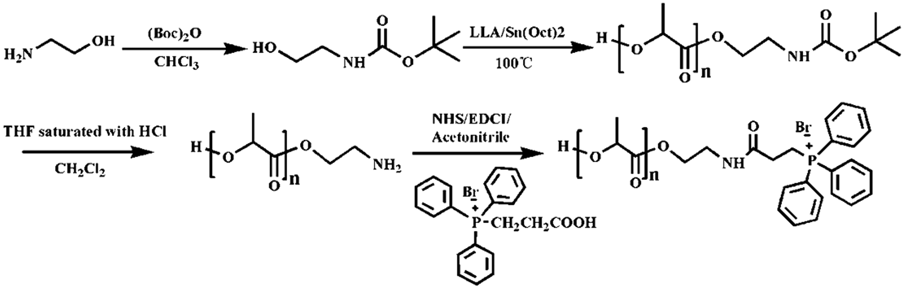
The synthesis process of TPP-PLLA.
The characterization of TPP-PLLA
1H NMR analysis
CDCl3 was used as the solvent to determine the hydrogen spectrum of the chemical structure (400 MHz; CDCl3; Me4Si; ppm).
FT-IR
After the dried samples were finely ground, an appropriate amount of dry potassium bromide powder was added and mixed evenly. The mixture was pressed into small pieces and scanned from 400 to 4000 cm−1. The dry tablet of potassium bromide was used as a blank background film.
UV
Firstly, dichloromethane was used for zero calibration. Secondly, TPP-PLLA was dissolved in dichloromethane and scanned at the wavelength of 200–800 nm to compare whether the maximum absorption wavelength was consistent with the characteristic absorption peak wavelength of TPP.
Preparation of 7-HC/TPP-PLLA NPs
7-HC/TPP-PLLA NPs were prepared by the emulsification-solvent evaporation method.26–28 The carrier material of TPP-PLLA (0.0100 g) was dissolved in dichloromethane (3 mL) containing 0.25 g of Span 80, which was used as the oil phase (O). 7-HC (0.0030 g) was dissolved in ethanol (0.5 mL), which was mixed with 1 mL of distilled water and used as the internal water phase (W1). The inner water phase was slowly added into the oil phase and the mixture was stirred for 3 min to form W1/O primary emulsion. Then, the obtained primary emulsion was slowly added into the SDS solution (5 mL, 3% m/v) and the mixture was followed by sonication for 15 min with a probe-type sonicator at 195 Watts to form a W1/O/W2 double emulsion. 7-HC/TPP-PLLA NPs were finally obtained after the organic solvent was removed by gentle stirring at room temperature overnight. The resulting NPs dispersions were centrifuged and re-dispersed in normal saline solution.
Characterization of 7-HC/TPP-PLLA NPs
An appropriate amount of diluted 7-HC/TPP-PLLA NPs was sprayed on a copper mesh coated with amorphous carbon and air-dried naturally. Then the sample was stained with 2% phosphotungstic acid and the morphology of 7-HC/TPP-PLLA NPs was observed by transmission electron microscope (Tokyo, Japan) under an acceleration voltage of 100 kV. The particle size and zeta potential of 7-HC/TPP-PLLA NPs were measured with a laser particle size analyzer (Malvern, Zetasizer Nano ZSE).
The hemolysis test of drug-free TPP-PLLA NPs
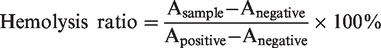
Fresh blood (2 mL) was taken from Sprague-Dawley (SD) rats and put into a centrifuge tube rinsed with heparin. The blood was centrifuged at 1000 rpm for 5 min to obtain a blood cell pellet. Then the blood cell pellet was washed with PBS several times until the supernatant was clear. The erythrocytes suspension was adjusted to 2% (v/v) by dilution with normal saline and placed evenly in centrifuge tubes, drug-free TPP-PLLA NPs solution (150 μL) were added to erythrocytes suspension and produced mixture at various concentrations (0.01%, 0.03%, 0.05%, 0.1%, 0.15%, 0.2%, v/v). After incubation at 37 °C for 1 h, the cells were centrifugated at 1000 rpm for 5 min and the supernatant was placed in a 96-well plate. Furthermore, normal saline was used as the negative control and double-distilled water was used as the positive control. The absorbance was measured at 540 nm with a spectrophotometer. The ratio of hemolysis was calculated by using the following equation.
Preparation of fluorescein/TPP-PLLA NPs
The preparation method of fluorescein/TPP-PLLA NPs was similar to that of 7-HC/TPP-PLLA NPs. The carrier material of TPP-PLLA (0.0100 g) was dissolved in dichloromethane (3 mL) containing 0.25 g of Span 80, which was used as the oil phase (O). Fluorescein (0.0030 g) was dissolved in ethanol (0.5 mL), which was mixed with 1 mL of distilled water and used as the internal water phase (W1). The other steps were the same as described in Preparation of 7-HC/TPP-PLLA NPs.
Mitochondrial targeting effects in vitro
C6 cells were inoculated in a small confocal dish and incubated overnight. The cells were treated with 4 µL of nanoparticle solution (fluorescein-labeled TPP-PLLA NPs). After incubation at 37 °C for 10 h, the cells were incubated with the Mito Tracker Mitochondrion-Selective Probes (red fluorescence) for 15–45 min. Then the culture medium was removed and the cells were washed with PBS. After that, the cells were fixed in a formaldehyde solution for 10 min and washed with PBS 3 times. Then the cells were stained with DAPI (blue fluorescence) for 10 min until the dye was aspirated and washed with PBS 3 times again. Finally, the cells were immediately observed and took pictures with a laser confocal microscope.29,30
Cell proliferation assay
The cytotoxicity of TPP-PLLA on rat C6 glioma cells was determined by the MTT assay. C6 cells at the logarithmic growth phase were inoculated into a 96-well plate at a density of 5 × 10³ cells/well and incubated overnight. Then the culture medium was discarded and the fresh medium (100 µL) containing 7-HC (the same amount of 7-HC as the 7-HC/TPP-PLLA NPS was dissolved in PBS), drug-free TPP-PLLA NPs and 7-HC/TPP-PLLA NPs (at concentrations of 0.01%, 0.03%, 0.05%, 0.1%, 0.15%, 0.2%, v/v) were added respectively. After 10 h of incubation, 20 μL of MTT (5 mg/mL) was added into each well and continued to incubate for another 4 h. Then, the supernatant was discarded, and 150 μL of DMSO was added into each well to dissolve the formazan crystals in the culture plate. The absorbance value was measured by a microplate reader (Bio-Tek Instruments, Winooski, VT, USA) at 490 nm.
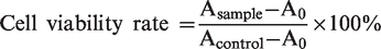
Asample: treatment group, Acontrol: control group, A0: zero adjustment group
Cell scratch test
C6 cells were inoculated into 6-well plates at a density of 7 × 10⁵ cells/well and incubated overnight. The scratch test can be performed when the cell density reaches about 90% confluency. Firstly, a 200 μL pipette tip was used to scrape the cells in a straight line. The debris was removed by washing with PBS. The cells were incubated with 10% FBS culture solution for 20 min and recorded under the microscope. Subsequently, the cells were treated with 7-HC/TPP-PLLA NPs (0.01%, 0.03%, 0.05%, 0.1%, 0.15%, v/v). After 10 h of incubation, the cells state was recorded under the microscope, and the images were further analyzed using Image J software. Each experiment was repeated three times.
Statistical analysis
Statistical analyses were performed using GraphPad Prism 8.0 software. When P < 0.05, the difference was statistically significant.
Results and discussion
1H NMR characterization of TPP-COOH
The 1H NMR spectroscopy of TPP-COOH was shown in Figure 2(a). The assignment of each peak was as follows, δ: 7.85–7.37 (15H, Ar-H), 3.81–3.74 (2H, -CH2), 3.08–3.01 (2H, -CH2).
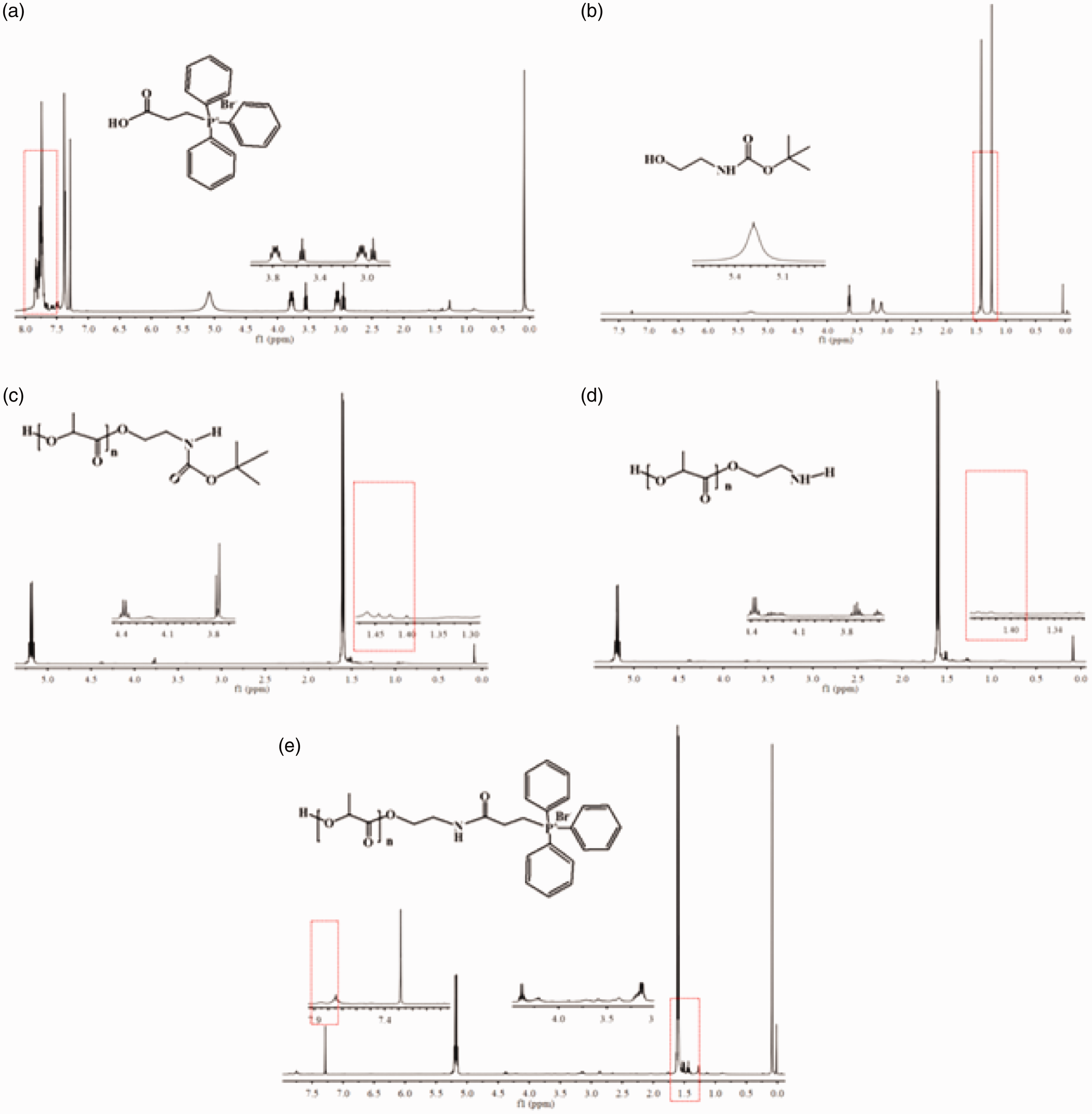
(a) The 1H NMR spectra of TPP-COOH. (b) The 1H NMR spectra of EABoc. (c) The 1H NMR spectra of Boc-NH-PLLA. (d) The 1H NMR spectra of PLLA-NH2. (e) The 1H NMR spectra of TPP-PLLA.
At δ = 7.85–7.37 × 10⁻⁶, there was the characteristic peak of the benzene ring group. At δ = 3.08–2.93 × 10⁻⁶, there was the characteristic peak of two methylene in bromopropionic acid, the two sets of methylene peaks corresponding to each other showed that there remained unreacted bromopropionic acid. During the synthesis, the amount of 3-bromopropionic acid should be appropriately reduced. After the reaction was completed, methods such as recrystallization should also be used to reduce the unreacted reactants.
Structural characterization of EABoc
The NMR spectroscopy of EABoc was shown in Figure 2(b). The assignment of each peak was as follows, δ:5.27 (1H, -NH-), 3.62–3.65 (1H, -OH), 3.22 (2H, -CH2), 3.08 (2H, -CH2), 1.41 (9H, -C(CH3)3).
Compared with the NMR spectrum of ethanolamine, the NMR spectrum of Boc-aminoethanol showed a characteristic peak of the Boc-group (tert-butyl) at δ = 1.41 × 10⁻⁶. The amino peak shifted from the initial δ = 2.76 × 10⁻⁶ to δ = 5.27 × 10⁻⁶ due to the decrease of surrounding electrons. Meanwhile, it could be seen from (a) in Figure 3(A) that EABoc had absorption peaks of carbonyl and secondary amines at 1689 and 1523 cm−1. The frequency-doubled absorption peak of secondary amine appeared at 3069 cm−1 and the hydroxyl peak appeared at 3386 cm−1. The cleavage peaks of the Boc-group (tert-butyl) appeared at 1367 and 1393 cm−1, however, the anhydride peaks of (Boc)2O at 1772 and 1811 cm−1 had disappeared in EABoc from (b) in Figure 3(A).
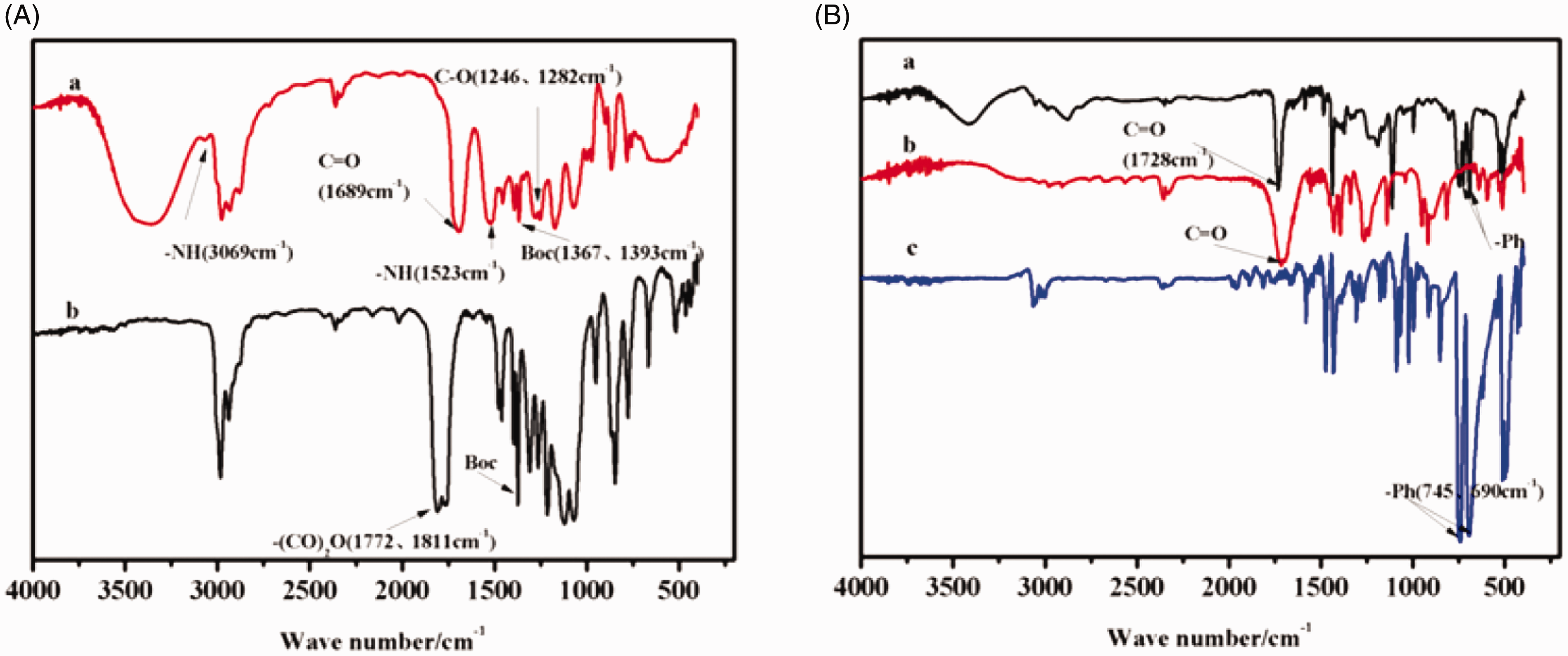
(A) The FT-IR spectra of (a) EABoc and (b) (Boc)2O. (B) The FT-IR spectra of (a) TPP-PLLA, (b) NH2-PLLA and (c) TPP-COOH.
1H NMR characterization of Boc-NH-PLLA and PLLA-NH2
The 1H NMR spectrum of Boc-NH-PLLA and PLLA-NH2 were shown in Figure 2(c) and (d), respectively, and the peaks of each compound were assigned as follows. Boc-NH-PLLA δ: 5.21–5.16 (1H, -CH), 4.40–4.35 (2H, -CH2), 3.78–3.76 (2H, -CH2), 1.61–1.59 (3H, -CH3), 1.46–1.40 (9H, -C(CH3)3). PLLA-NH2 δ: 5.21–5.16 (1H, -CH), 4.40–4.35 (2H, -CH2), 3.77–3.70 (2H, -CH2), 1.61–1.59 (3H, -CH3).
Compared with Boc-NH-PLLA, the characteristic peak of Boc-methyl group at δ = 1.46–1.40 × 10⁻⁶ was disappeared after deprotection of the amino group. Meanwhile, the characteristic peak of the methylene group associated with -NH- moved from δ = 3.78–3.76 × 10⁻⁶ to δ = 3.77–3.70 × 10⁻⁶ due to the decrease of the surrounding electron density. The other characteristic peak displacements and the relative area ratio had not changed obviously, which indicated that the deprotection was successful.
Structural characterization of TPP-PLLA
The TPP-PLLA NMR spectrum was shown in Figure 2(e). The peaks were assigned as follows, δ: 7.88–7.47 (15H, Ar-H), 5.20–5.15 (1H, -CH), 4.40–4.32 (2H, -CH2), 3.23–3.11 (2H, -CH2), 1.61–1.59 (3H, -CH3).
In the final product of synthesis, the peak shifts of benzene ring group, -CH and -CH3 of PLLA had not changed significantly, which might be related to the little change in the chemical environment before and after the synthesis of the three compounds. Due to the formation of amide bonds, the two methylene peaks in the middle moved to the same position, while the other two were not significantly transferred. The characteristic peaks of the benzene ring of TPP-PLLA and TPP-COOH appeared at 744 cm−1 of (a) and 688 cm−1 of (c) in Figure 3(B), respectively. Through the ultraviolet wavelength scanning, the material had an obvious maximum absorption wavelength of TPP at 260 nm (Figure 4). The above results showed the successful synthesis of TPP-PLLA.
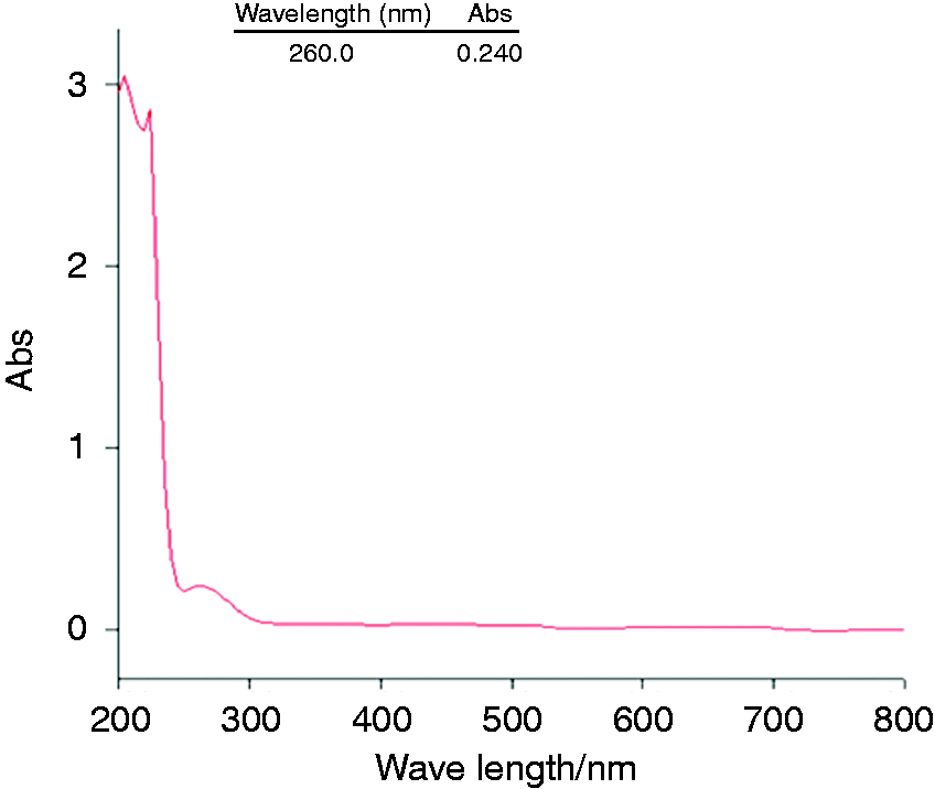
The UV wavelength scan chart of TPP-PLLA.
Prescription optimization of 7-HC/TPP-PLLA NPs
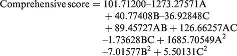
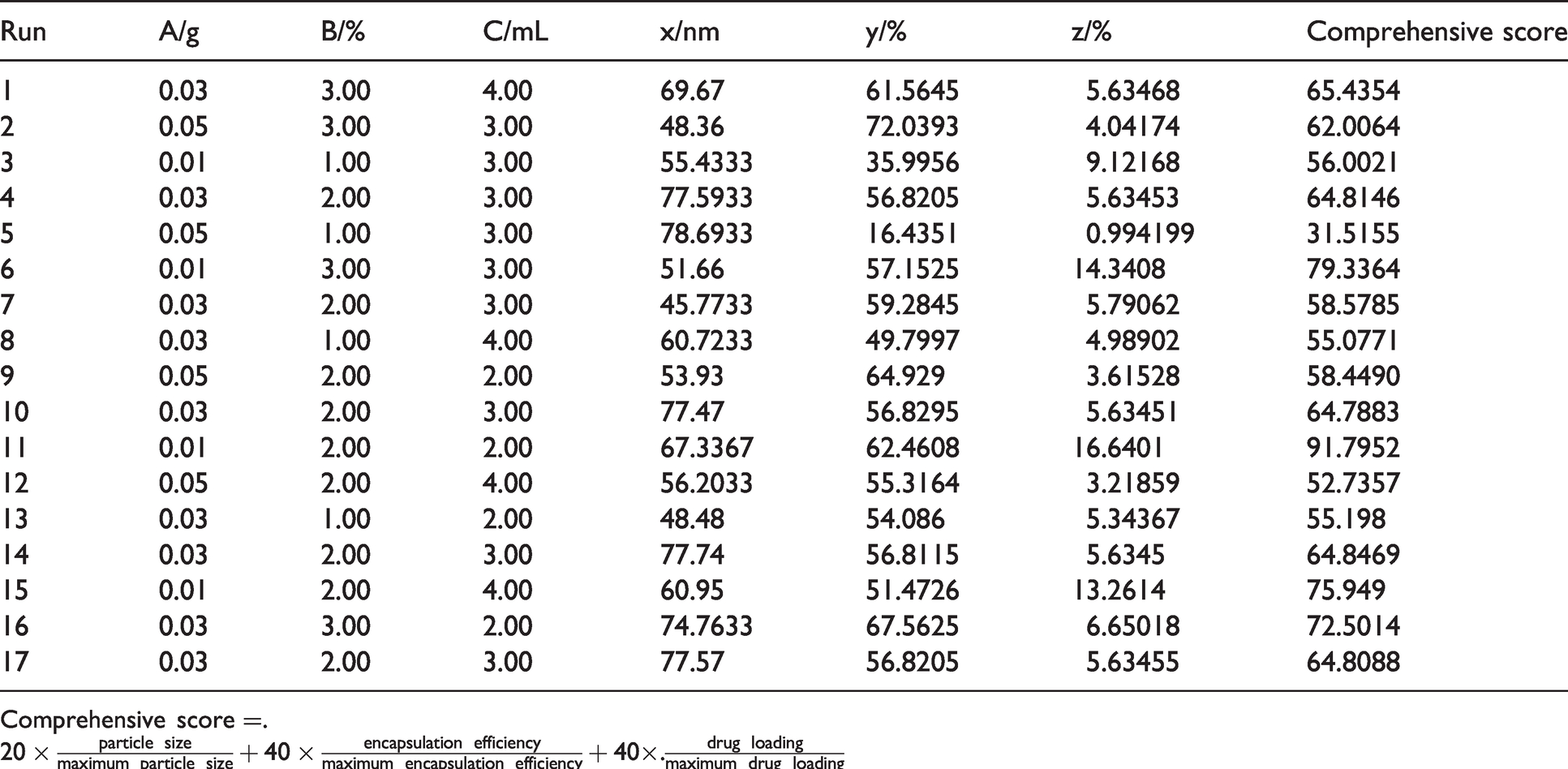
The Box-Behnken response surface method was used to get the most optimized prescription. The weight of the carrier material (A), the concentration of SDS (B), and the volume of the organic phase (C) were used as the independent variables, and the particle size (X), encapsulation efficiency (Y) and drug loading (Z) were considered as the dependent variables. The Box-Behnken design was used to conduct response surface tests with three factors and three levels. The factors and levels were shown in Table 1, and the experimental design and results were shown in Table 2. The regression equations from the design expert 8.0.6 software were as follows:
Box-Behnken response surface design.
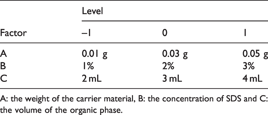
A: the weight of the carrier material, B: the concentration of SDS and C: the volume of the organic phase.
Design and results of Box-Behnken test.
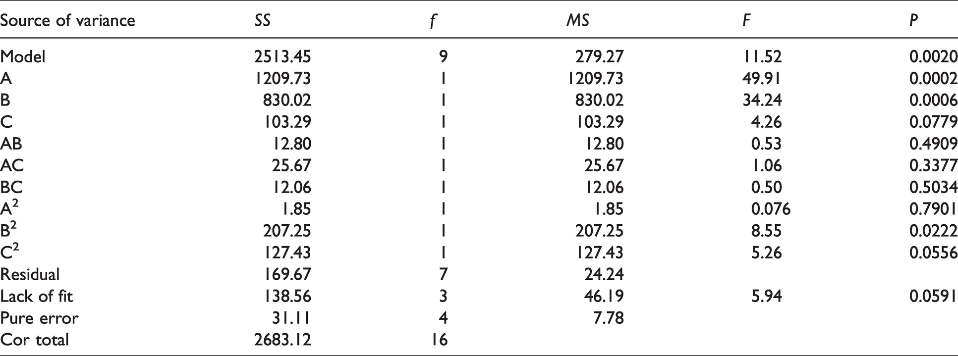
Statistical analysis results of the regression model were shown in Table 3, the model of the regression equations was significant with P value of 0.002, P value of the lack of fit was 0.0591, and the coefficient of R was fine.
Statistical analysis results of Box-Behnken test.
From the design response surface experiment, the optimum prescription was obtained and the composition was carrier material (0.01 g), SDS (2.5%) and the organic phase (2 mL). The experimental values of the encapsulation efficiency were 68.24 ± 0.15%, and the drug loading was 16.30 ± 0.11%.
The characterization of 7-HC/TPP-PLLA NPs
7-HC/TPP-PLLA NPs were prepared by a double emulsion (W1/O/W2) process. The outer shell of the nanoparticles was made of polylactic acid (PLLA), and the core of the nanoparticles was 7-hydroxyl coumarin, which was wrapped with the outer layer of PLLA, and a core-shell structure was developed. The obtained nanoparticle solution had a light blue opalescence (Figure 5(a)). The mean diameter of 7-HC/TPP-PLLA NPs was 67.6 ± 0.19 nm, with the polydispersity index (PDI) of 0.34 ± 0.006 (Figure 5(b)), revealing the uniform size and good dispersion. Due to the existence of the phosphorus group in TPP, the coupling of TPP in 7-HC/TPP-PLLA NPs gives a permanent positive charge to the nanoparticles. The zeta potentials of TPP-COOH and TPP-PLLA NPs were all positive except for PLLA NPs (Figure 5(c)). Transmission electron microscope (TEM) was further applied to study the morphology of 7-HC/TPP-PLLA NPs. The results showed that 7-HC/TPP-PLLA NPs were highly dispersed and had a spherical shape (Figure 5(d)).
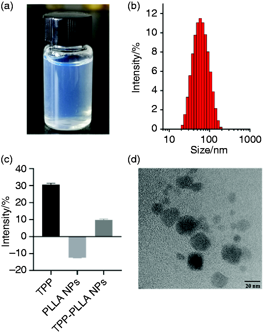
The characterization of 7-HC/TPP-PLLA NPs. (a) Appearance. (b) Particle size distribution. (c) Zeta potential of TPP-COOH, PLLA NPs, TPP-PLLA NPs. (d) TEM image of 7-HC/TPP-PLLA NPs.
The hemolysis test of drug-free TPP-PLLA NPs
Polylactic acid (PLLA) is an environmental protection substance, which has been approved by the U.S. FDA in 1997 for use as a suture thread for medical surgery and as a carrier material for injection microcapsules, microspheres, implants.31–33 PLLA can be degraded into non-toxic products such as CO2 and H2O in vitro by microorganisms, light or digested by enzymes in vivo. PLLA and its derivatives have been widely used as carrier materials due to their good biocompatibility.34–36 To explore the biological safety of TPP-PLLA materials, the hemolytic test was performed. The results showed that the hemolysis rate value of all testing samples (drug-free TPP-PLLA NPs) was less than 5% (Figure 6), indicating that the carrier material had no obvious effect on the hemolysis rate.
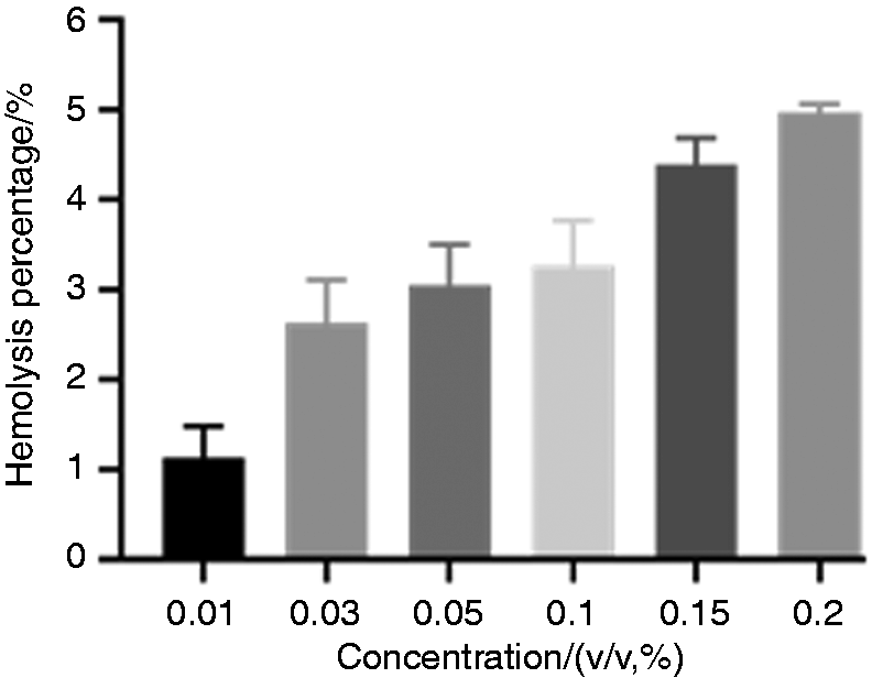
Hemolysis percentage of drug-free TPP-PLLA NPs, n = 5.
Mitochondrial targeting
The fluorescein-labeled TPP-PLLA NPs were functionalized with mitochondrial targeting ligand TPP, which was used to facilitate nanoparticles penetration through the mitochondrial membrane. The mitochondrial targeting properties of fluorescein-labeled TPP-PLLA NPs to C6 cells were observed by a confocal laser scanning microscope with Mito tracker red staining methods. Fluorescein and Mito tracker red emit green and red fluorescence, respectively. The confocal images of C6 cells showed that co-localization of the green fluorescence of the nanoparticles and the red fluorescence of the stained mitochondrial appeared yellow. The comparative intensity of yellow fluorescence indicated that a significantly greater overall uptake of TPP-PLLA NPs than PLLA NPs in C6 mitochondria. The results showed that TPP-PLLA NPs were mostly co-localized with mitochondria (red) while PLLA NPs were disorderly distributed in the cells after 10 h of cell uptake (Figure 7), indicating that TPP-PLLA NPs had significant mitochondria targeting properties and effectively delivered drugs to the mitochondria. This may be due to the highly lipophilic ligand of TPP with three phenyl groups and a positive charge on phosphorous, which enhanced its cell association and mitochondrial targeting.
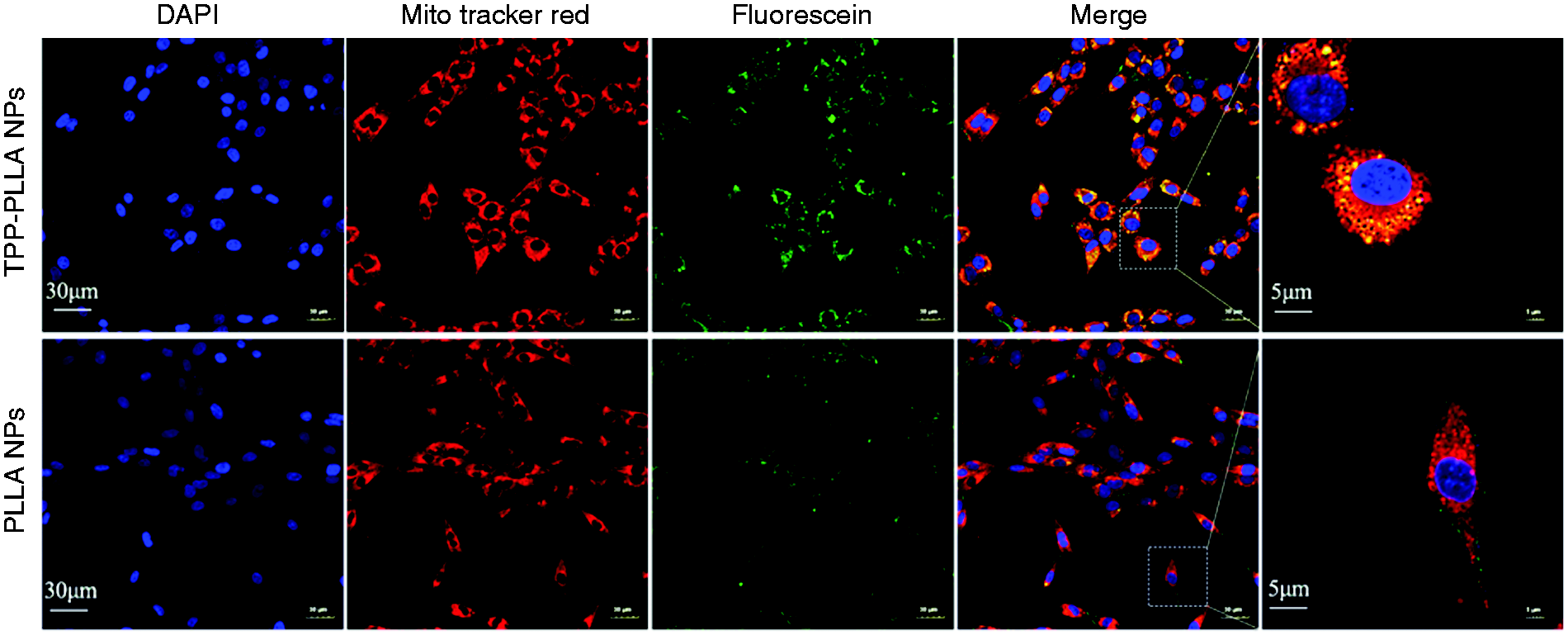
The distribution of nanoparticles in C6 cells. C6 cells were incubated in TPP-PLLA NPs or PLLA NPs solution (fluorescein-labeled) at 37 °C for 10 h. Mitochondria were stained with Mito Tracker Red, and nuclei were stained with DAPI. The cells were observed under a confocal laser scanning microscope. The overlap between the fluorescence of fluorescein (green) and MitoTracker Red (red) appeared as yellow which represented the distribution of nanoparticles in the mitochondria of cells.
Cell proliferation assay
It has been reported that DOX-TPP-HA, which is formed by the combination of TPP and tumor targeting molecule hyaluronic acid (HA), can complete the multi-stage delivery from cells to mitochondria and then to the nucleus, thereby enhancing the anti-tumor effect of doxorubicin. 12 In addition, Xing et al. showed that TPP chemically linked amphiphilic drug quercetin could self-assemble to form mitochondrial-targeted nanoparticles, achieving better anti-tumor effect than quercetin alone. 37 In order to investigate the anti-tumor effect of TPP-modified 7-HC/PLLA NPs, MTT method was used to detect the effects of drug-free TPP-PLLA NPs, 7-HC and 7-HC/TPP-PLLA NPs on cell proliferation. At the evaluated concentration range (0.01%–0.20%, v/v) of drug-free TPP-PLLA NPs, there were no apparent effects on the cell viability of C6, which showed that the drug-free TPP-PLLA NPs had little cytotoxicity to the C6 cells. However, with the increase of concentration, the cell viability of both 7-HC and 7-HC/TPP-PLLA NPs treatment were decreased, and the latter was more obvious (Figure 8). The results of the anti-tumor test in vitro showed that 7-HC/TPP-PLLA NPs had a dose-dependent inhibitory effect on glioma (C6) cells, with an IC50 of 2.7705 µM, which was much lower than that of raw material (23.930 µM).
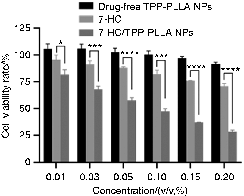
The anti-tumor (glioma) activity of drug-free TPP-PLLA NPs, 7-HC and 7-HC/TPP-PLLA NPs in vitro. (*P < 0.05, **P < 0.01, ***P < 0.001 vs. 7-HC.)
Cell migration rate of 7-HC/TPP-PLLA NPs
Migration capability is the basic biological characteristics of tumor cells. The effect of drugs on cell migration is usually reflected by observing the growth of cells before and after drug administration through the scratch test. In order to investigate the effect of 7-HC/TPP-PLLA NPs on the migration of C6 cells, the pre-scraped cells were treated with the drug-loaded NPs at the concentrations of 0.01%, 0.03%, 0.05%, 0.1%, 0.15% (v/v), respectively. The results showed that, at 10 h, as the concentration of drug-containing nanoparticles increased, the scratch width of C6 cells became larger and larger, until the width was close to that of 0 h (Figure 9(a) and (b)). Further analysis revealed that the migration rate of C6 cells was significantly reduced after 7-HC/TPP-PLLA NPs treatment (P < 0.05) (Figure 9(c)). The results indicated that the presence of drug-containing nanoparticles affected the migration ability of C6 cells and had a concentration inhibitory effect, which was consistent with the trend of cell proliferation experiment results.
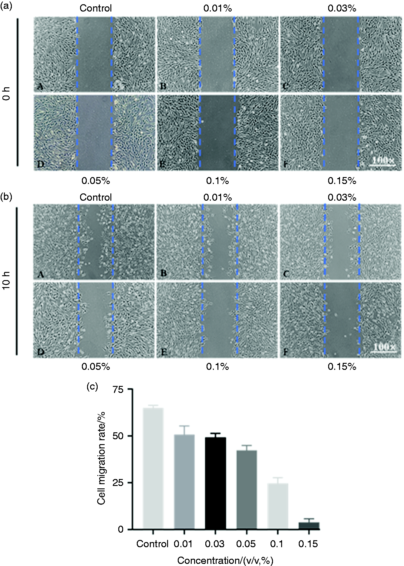
The effects of 7-HC/TPP-PLLA NPs on cell migration. Cell migration picture in the presence or absence of 7-HC/TPP-PLLA NPs at (a) 0 h and (b) 10 h. (c) Cell migration rate at 10 h. (*P < 0.05, **P < 0.01, ***P < 0.001 vs. Control.)
Conclusion
Mitochondria of tumor cells have different activities and transmembrane potentials from normal cells, making them potential targets for chemotherapy drug delivery. In this study, TPP was selected as the mitochondrial target compound because the three benzene rings of TPP form a delocalized positive charge, making it easy to pass through the mitochondrial double hydrophobic membrane and accumulate in the mitochondria. Our results showed that TPP-PLLA NPs had good biocompatibility and mitochondrial targeting, and 7-HC loaded TPP-PLLA NPs exhibited higher anti-tumor activity, indicating that the development of mitochondrial targeting agents was helpful to improve the therapeutic effect of chemotherapy drugs.
Footnotes
Declaration of conflicting interests
The author(s) declared no potential conflicts of interest with respect to the research, authorship, and/or publication of this article.
Funding
The author(s) disclosed receipt of the following financial support for the research, authorship, and/or publication of this article: This work was financially supported by the Funds of Science and Technology Department of Hubei Province (2018CFB533); Health and Family Planning Commission Project of Hubei Province (WJ2017Q042); Initial Scientific Research Fund of Ph.D. in Hubei University of Science and Technology (BK202120), Research Fund of Cultivation in Hubei University of Science and Technology (2020-21X23).