






Abstract

Poor solubility and non-specific biological distribution are critical obstacles for applying Juglone (Jug) into clinical practice. To improve the antitumor efficacy of Jug, an iRGD-modified red blood cell membrane (RBCm) platform was developed to package Jug and Oxaliplatin (Oxa). The stability and release of iRGD-modified RBCm nanoparticles loaded with Jug and Oxa (RBCm-(Jug, Oxa)-iRGD-NPs) were evaluated by Dynamic light scattering (DLS) and ultraviolet spectrophotometer, respectively. The in vitro uptake and cytotoxicity of such nanoparticles were detected by immunofluorescence microscope and Methyl Thiazolyl Tetrazolium ( MTT), respectively. Near-infrared imaging was utilized to confirm the tumor-targeting. The in vivo antitumor activity was evaluated in a xenografted nude mice model stablished by HCT-116 cells. In vitro studies, iRGD promoted the uptake of NPs by tumor cells, as shown by high fluorescence intensity and low cell viability. Also, the NPs retained higher amount of drug at the tumor site for a long time, while in vivo study indicated that the RBCm-(Jug, Oxa)-iRGD-NPs exhibited higher efficacy to eradicate human colorectal cancer (CRC) xenografted tumors than RBCm-(Jug, Oxa)-NPs and free Jug/Oxa. The RBCm-(Jug, Oxa)-iRGD-NPs significantly enhanced tumor targeting and antitumor effect. Thus, this innovative nanoplatform offers a potent strategy to improve anti-CRC efficacy.
Introduction
Following lung and breast cancer, colorectal cancer (CRC) is the third life-threatening neoplastic diseases worldwide.1,2 Although the screening methods and treating modalities for CRC have been improved,3,4 the morbidity and mortality are still increasing. 5 Chemotherapy remains the main therapeutic strategy for patients with advanced CRC.6,7 However, the most widely-used chemotherapy agent, Oxaliplatin (Oxa), is also restricted by the side effects, including short terminal half-life and severe normal organ damage.8–10
Juglone (Jug, 5-hydroxy-1, 4-naphthoquinone), a natural compound isolated from Juglans mandshurica Maxim, has antiviral, antibacterial, and antifungal effects. 11 Peptidyl-prolyl isomerase (Pin1) is overexpressed in several malignant diseases and is hypothesized to be a therapeutic target. It has been demonstrated that Jug elicits the antitumor activity by inhibiting Pin1.12–15 Nevertheless, the clinical use of this drug is hindered by poor solubility, non-specific biological distribution, and toxicity to normal tissues. 16
The nanotechnology-based approach offers an effective delivery of drugs to the targeted tissues in a safe and controlled manner. 17 Red blood cell membrane (RBCm) is the earliest and the most widely explored material for synthesizing biomimetic nanocarriers due to its simple structure and abundance in vivo.18–26 As a drug carrier, RBCm has unique advantages such as high biocompatibility, long half-life, low immunogenicity, and targeting,27,28 which can significantly reduce immune-related reactions and improve the bioavailability of drugs. In recent years, red blood cells not only have been used to contain some small or large molecules drugs, but also used to explore gene engineering RBC, 29 non-gene engineering RBC, and RBCm coating biological nanoparticles. RBC and RBCm play a major role in a variety of biomedical fields. Therefore, RBCm-coated drugs with active targeting ligands or peptide are likely to be a good option for biomimetic NPs in antitumor therapy.
IRGD is composed of vascular targeting sequence, CendR sequence motif, and protease recognition site.30–33 It could be cleaved by protease in the tumor tissue to obtain the residual peptide CRGDK/R and CendR sequence motif, followed by binding to the neuropilin-1 receptor (NRP-1) on the surface of tumor cells and tumor vascular endothelial cells.34–39 Furthermore, NRP-1 is overexpressed in CRC cells. 37 The NRP-1-mediated pathway regulates drug penetration into tumor parenchyma. As a result, iRGD-modified nanocarriers enable loaded drugs to penetrate the tumor essence and improve the treatment outcome of chemotherapy. Thus, it can be seen iRGD-modified RBCm NPs are expect to achieve better tumor-targeting ability and antitumor efficiency.
In the present study, we developed an iRGD-modified RBCm nanoplatform to deliver and sustain the release of Jug and Oxa into the tumor site (Figure 1(a)). This potent strategy was explored in a CRC-xenografted mice model. (A) Schematic representation of preparations of RBCm-(Jug, Oxa)-iRGD-NPs. (B) Chemical equation of synthesizing DSPE-PEG-iRGD (a) and the structural formula of Juglone (b) and Oxaliplatin (c). (C)-(D) MALDI-TOF MS analysis of DSPE-PEG-iRGD. (E) Flow cytometry graph of RBC alone (red) and the cells incubated with iRGD-FAM (blue) and DSPE-PEG-iRGD-FAM (yellow). (F) Correlation between surplus dose and input dose of DSPE-PEG-iRGD-FAM. (G)-(H) Cross-sectional SEM micrographs of RBCm-(Jug, Oxa)- NPs and RBCm-(Jug, Oxa)-iRGD-NPs (Scale Bar=100 nm). Abbreviations: Jug, Juglone; Oxa, Oxaliplatin; RBCm, red blood cell membrane; NPs, nanoparticles; RBCm-(Jug, Oxa)-iRGDNPs, iRGD modified red blood cell membrane nanoparticles packaging Juglone and Oxaliplatin; MALDI-TOF MS, matrix-assisted laser desorption/ionization-time of flight mass spectrometry; SEM, scanning electron microscope; RBCm-(Jug, Oxa)-NPs, red blood cell membrane nanoparticles packaging Juglone and Oxaliplatin.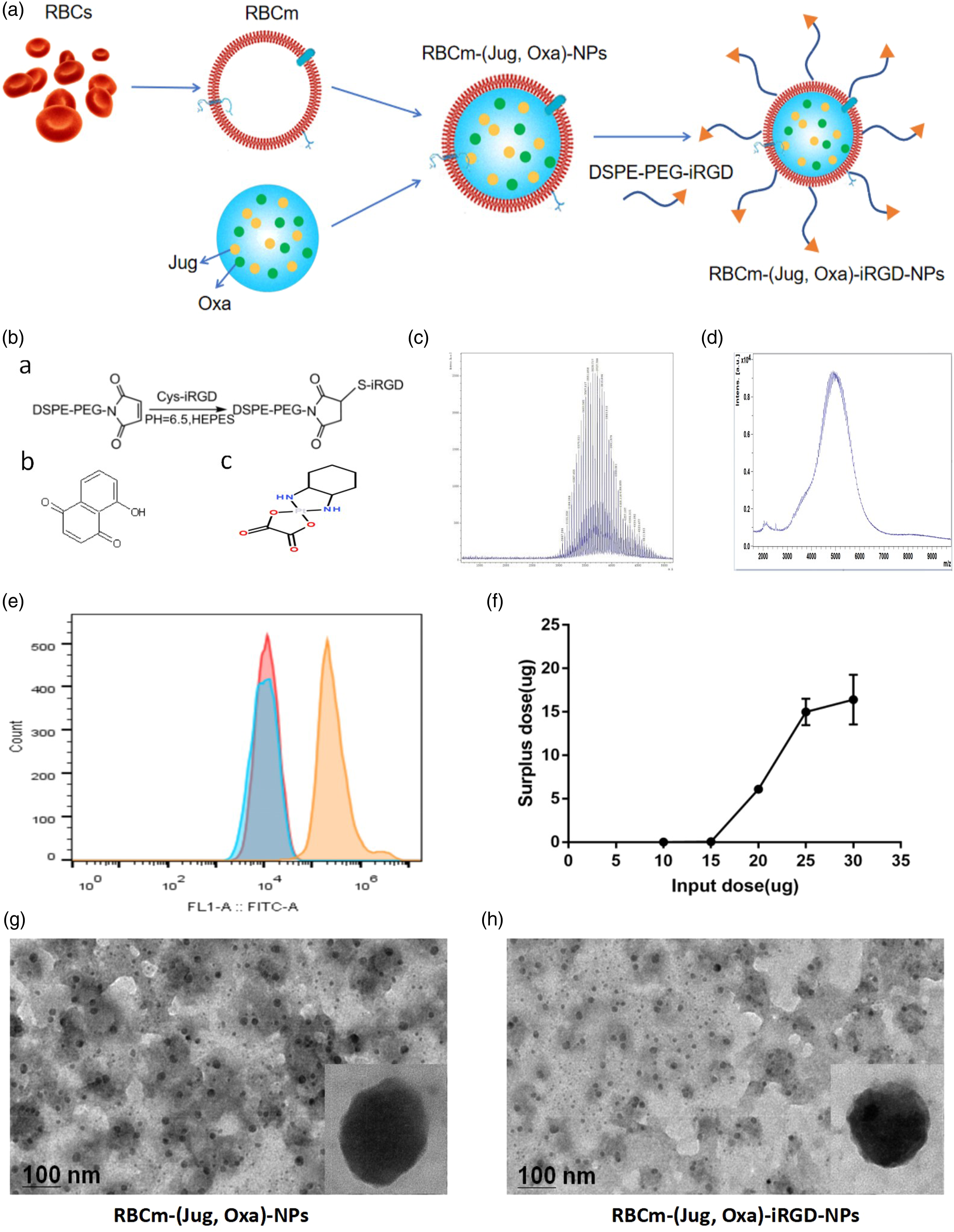
Material and methods
Reagents
Jug was purchased from Aladdin (Shanghai, China), while Oxa was purchased from Sanofi-Aventis (Hangzhou, China). C-iRGD and C-iRGD-FAM were purchased from Shanghai Top-Peptide Biotechnology (Shanghai, China). DSPE-PEG-Mal was obtained from Laysan Bio (USA). The crystal violet staining solution was purchased from Beyotime Biotechnology (Shanghai, China). 4′,6-diamidino-2-phenylindole (DAPI) was purchased from Beijing Solarbio Science and Technology (Beijing, China). 1, 1-Dioctadecyl-3, 3, 3, 3-tetramethylindocarbocyanine (DiR) was procured from ATT Bioquest (USA). Other reagents were used without further purification.
Cell lines and mice
Human colon cancer cell lines HCT-116 and HT-29 were purchased from the Cell Bank of the Chinese Academy of Sciences (Shanghai, China). The cells were cultured in RPMI-1640 medium (Gibco, USA) with 10% fetal bovine serum (Gibco) at 37°C with 5% CO2 in a humidified incubator. Male BALB/c mice (weight: 18–24 g, age: 5–6 weeks) were purchased from the Department of Experimental Animals, Yangzhou University, China. All animal experiments were carried out in accordance to the Guide for the Care and Use of Laboratory Animals and were approved by the Ethics Review Board for Animal Studies of Nanjing Drum Tower Hospital, Medical School of Nanjing University, China.
Synthesis of DSPE-PEG-iRGD
As described previously, 40 DSPE-PEG-Mal and C-iRGD or C-iRGD-FAM were mixed in a 1:1 M ratio in HEPES buffer (0.5 mM, pH 6.5) at room temperature for at least 48 h under nitrogen gas. Then, the reaction mixture was dialyzed (molecular weight cutoff = 3500 Da) against deionized water that was changed every 8 h, at least six times to remove free iRGD or iRGD-FAM. Finally, the product in the dialysis bag was lyophilized and stored at −20°C. The final product was analyzed by matrix-assisted laser desorption/ionization-time-of-flight-mass spectrometry (MALDI-TOF MS).
Preparation of RBCm NPs
According to the protocol of Jaya et al., 41 fresh whole blood was withdrawn from healthy individuals, washed with normal saline (0.9% NS) at 4°C, and the supernatant was collected by centrifugation at 3000 r/min for 8 min at 4°C. This process was repeated three times, the supernatant discarded, and the red blood cell pellet dispersed in hypotonic (0.25×) NS at 0°C. Then, the red blood cells were lysed in hypotonic medium and hemoglobin released, followed by centrifugation at 12,000 r/min for 10 min. The upper hemoglobin was discarded and the RBCm at the bottom was retained, repeatedly adding hypotonic (0.25×) NS until it was clarified. Then, RBCm was sonicated for 10 min by a bath sonicator at a frequency of 37 kHz and a power of 60 W. Finally, the RBCm NPs were prepared, lyophilized, and maintained at −20°C. NPs were extracted from per 800 μL red blood cells as a piece of RBCm NPs.
Preparation of RBCm NPs loaded with Jug and Oxa (RBCm-(Jug, Oxa)-NPs)
RBCmNPs were solubilized in 3 mL of 0.9% NS, sonicated for 10 min by a bath sonicator at a frequency of 37 kHz and a power of 60 W, mixed for 3 min on a magnetic stirring apparatus while adding different proportions of Jug and Oxa (Jug:Oxa = 1:4 and = 1:2, respectively) to a total volume of 300 μL. Subsequently, the unloaded drugs retained at the bottom of the mixture were removed by centrifugation at 3000 r/min for 5 min and repeated at 14,000 r/min for 30 min. Finally, RBCm-(Jug, Oxa)-NPs which were retained at the bottom were lyophilized and stored at −20°C. The morphology of RBCm-(Jug, Oxa)-NPs was observed by transmission electron microscopy (TEM, JEM-100 S, JEOL, Japan). The drug loading and encapsulation efficiency (EE) of Jug and Oxa were analyzed by an ultraviolet spectrophotometer. The standard curve of Jug was A2 = 20.27 × C2 + 0.0928, R2 = 0.9974, while that of Oxa was A2 = 5.9093 × C2 + 0.0885, R2 = 0.9889. (C2 is the concentration of the drug in the medium, A2 is the absorbance, and R2 indicated coefficient of association; the characteristic peak of Jug and Oxa was 426 and 250 nm, respectively, in acetonitrile. The drug loading content (DL) and EE were calculated by the following equations, equations (1) and (2), respectively
Preparation of RBCm NPs loaded with Jug and Oxa and DSPE-PEG-iRGD (RBCm-(Jug, Oxa)-iRGD-NPs)
RBCm-(Jug, Oxa)-NPs and DSPE-PEG-iRGD or DSPE-PEG-iRGD-FAM were incubated for 30 min in a 37°C water-bath to achieve the modification of iRGD or iRGD-FAM on the surface of RBCms, followed by centrifugation at 14,000 r/min for 30 min; the NPs were collected from the bottom, lyophilized, and stored at −20°C.
DSPE-PEG-iRGD-FAM and iRGD-FAM were incubated with RBC overnight, respectively. Next day, the cells were collected, washed with 0.9% NS for three times, and the proportion of positive cells was determined by flow cytometry.
The dose of DSPE-PEG-iRGD or DSPE-PEG-iRGD-FAM was decided by analyzing the surplus DSPE-PEG-iRGD-FAM in the supernatant post-centrifugation, using microplate reader to detect the fluorescent of FAM. The standard curve of DSPE-PEG-iRGD-FAM was A2 = (2 × 10⁶) × C2 + 6292.6, R2 = 0.996 (C2 is the concentration of the drug in the medium, A2 is the absorbance, R2 is the coefficient of association, the excitation wavelength was 485 nm, and the emission wavelength was 535 nm).
Characterization of RBCm-(Jug, Oxa)-NPs and RBCm-(Jug, Oxa)-iRGD-NPs
The composite structure of the RBCm-(Jug, Oxa)-NPs and RBCm-(Jug, Oxa)-iRGD-NPs was examined using a TEM. Drops of the NPs suspension were deposited onto the copper grid and air-dried at room temperature. Then, the sample was stained with 1% uranyl acetate before observation.
The diameter and polydispersity of the RBCm-(Jug, Oxa)-NPs and RBCm-(Jug, Oxa)-iRGD-NPs were measured by dynamic light scattering (DLS) using a Mastersizer 2000 laser particle size analyzer (Malvern Instruments, Malvern, UK). To evaluate the stability, RBCm-(Jug, Oxa)-NPs and RBCm-(Jug, Oxa)-iRGD-NPs were dispersed in 0.9% NS and 0.9% NS with 10% FBS, respectively, and maintained at 4°C, and the diameter and polymer dispersity index (PDI) were determined by DLS daily for 7 days.
In order to study drug release, RBCm-(Jug, Oxa)-NPs and RBCm-(Jug, Oxa)-iRGD-NPs were dispersed in 0.9% NS, placed in a dialysis bag (molecular weight cutoff =14 kDa), immersed in 10 mL slow-release phosphate-buffered saline (PBS containing 0.1% Tween-80, pH 7.4), and stored at 37°C in the dark. The slow-release liquid was collected at 0.5, 1, 2, 4, 8, 12, 24, 48, 72, 96, 120, 144, and 168 h while adding a new 10 mL volume of slow-release liquid into the mixture. The cumulative release of Jug and Oxa was calculated by analyzing the absorbance of the slow-release liquid at 418 nm and 210 nm by ultraviolet spectrophotometer, respectively. The standard curve of Jug was A2 = 13.62 × C2 + 0.0806, R2 = 0.9926 and that of Oxa was A2 = 8.505 × C2 + 0.2107, R2 = 0.9648 (C2 is the concentration of the drug in the medium, A2 is the absorbance, R2 is the coefficient of association, the characteristic peak of Jug was 418 nm, and the characteristic peak of Oxa was 210 nm in the slow-release liquid).
In vitro cytotoxicity
Methyl Thiazolyl Tetrazolium (MTT) assays were used to test the in vitro cytotoxicity of Jug, Oxa, free Jug and Oxa, RBCm-(Jug, Oxa)-NPs, and RBCm-(Jug, Oxa)-iRGD-NPs against HCT-116 and HT-29 cells. The cells were seeded in 96-well plates at a density of 5000 cells/well and incubated at 37°C with 5% CO2 in a humidified incubator for attachment. Subsequently, the cells were exposed to various concentrations of Jug, Oxa, free Jug and Oxa, RBCm-(Jug, Oxa)-NPs, and RBCm-(Jug, Oxa)-iRGD-NPs for 24 h and 48 h, followed by addition of 20 μL of 5 mg/mL MTT solution to each well and incubation for an additional 4 h. Finally, the supernatant was discarded, and 150 μL of dimethyl sulfoxide (DMSO) was added to dissolve the formazan crystals. The absorption was measured at 490 nm using a Multiskan spectrum microplate reader (Thermo Fisher Scientific), and cell inhibition rate was calculated by the following equation
Apoptosis assay
Apoptosis was assessed using the Annexin V-FITC/PI apoptosis kit according to the instructions. After exposure to various concentrations of control, Jug, Oxa, free (Jug, Oxa), RBCm-(Jug, Oxa)-NPs, and RBCm-(Jug, Oxa)-iRGD-NPs for 48 h, 5 × 10⁵ HCT-116 and HT-29 cells were collected and resuspended in 500 μL of 1× binding buffer, following by 5 μL Annexin V-FITC and 10 μL PI per were added to each sample and incubated in the dark for 5 min at room temperature. The apoptosis of HCT-116 and HT-29 cells was analyzed by flow cytometry (EMD Millipore, Billerica, MA, USA).
Cell-cycle analysis
Cell-cycle was detected using the cell-cycle analysis kit according to the manufacturer’s instructions. After exposure to various concentrations of control, Jug, Oxa, free (Jug, Oxa), RBCm-(Jug, Oxa)-NPs, and RBCm-(Jug, Oxa)-iRGD-NPs for 48 h, 5 × 10⁵ HCT-116 and HT-29 cells were collected and washed with PBS, followed by addition of 1 mL DNA staining solution and 10 μL permeabilization solution into each sample, which was incubated in the dark for 30 min at room temperature. Subsequently, the samples were analyzed by flow cytometry.
In vitro uptake
HCT-116 and HT-29 cells were seeded in six-well plates at a density of 5 × 10⁵ cells/well. After adherence, 500 μL DiI-RBCm-(Jug, Oxa)-NPs and DiI-RBCm-(Jug, Oxa)-iRGD-FAM-NPs solution was added into each well, respectively. After 6 h, the supernatant was removed, the plates were washed three times with 0.9% NS, fixed for 10 min with 4% paraformaldehyde, and stained with DAPI for 15 min. The fluorescence intensity of the cells was detected by immunofluorescence microscope.
In vivo near-infrared imaging
In vivo near-infrared imaging was used to investigate the tumor-targeting efficiency of RBCm-(Jug, Oxa)-NPs and RBCm-(Jug, Oxa)-iRGD-NPs in tumor-bearing mice. RBCm-(Jug, Oxa)-NPs and RBCm-(Jug, Oxa)-iRGD-NPs were resuspended in 1 mL 0.9% NS with 10 μL DIR (10 mg/mL) and incubated for 15 min in room temperature, respectively. Three different groups (RBCm-(Jug, Oxa)-NPs, RBCm-(Jug, Oxa)-iRGD-NPs, and 0.9% NS group) were injected 100 μL RBCm-(Jug, Oxa)-NPs loaded with DIR, RBCm-(Jug, Oxa)-iRGD-NPs loaded with DIR, and 0.9% NS loaded with equivalent DIR via the tail vein. The fluorescence signals were examined at different time points (12, 24, 48, 72, and 96 h) and recorded using a Maestro EX in vivo fluorescence imaging system CRi (Hopkinton, MA, USA). At the end of the test, the tumor-bearing mice were sacrificed to collect tumors and vital organs, including hearts, livers, spleens, lungs, and kidneys, to assess the biodistribution in vivo.
In vivo antitumor efficacy and safety studies
An equivalent of 5 × 10⁶ HCT-116 cells was implanted subcutaneously into the left inguinal region of BALB/c (nu/nu) mice. The tumors were allowed to grow until the volume reached about 100 mm3. Then, the HCT-116 tumor-bearing nude mice were randomly divided into four groups (n = 5) and injected with 0.9% NS (NS group), free (Jug, Oxa), RBCm-(Jug, Oxa)-NPs, and RBCm-(Jug, Oxa)-iRGD-NPs, respectively, via the tail vein every 5 days for a total of three times. The dose of Jug and Oxa was 3 and 2.625 mg/kg, respectively, while the NS group was administered 0.9% NS. The tumor size and mouse weight were measured every 3 days. The tumor volume was calculated as V = D × d2/2, wherein D and d were the longest and shortest diameter of the tumor in mm, respectively. In order to reduce the impact of difference in the initial weight and tumor volume among different groups, relative weight and tumor volume were calculated using the following equations
W represents the absolute weight of mice, W0 represents the weight of mice on day 1 of treatment, V represents absolute tumor volume, and V0 represents tumor volume on day 1 of treatment.
On day 15, the mice were sacrificed, the tumor tissues and vital organs, including hearts, livers, spleens, lungs, and kidneys, of each mouse were collected for histological observation. The Ki-67 immunohistochemistry was used to evaluate the proliferation of the cells in the tumors, and systemic toxicity was assessed by staining the organ slices with hematoxylin and eosin (H&E).
Statistical analysis
The SPSS software 25.0 and GraphPad Prism7 were used for statistical analysis. The data are presented as mean ± SD (standard deviation), unless otherwise indicated. Student’s t-test or ANOVA was used to estimate the differences among various treatments. The statistical significance of the difference was set at p < .05.
Results
Characterization of RBCm-(Jug, Oxa)-iRGD-NPs
The DL, EE, Diameter, PDI and Zeta of RBCm-(Jug, Oxa)-NPs at the ratio of Jug to Oxa is 1–4

DL, drug-loading content; EE, encapsulation efficiency; PDI, polymer dispersity index.
The DL, EE, Diameter, PDI and Zeta of RBCm-(Jug, Oxa)-NPs at the ratio of Jug to Oxa is 1–2

DL, drug-loading content; EE, encapsulation efficiency; PDI, polymer dispersity index.
DSPE-PEG-Mal was utilized to insert iRGD into RBCm for enhancing the accumulation of NPs in the tumor region. MALDI-TOF MS analysis showed a successful connection of DSPE-PEG-Mal and iRGD, which was based on the difference in those two molecular weight (Figure 1(b)–(d)).
DSPE-PEG-iRGD was inserted into RBC, because erythrocyte membrane nanoparticles were too small to observe by flow cytometry. DSPE-PEG-iRGD-FAM and iRGD-FAM were incubated with RBC at the same condition, and the cells after reaction were detected by flow cytometry. As shown in Figure 1(e), DSPE-PEG-iRGD can be modified on the surface of the red blood cells, while iRGD cannot.
RBCm-(Jug, Oxa)-iRGD-FAM-NPs were prepared with different doses of DSPE-PEG-iRGD-FAM (10, 15, 20, 25, and 30 μg). As shown in Figure 1(f), when DSPE-PEG-iRGD-FAM was 15 μg, the surplus dose was about 0 μg. Strikingly, 15 μg DSPE-PEG-iRGD or DSPE-PEG-iRGD-FAM was fully integrated with RBCm-(Jug, Oxa)-NPs.
The structure of RBCm-(Jug, Oxa)-NPs and RBCm-(Jug, Oxa)-iRGD-NPs was visualized by TEM. As shown in Figures 1(g) and (h), the two NPs have similar morphology with uniform size. However, because the NPs are in a dry state, the particle size display was slightly smaller than the particle size measured by DLS. The scattered black dots in the class round were considered as Jug.
Either in 0.9% NS or 0.9% NS with 10% FBS, the size and PDI of both NPs with or without iRGD modification remained stable within 5 days, following which, RBCm-(Jug, Oxa)-NPs experienced a constant upsurge (Figure 2(a)–(d)). The above findings indicated that RBCm-(Jug, Oxa)-iRGD-NPs were more stable than RBCm-(Jug, Oxa)-NPs. Changes in size and PDI of RBCm-(Jug, Oxa)-NPs and RBCm-(Jug, Oxa)-iRGD-NPs in 0.9% NS (A)- (B) and 0.9% NS with 10% FBS (C)-(D) at 4 °C within 7 days. The accumulative diffusion profiles of Jug (E) and Oxa (F) through RBCm-(Jug, Oxa)-NPs and RBCm-(Jug, Oxa)-iRGD-NPs. Abbreviations: RBCm-(Jug, Oxa)-NPs, red blood cell membrane nanoparticles packaging Juglone and Oxaliplatin; RBCm-(Jug, Oxa)-iRGD-NPs, iRGD modified red blood cell membrane nanoparticles packaging Juglone and Oxaliplatin; Jug, Juglone; Oxa, Oxaliplatin; 0.9% NS, normal saline; FBS, fetal bovine serum.
Figure 2(e) showed the accumulative diffusion profiles of Jug through RBCm-(Jug, Oxa)-NPs and RBCm-(Jug, Oxa)-iRGD-NPs. The two well-overlapped curves displayed a slow and sustained release. The accumulative diffusion profiles of Oxa were similar to that of Jug in changing patterns (Figure 2(f)). Approximately 70% of the encapsulated drug was released over 7 days.
In vitro effects of NPs on the CRC cells
MTT assays were used to evaluate the cytotoxicity of NPs on CRC cells, including HCT-116 and HT-29 cells. After incubation for 24 h, the tumor cell inhibition rate of free (Jug, Oxa) group was markedly higher than that of the other four groups (*p < .001, n = 3) (Figure 3(a)), since only a small portion of the drug was released from the two NPs. Hence, after incubation for 48 h, the RBCm-(Jug, Oxa)-iRGD-NPs demonstrated better efficacy than the other four groups (**p < .001, ***p = .01, ****p = .04; n = 3), while no significant difference was detected between the free (Jug, Oxa) and RBCm-(Jug, Oxa)-NPs groups (*****p = .283; n = 3). A similar phenomenon is illustrated in Figure 3(b) (***p = .004, ****p = .014; n = 3, *****p = .427; n = 3). Similar to the results of MTT assay, RBCm-(Jug, Oxa)-iRGD-NPs had the highest total apoptosis (early apoptosis plus late apoptosis) than the remaining five groups (*p < .001; n = 3) in both HCT-116 (Figure 3(c) and (e)) and HT-29 (Figure 3(d) and (f)) cells. In vitro cytotoxicity of RBCm-(Jug, Oxa)-iRGD-NPs. Cell inhibition rate of HCT-116 (A) cells and HT-29 (B) cells after incubation with Jug, Oxa, free (Jug, Oxa), RBCm-(Jug, Oxa)-NPs, and RBCm-(Jug, Oxa)-iRGD-NPs for 24 h and 48 h. Apoptosis rates of HCT-116 cells (C), (E) and HT-29 cells (D), (F) after incubation with NS, Jug, Oxa, free (Jug, Oxa), RBCm-(Jug, Oxa)-NPs, and RBCm-(Jug, Oxa)-iRGD-NPs. Abbreviations: RBCm-(Jug, Oxa)-iRGD-NPs, iRGD modified red blood cell membrane nanoparticles packaging Juglone and Oxaliplatin; RBCm-(Jug, Oxa)-NPs, red blood cell membrane nanoparticles packaging Juglone and Oxaliplatin; Jug, Juglone; Oxa, Oxaliplatin; free (Jug, Oxa), free Juglone and free Oxaliplatin; NS, normal saline.
To illustrate the possible mechanism of cytotoxicity in vitro, further analysis of the cell cycle was also conducted. As shown in Figure 4(a)–(d), groups combining Jug and Oxa had significantly increased G0/G1 phase ratio and decreased S phase ratio as compared to either one drug (p < .01; n = 3). Also, no marked difference was detected among the formulations of combining groups. The results of the cell cycle analysis indicated that G0/G1 blocking could account for the synergistic effects of Jug and Oxa. Synergistic effects of Jug and Oxa. G0/G1 phase ratio, S phase ratio, and G2/M phase ratio of HCT-116 cells (A), (C) and HT-29 cells (B), (D) after incubation with NS, Jug, Oxa, free (Jug, Oxa), RBCm- (Jug, Oxa)-NPs, and RBCm-(Jug, Oxa)-iRGD-NPs. Abbreviations: Jug, Juglone; Oxa, Oxaliplatin; free (Jug, Oxa), free Juglone and free Oxaliplatin; NS, normal saline; RBCm-(Jug, Oxa)-NPs, red blood cell membrane nanoparticles packaging Juglone and Oxaliplatin; RBCm-(Jug, Oxa)-iRGD-NPs, iRGD modified red blood cell membrane nanoparticles packaging Juglone and Oxaliplatin.
iRGD enhances the uptake of NPs by tumor cells
After RBCm-(Jug, Oxa)-NPs and RBCm-(Jug, Oxa)-iRGD-NPs were co-incubated with tumor cells for 6 h, respectively, the fluorescence intensity of tumor cells in both groups was measured by immunofluorescence microscope. As shown in Figure 5(a), iRGD, RBCm, and nucleus exhibited green puncta (FAM dye), red puncta (DiI dye), and blue puncta (DAPI dye), independently and overlapping each other. The fluorescence intensity of tumor cells in RBCm-(Jug, Oxa)-iRGD-NPs group was higher, compared to the RBCm-(Jug, Oxa)-NPs, indicating that iRGD promoted the uptake of NPs by tumor cells. In vitro and in vivo uptake of NPs. (A) Immunofluorescence images of NPs uptake by tumor cells (Scale Bar=100 μm); (B) Near-infrared imaging of the distribution of DIR-loaded different systems following intravenous administration. (C) Fluorescence images of the subcutaneous tumor and main organs (hearts, livers, spleens, lungs, and kidneys) taken out after 96 h. Abbreviations: NPs, nanoparticles.
In vivo real-time imaging
The drug-incorporated NPs can deliver and continuously release Jug and Oxa into tumors and other tumor-draining sites to maintain high local drug concentration. During the current study, in vivo real-time biodistribution of free drugs and different NPs were examined through a non-invasive near-infrared fluorescence imaging by binding DIR to the drugs. After NPs-dye or free drug-dye was intravenously injected, mice were scanned at 12, 24, 48, 72, and 96 h to obtain the DIR fluorescence signals and observe the in vivo distribution of different dye formations. The signals from free drug dye were extremely weak to be detected in tumor sites (Figure 5(b)). The other two groups, RBCm-(Jug, Oxa)-NPs and RBCm-(Jug, Oxa)-iRGD-NPs, exhibited similar changing trends of fluorescence intensity; that is, a rise in the first 3 days, followed by a decline with the passage of time. Yet, conspicuously, the latter showed stronger signals compared to the former. After 96 h, the tumor tissues and vital organs (hearts, livers, spleens, lungs, and kidneys) of each group were excised for imaging. As shown in Figure 5(c), the fluorescence intensity of subcutaneous tumor in RBCm-(Jug, Oxa)-iRGD-NPs group was significantly higher than that of the other two groups, suggesting that iRGD-modified NPs preferred to concentrate in the tumor regions.
Anticancer effect in CRC-xenografted nude mice
To explore the in vivo antitumor efficacy of different Jug and Oxa formulations, the mice, subcutaneously implanted with human CRC HCT-116 cells, were used as a mice model (Figure 6(a)). Mice treated with NS were used as controls. RBCm-(Jug, Oxa)-iRGD-NPs significantly inhibited the tumor volume and reduced the tumor weight as compared to the control group, free (Jug, Oxa) group, or RBCm-(Jug, Oxa)-NPs group (Figure 6(c) and (d)). Figure 6(e) did not show any significant weight loss in RBCm-(Jug, Oxa)-NPs and RBCm-(Jug, Oxa)-iRGD-NPs group as compared to the control and free (Jug, Oxa) groups. Figure 6(b) showed the image of excised tumors from each treatment groups. The tumor volumes of mice treated by RBCm-(Jug, Oxa)-iRGD-NPs were smallest compared to the other three groups. In vivo antitumor efficacy of RBCm-(Jug, Oxa)-iRGD-NPs on BALB/c nude mice bearing HCT-116 colorectal cancer cells. (A) Flowchart of mice experiments. Photo (B) and tumor weight (C) of excised tumors on day 15. (*P<0.001, **P=0.015, ***P=0.048, n=5 each group). (D) Changes in tumor volume in each group. (E) Weight changing of mice following different treatments. Abbreviations: RBCm-(Jug, Oxa)-iRGD-NPs, iRGD modified red blood cell membrane nanoparticles packaging Juglone and Oxaliplatin.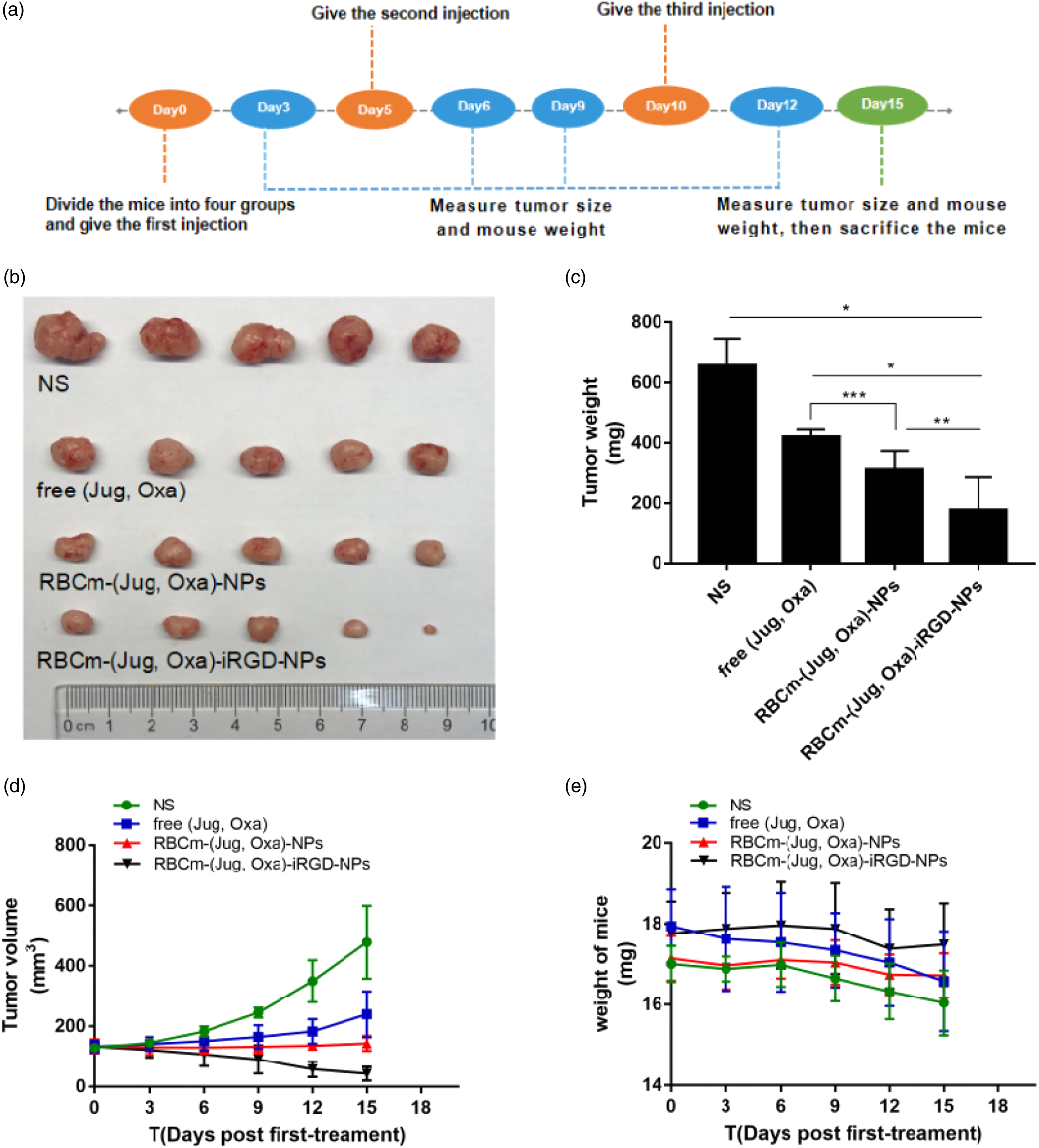
To investigate the mechanisms underlying the potent antitumor effects, we performed histological changes of tumor tissues for the mice treated with different groups. Figure 7(a) shows the Ki67 staining of the excised tumors treated with normal saline, free (Jug, Oxa), RBCm-(Jug, Oxa)-NPs, and RBCm-(Jug, Oxa)-iRGD-NPs after 15 days post-injection. Figure 7(b) shows that the number of positive tumor cells with high proliferation ability in RBCm-(Jug, Oxa)-iRGD-NPs-treated group was much lower than that of the other groups (*p < .001, **p = .02, ***p = .01). Moreover, H&E-stained section images of the major organs in each group did not show any noticeable signal of organ damage (Figure 7(c)), indicating that RBCm-(Jug, Oxa)-iRGD-NPs therapy exerted little systemic toxicity in vivo. Tissue staining. (A) Ki67 staining of the excised tumors (scale bar=200 mm) and (B) Ki67 positive cells. (*P<0.001, **P=0.02, ***P=0.01). (C) H&E-stained section images of the major organs in each group (scale bar=200 mm). Abbreviations: H&E, hematoxylin-eosin.
Discussion
Erythrocyte membrane is derived from autologous cells (red blood cells), which, as a drug carrier, has the unique advantages of high biocompatibility, no immunogenicity, and degradability.42–44 However, the direct use of red blood cells as drug carriers has some defects, such as off-targeting property and fast release rate of the loaded drug. 43 Nanoscale erythrocyte membrane carrier made from RBCm not only retains the advantages of red blood cells but also has the characteristics of tumor-targeting and slow-release.18,45 iRGD is a small molecular tumor-targeting penetrating peptide that promotes extravasation and tumor-specific penetration of small molecules and nanoparticles via RGD domain and CendR motif. iRGD can be used for tumor-specific targeted delivery of small-molecule drugs, monoclonal antibodies (trastuzumab), and nanoparticles (albumin paclitaxel nanoparticles and doxorubicin liposome) to improve tumor penetration and antitumor effects of drugs.32,35,46–48 Based on the RBCm-NP platform with optimal stability and biocompatibility, 49 the modification of DSPE-PEG-iRGD endowed the active tumor targeting ability for this biomimetic nanocarrier. The physical insertion of iRGD into RBCm maintained the integrality of the original structure. The current study demonstrated that RBCm-(Jug, Oxa)-iRGD-NPs had a drug-release profile similar to RBCm-(Jug, Oxa)-NPs, but were more stable. Intriguingly, iRGD modification promotes the stability of NPs.
CRC is the third leading cause of cancer-related mortality worldwide. Oxa, the third generation of platinum anticancer drugs, is widely applied for the treatment of CRC. However, its application is restricted by side effects, including short terminal half-life and severe normal organ damage, especially neurotoxicity. J. mandshurica Maxim, a potential traditional Chinese medicine, has been clinically utilized for a thousand years owing to its anti-inflammatory, antifungal, antitumor, antioxidation, and other pharmacological effects. Jug, the main antitumor component of J. mandshurica Maxim, has a good inhibitory effect on malignant tumors, such as liver cancer, 50 nasopharyngeal cancer, 51 and cervical cancer. 52 Thus, Jug could be deemed as a broad spectrum antitumor drug. However, poor solubility, non-specific biological distribution, and toxicity to normal tissues limit its clinical use. Jug NPs have been prepared from a variety of materials, such as Jug liposomes 16 and Jug-plga-NPs modified with folic acid; however, only a few studies have been conducted using RBCm-NPs and on CRC. Previous experiments revealed a synergistic effect of Jug and Oxa based on the Q value. Herein, the synergistic effect of Jug and Oxa was proved by the results of cytotoxicity, apoptosis, and cell cycle analysis. Hence, RBCm-(Jug, Oxa)-iRGD-NPs not only resolved the problem about the application of Jug with poor water solubility but also enhanced the antitumor effect by tumor-targeting characteristics and synergistic effects.
The tumor-targeting ability and antitumor efficiency of RBCm-(Jug, Oxa)-iRGD-NPs were both examined on CRC cells in vitro and CRC-xenografted nude mice in vivo. The in vitro tumor cell uptake experiments showed that iRGD significantly promoted the uptake of NPs by tumor cells. The infiltration ability of NPs differed in the near-infrared fluorescence intensity in various groups. A large number of iRGD-modified NPs penetrated into the core site of tumor tissue as compared to free drugs and NPs without iRGD. This phenomenon could be attributed to the osmotic effect of iRGD.
In terms of the antitumor efficiency in vitro and in vivo, RBCm-(Jug, Oxa)-iRGD-NPs exhibited cytotoxicity, apoptosis, and cell cycle and tumor inhibition. RBCm-(Jug, Oxa)-iRGD-NPs showed the highest cytotoxicity for 48 h at the same dose in vitro, while free (Jug, Oxa) showed a similar effect for 24 h, since only a small part of the drug was released from the NPs for 24 h. Moreover, RBCm-(Jug, Oxa)-NPs had almost the same cytotoxicity as free (Jug, Oxa) for 48 h due to the same dose and the prolonged circulation of NPs. Simultaneously, the cell apoptosis of RBCm-(Jug, Oxa)-iRGD-NPs was maximal. The results of the cell cycle indicated that the combination of Jug and Oxa could block the G0/G1 phase, irrespective of the formulations. Although this could be a putative mechanism of the synergistic effect of Jug and Oxa, additional studies are needed in the future. The difference in antitumor activity in vivo among the groups could be explained by the results of near-infrared imaging. Compared to the control group, the free (Jug, Oxa) group showed mild antitumor activity, which was due to the rapid elimination of Jug in vivo. RBCm-(Jug, Oxa)-iRGD-NPs group had the strongest antitumor effect because of abundant Jug and Oxa infiltration into the tumor sites. The results of Ki67 staining in the tumor tissues were consistent with the trend of antitumor experiment, which further confirmed the pathology of the antitumor activity of each group. Finally, we concluded that RBCm-(Jug, Oxa)-iRGD-NPs exert an antitumor effect as follows: retention effect, unlike naked drugs, which could be quickly cleared in vivo, and tumor-targeting characteristic that increases the drug concentration at tumor sites.
iRGD-modified nanocarriers not only increase the antitumor effect of drugs but also have biosafety. In our safety assessment experiments, no obvious side effects, such as organ damage and mice weight loss, were detected with respect to Jug, Oxa, and their formulations.
In the current study, we built a new nanoplatform made of RBCm with iRGD modified on the surface. The current findings verified that iRGD-modified NPs had increased stability and preferable ability to target tumor. Jug and Oxa exhibited a synergistic effect. RBCm-(Jug, Oxa)-iRGD-NPs retained the activity and sustainably released Jug and Oxa, such that it would be detrimental to tumor cells. The prolonged retention time of iRGD and the tumor penetration of NPs promoted the antitumor effect of RBCm-(Jug, Oxa)-iRGD-NPs without obvious side effects. Thus, this innovative nanoplatform served as a promising delivery system to improve anti-CRC efficacy.
Footnotes
Declaration of conflicting interests
The author(s) declared no potential conflicts of interest with respect to the research, authorship, and/or publication of this article.
Funding
The author(s) disclosed receipt of the following financial support for the research, authorship, and/or publication of this article: This work was supported by the Health and Family Planning Commission key program of Nanjing under Grant number ZKX17012; and Health and Family Planning Commission development program of Jiangsu Province under Grant number H2817042; Livelihood Science and Technology - Basic research on Medical and Health application program of Suzhou under Grant numer SYSD2020035.
