
Abstract
This research involved the synthesis of secondary nano copper oxide nanoparticles (CuO NPs) at a pH of 9 via the hydrothermal method. Subsequently, the CuO NPs were integrated with a polymer mix of polyacrylonitrile (PAN) and polymethyl methacrylate (PMMA) using the electrospinning technique to produce fibres from the resulting composite material, which exhibited improved properties and achieved commendable homogeneity between the inorganic and organic phases. The synthesised nanofibers were analysed utilising advanced techniques, including Field Emission Scanning Electron Microscopy (FESEM), to examine the surface structure, morphological characteristics, and nanoparticle distribution within the fibres. The results displayed an excellent homogeneous fibrous structure and efficient dispersion of CuO particles within the prepared fibers. Energy-Dispersive X-ray (EDX) an study was conducted to verify the presence of encapsulated CuO, Cu, and O elements, in the PAN-PMMA nanofibers, with other constituent elements. Furthermore, FTIR spectra was conducted to detect functional groups and ascertain potential interactions between the CuO nanomaterial and the PAN-PMMA polymer blend. The results displayed Nanofibers have garnered significant attention over the last two decades owing to their a slight shift in the positions of some peaks, confirming the presence of interactions between the composite components. The optical characteristics of the synthesised fibres were examined by UV-Vis spectroscopy to assess the absorption behaviour and energy gap. The antibacterial efficacy of the synthesised fibres was assessed against Klebsiella pneumoniae and Staphylococcus aureus, revealing significant inhibitory activity against both species. This phenomenon is ascribed to the CuO NPs, which generate reactive oxygen species (ROS) that undermine the bacterial cell wall and other cellular components. The PAN–PMMA matrix facilitated superior nanoparticle distribution and augmented the effective surface area, hence increasing interaction with bacterial cells. These findings suggest significant potential for these fibres in antibacterial applications.
Keywords
Introduction
Nanofibers have attracted considerable interest during the past two decades because to their advantageous and distinctive features applicable across numerous domains. 1 Nanofibers exhibit a substantial specific surface area, favourable porosity, and superior mechanical qualities, facilitating their fabrication and surface modification.2,3 A variety of techniques for nanofiber production have been documented in scientific literature, encompassing both sequential and iterative procedures, including stretching. 4 Die-casting. 5 Self-assembly. 6 Microphase separation. 7 Dry spinning. 8 Melt spinning. 9 Blow spinning. 10 Wet spinning. 11 Centrifugal spinning. 12 Microfluidic spinning. 13 Electrospinning. 14 Despite its simplicity, electrospinning is the easiest and most widely used method, allowing for significant optimization of system components.15,16 This allows for substantial changes in the morphology of the produced nanofibers.17,18 Electrospinning is considered a step-by-step method for manufacturing nanofibers. 19 It is among the most cost-effective and efficient techniques for nanofiber production and is seeing growing popularity. With the growing number of relevant scientific publications, it has garnered significant attention. Compared to other fibers, it offers greater control over the process. The interior pore architecture of electrospun nanofibers. 20 The fibre content and structure can be altered to meet performance specifications.21,22 The mechanical efficiency of electrospun nanofibers is a crucial factor influencing the structural characteristics of nanofiber mats, such as fibre diameter.23,24 Orientation of fibres and porosity of the mat.25,26
To enhance the mechanical characteristics of a nanofiber mat, it is necessary to either strengthen the bonds between the fibers or incorporate supplementary structural supports to accommodate the load in the transverse direction One of the biggest problems with making these nano-bonds stronger is finding the right polymer that can make strong bonds without changing the properties of the nanofibers.27,28 Dimethylformamide and formic acid are two common industrial solvents that can change the properties of nanofibers. This is because leftover solvents can change the fibres’ elasticity, which can change their mechanical strength and thermal stability attributable to their influence on fibre elasticity.29,30 Reneker and Chun provided the first report on the production of PAN nanofibers by electrospinning in 1996. 31 Polyacrylonitrile (PAN) fibers, a synthetic polymer, have become increasingly valuable due to their excellent mechanical properties. 32 Polyacrylonitrile has much greater viscosity compared to alternative polymer solutions. Acrylic fibres, or polyacrylonitrile (PAN) fibres, can be produced using gel spinning, wet spinning, and electrospinning. The flexible polyacrylonitrile polymer is utilised to manufacture oxidised PAN fibres, textile fibres, and hollow filter fibres. 33 Including ultrafiltration membranes. 34 Electrospinned polyacrylonitrile (PAN) nanofibers are fundamental to interconnected carbon nanofibers.35,36 Optical and sensing materials.37,31 Tissue structures. 38 Drug delivery systems. 39 and protective equipment. 40 Healing Injuries. 41 Smart textiles. 42 and nanocomposites. 31 Numerous literatures have referenced the use of PAN-PMMA blends as a precursor for the fabrication of electrically spun carbon nanofibers with an advantageous structure. 43 Polymethyl methacrylate (PMMA) is a thermoplastic polymer, an amorphous and transparent polymer (9) with excellent properties, particularly its high resistance to harsh cutting conditions and its stability in the presence of acids and bases. It is utilised in several applications, including lenses.44,45 However, a critical challenge in developing antimicrobial nanofiber mats is achieving a balance between the high mechanical strength of PAN and the chemical stability and processing flexibility of PMMA. While individual polymers have been studied, the specific integration of tailored metal oxide nanoparticles into a hybrid PAN-PMMA matrix remains under-explored, particularly regarding the enhanced effect of the blend on nanoparticle dispersion and subsequent biological activity.46,47
To produce good nanofibers, The distance between the spindle and the collector must be taken into account, as it influences the configuration and alignment of the fibres. 48 The rotational velocity of the collector drum influences the characteristics of the nanofibers; increased speeds promote the formation of thinner and more aligned fibres. The temperature and humidity conditions during the electrospinning process can affect both the procedure and the properties of the fibres. 49 The solvent affects the solution’s viscosity and flow rate; therefore, dimethylformamide (DMF) is the most common solvent for dissolving and electrospinning both PAN and PMMA polymers owing to its excellent solubility, low viscosity, superior conductivity, elevated boiling point, and accessibility.50,51 To overcome the limitations of conventional antimicrobial agents, CuO NPs synthesized via a hydrothermal method at a precisely controlled pH of 9 were incorporated into the fibers. The choice of this specific synthesis route and pH environment is pivotal, as it directly influences the nanoparticles’ crystallinity and optical energy gap factors that are fundamental to their ability to generate reactive oxygen species (ROS) for bacterial inactivation.52,53 In this work, a detailed investigation of a PAN-PMMA hybrid system reinforced with these hydrothermally-tailored copper NPs to produce nanofibers. Unlike existing studies that focus on commercial or simple metal-polymer mixtures, this research provides a comprehensive analysis of the morphological, structural, optical, and electrical properties, correlating these physical characteristics with the bacterial inhibition effects. 54 Two bacterial strains, Klebsiella and Staphylococcus aureus, were used to evaluate the enhanced antimicrobial performance of the resulting nanocomposite.
Although there is increasing attention towards polymer nanocomposites, few efforts have been reported on studying the effect of blending PAN–PMMA and the hydrothermal synthesis of CuO nanoparticles on morphology and antibacterial properties. In this regard, the current study contributes to the literature by presenting a thorough investigation of the effect of conditions during nanoparticle synthesis on nanofiber morphology and antibacterial property.
Methodology
Materials
Cupric sulphate pentahydrate (CuSO4·5H2O, purity ≥99%, Mw = 249.68 g/mol) and hydrochloric acid (HCl, 35–38%, Mw = 36.46 g/mol) were purchased from Thomas Baker Laboratory Reagent Co., Ltd (India). Sodium hydroxide (NaOH, purity ≥99%, Mw = 40 g/mol) was obtained from Himedia Pvt. Ltd (India).
Poly (methyl methacrylate) (PMMA, Mw = 20,000 g/mol, purity ≥99%) was supplied by Thomas Baker Laboratory Reagent Co., Ltd (India), while polyacrylonitrile (PAN, Mw = 150 × 103 g/mol, purity ≥99%) was procured from Sigma-Aldrich (Merck KGaA, Darmstadt, Germany).
N,N-dimethylformamide (DMF, purity ≥99.7%, Mw = 73.09 g/mol) was obtained from Alpha Chemika (India) and used as a solvent. Ethanol and ultra-pure water (UPW) were also used as solvents.
All chemicals were used as received without further purification.
CuO NPs Synthesis
The CuO nanoparticles were prepared using the hydrothermal route. First, 4 g of cupric sulphate pentahydrate (CuSO4·5H2O) were dissolved in 80 mL of distilled water and stirred at room temperature for 15 minutes. Next, 2 g of sodium hydroxide (NaOH) were dissolved in 80 mL of distilled water while being stirred continuously for 15 minutes.
Afterwards, the NaOH solution was slowly dripped into the copper sulphate solution while stirring. The mixture was stirred for 40 minutes to ensure homogeneity in the solution. Finally, the pH of the solution was set to 9 through the slow addition of hydrochloric acid (HCl).
In the next step, the temperature of the mixture was raised to 70°C and stirring was done for 4 hours to obtain a dark brown solution. Next, the solution was poured into a sealed autoclave and treated with a temperature of 120°C for 5 hours. At the end of the experiment, the autoclave was cooled down to room temperature.
The resulted precipitate was rinsed three times in ethanol and three times in distilled water using centrifugation at a speed of 4000 r/min to get rid of any contamination. Then, the product was dried in an oven in the range of temperatures between 60°C and 70°C for 2-3 hours.
Lastly, the dry sample was annealed at a temperature of 500°C for 2.5 hours and black CuO nanoparticles were synthesized. and final step, the resulting material was ground to obtain fine black, as shown in Figure 1. Illustrates the procedures for synthesizing CuO NPs and fabricating PAN-PMMA/CuO fibers.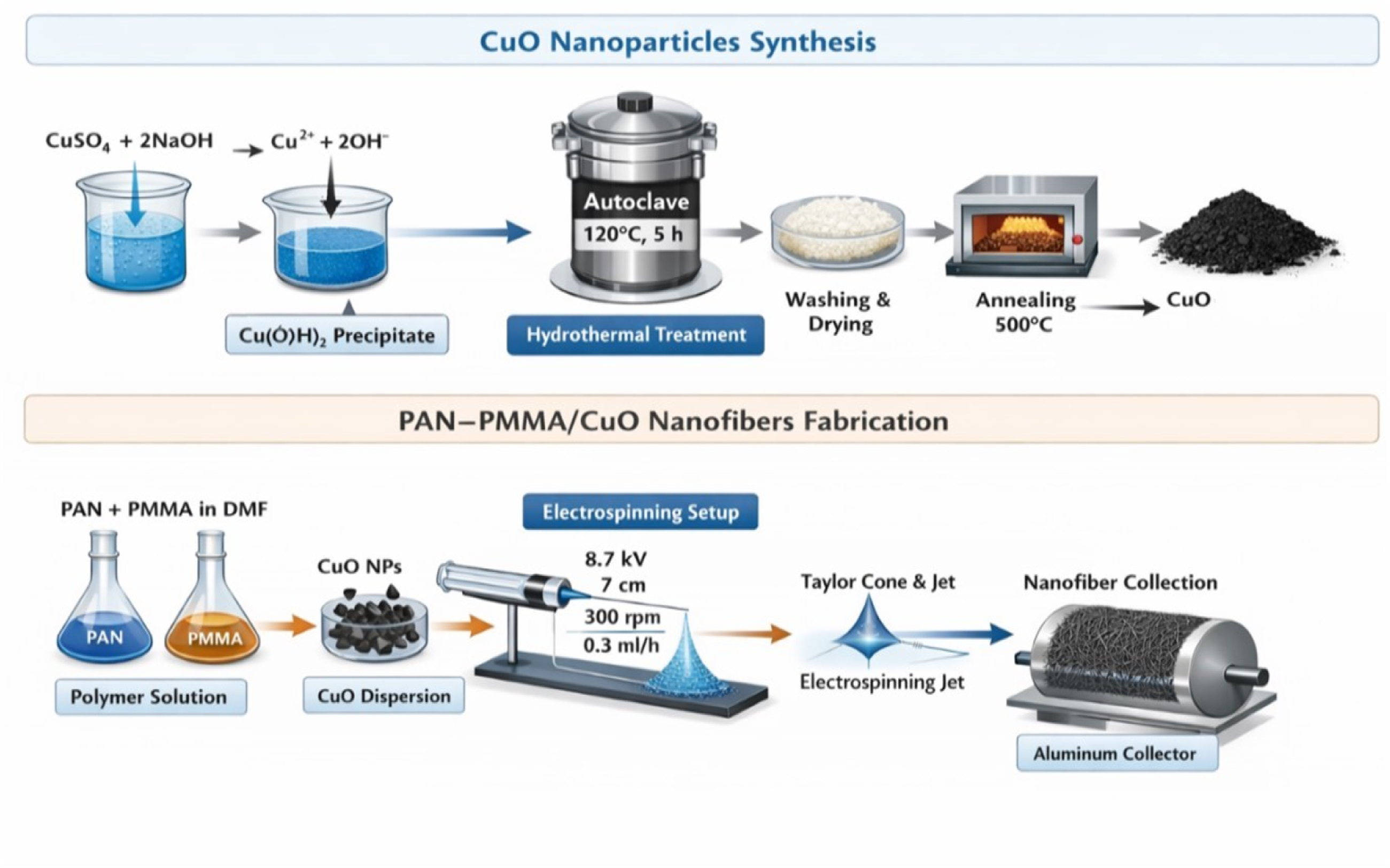
Polymer Blending and Electrospinning
First, 0.85 g of polyacrylonitrile (PAN) was dissolved in DMF solvent of 8 mL in magnetic stirring at ambient conditions for 5 hours. Later, 0.15 g PMMA was added in the PAN solution, which was further stirred for 5 hours in order to achieve a homogeneous polymer mixture.
CuO nanoparticles (0.02 g, 0.04 g, and 0.06 g) were then gradually mixed in PAN-PMMA solution in order to prepare different formulations of the nanocomposites. In each case, the samples were subjected to stirring for 30 minutes at ambient conditions and further sonicated for 3 minutes.
All prepared solutions were loaded into a 5 mL syringe having stainless steel needles with an inner diameter of 1.2 mm for electrospinning purposes. During electrospinning, the following parameters were used: applied voltage of 8.7 kV, tip to collector distance of 7 cm, solution feeding rate of 0.3 mL/hour and 2 hours of spinning time.
For fiber collection during electrospinning, a rotating aluminum covered drum collector of 10 cm by 30 cm dimensions was used at the rate of 300 revolutions per minute. On the other hand, glass slides of dimension 2.5 cm by 7.5 cm were attached on the collector to measure their optical properties.
Nanofiber samples were prepared with different CuO loadings corresponding to 0, 2.2, 4.4, and 6.6 wt% relative to the polymer matrix. The PAN:PMMA ratio of 85:15 wt% was determined based on prior research and first experiments to guarantee appropriate spinnability and fiber development. , as displayed in Figure 1.
Characterisation Techniques
Crystal structure analysis of synthesized CuO nanoparticles was carried out by XRD using Cu Kα radiations (λ = 1.5406 Å) having a voltage of 40 kV in the range of 2θ = 20-80°.
Functional group analysis of CuO nanoparticles and PAN-PMMA/CuO nanofibers was performed through FTIR spectroscopy in the region of 500-4000 cm−1.
Field emission scanning electron microscope (FESEM) MIRA III, TESCAN, Czech Republic along with energy dispersive X-ray spectroscopy (EDX) was employed for morphological and chemical analysis of the samples.
Average particle size and fiber diameter of the particles/fibers were calculated by ImageJ software through random selection of at least 100 particles/fibers from SEM images.
Optical properties of the samples were studied through UV-Vis spectroscopy (Shimadzu UV-1900, Japan) in the wavelength range of 200-1100 nm.
Antibacterial Test
The antibacterial test of the synthesized nanofibers was performed on Gram-negative bacteria K. pneumonia and Gram-positive bacteria S. aureus by the diffusion method through agar medium. The nanofibers samples containing various amounts of CuO content (0%, 2.2%, 4.4%, and 6.6% wt) were subjected to the test under identical conditions.
Statistical Analysis
The effectiveness was estimated based on the measurement of inhibitory zones. Representative data are shown for each extract; measurements were repeated to check reproducibility. Statistical analysis was not carried out.
Results and Discussion
UV–Vis Analysis
Optical Properties of CuO Nanoparticles
For the purpose of studying the optical absorption behaviour as a function of wavelength, a solution consisting of 0.021 g of CuO particles in 10 mL of deionized water was prepared, the mixture was stirred for 15 minutes, and then the solution was exposed to ultrasound waves for 3 minutes. The spectrum of absorption of the solution was recorded using a UV-Vis spectrometer (Shimadzu, UV-1800Ao model, Japan) between 300 and 1200 nm, as shown in Figure 2(a). The results on the optical spectrum edge indicate that maximum absorption occurs in the UV region near visible light at around 350 nm, followed by a progressive decrease with increasing wavelength, it is compatible with
55
. Figure 2(b) exhibits the direct band gap energy (a), (b) and (c) Absorbency, energy gap (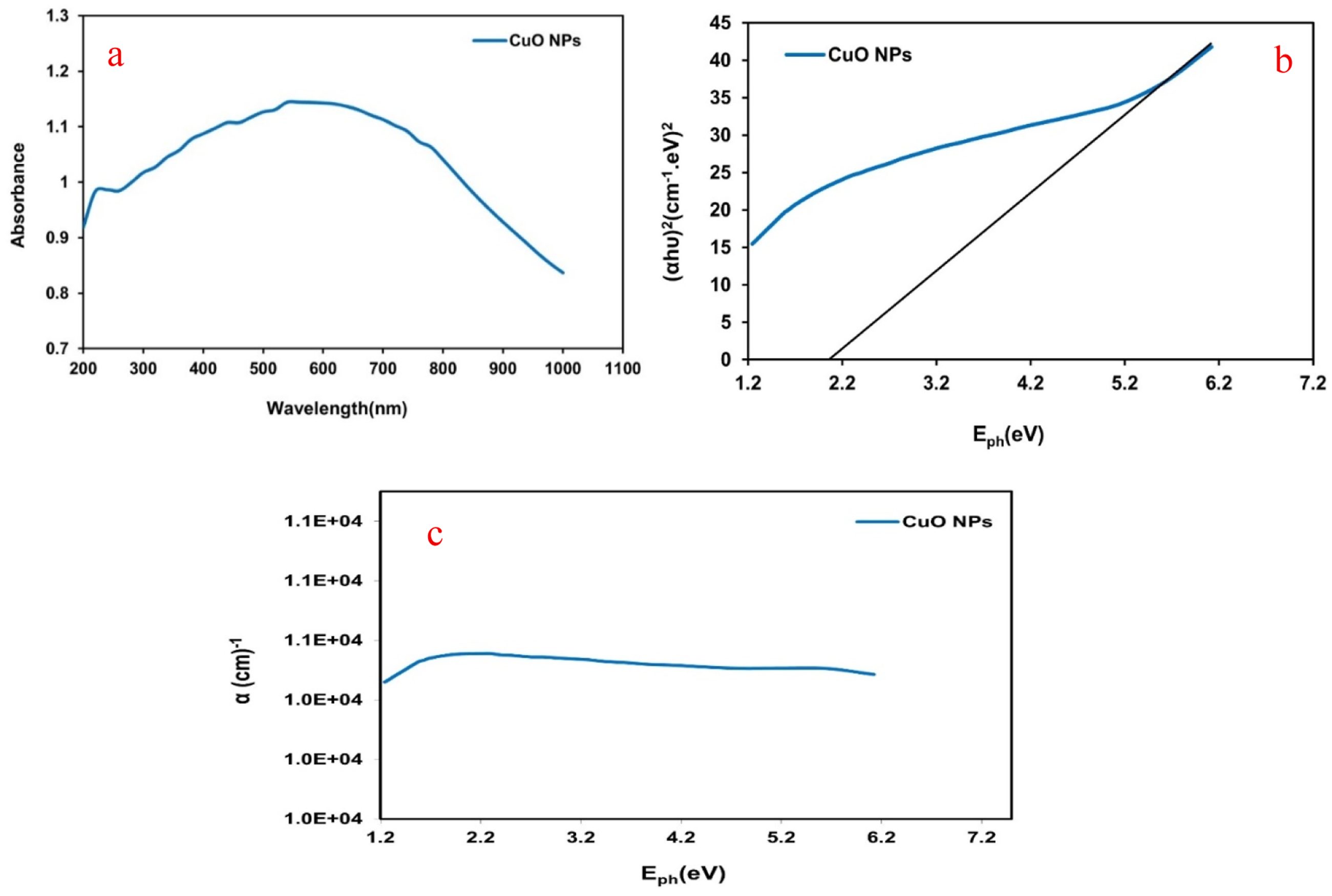
Optical Properties of PAN–PMMA/CuO Nanofibers
Figure 3(a) and (b) depicts the optical absorbance and optical indirect energy gap of PAN-PMMA nanofibre and PAN-PMMA/CuO nanofibre NCFs containing 2.2, 4.4, and 6.6 wt% CuO, with a thickness of 10 µm on a glass substrate, across a wavelength range of (300–1200) nm. All measurements were conducted using a double-beam spectrophotometer to analyse the web nanofibers at ambient temperature. The absorption spectra of the nanofibers demonstrate enhanced light absorption with the incorporation of CuO NPs, especially within the UV region of 300 to 450 nm. This resulted in ascribing sufficient energy to these photons to engage with atoms. The augmentation in nanoparticle concentration with a fine distribution within the matrix was accountable for the rise in the quantity of charge carriers, corroborating findings documented in the literature.
58
When calculating the values using equation (1) (Tauc relation), it is observed that the absorption effect extends into the visible spectrum, indicating an effective Reduction in the optical indirect band gap from 3.3 to 2.8 eV as CuO concentrations rise from 2.2% to 6.6% wt%, respectively. The reduction in the band gap indicates an enhancement in the compound’s shape, as well as its optical and electrical properties, rendering it more appropriate for antibacterial applications. These findings align with prior research.59,60 Figure 3(c) illustrates the absorption coefficient (α) of PAN-PMMA nanofibre and PAN-PMMA nanocomposite nanofibre as a function of photon energy. The absorption coefficient demonstrated a consistent increase in values with rising photon energy, reaching a peak at 4.1 eV. This may pertain to the lower transition of the electron, when the input photon energy was insufficient to elevate the electron from the valence band to the conduction band. Above 4.1 eV, the absorbance coefficient exhibits a rapid increase across all samples due to significant electron transitions to the conduction band. At 4.1 eV, the results demonstrated a notable increase in the absorption coefficient of 24%, 44%, and 127% with the augmentation of CuO concentrations in the nanocomposites to 2.2, 4.4, and 6.6 wt %, respectively. The increase to 127% indicates the success of the structural modification process caused by the nanomaterial on the fibers, which helped to increase the efficiency of the fiber network in absorbing photons in the ultraviolet (UV), where 4.1 eV falls within this range. Figure 9(d) depicts the refractive index curves of PAN-PMMA fibres and PAN-PMMA/CuO NCFs as a function of wavelength. The refractive index of the samples was enhanced by the introduction of CuO NPs into the nanocomposite fibres. This behaviour may be ascribed to the augmentation of nanocomposite density. At 500 nm, the results exhibited substantial improvements in the refractive index of up to 17%, 25%, and 32% with the incorporation of CuO NPs at concentrations of 2.2, 4.4, and 6.6 wt % in the nanocomposites. Tauc relation.
61
o (a), (b),(c) and (d) Absorbance, energy gap (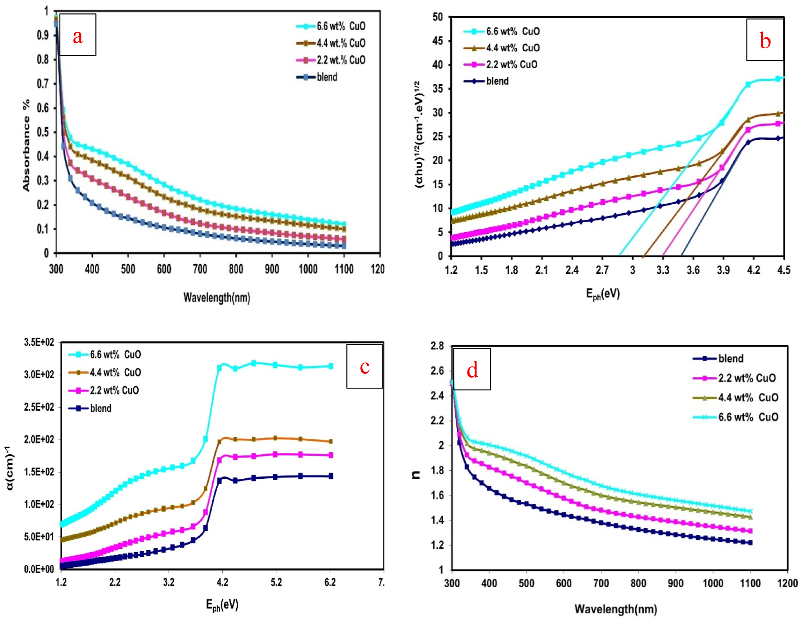

FTIR Analysis
Figure 4 presents the FTIR spectra for CuO NPs, PAN-PMMA mixture, and PAN-PMMA/CuO nanofibers within the wavenumber range from 4000 cm−1 to 500 cm−1. The broad band appearing at about 3425 cm−1 in the FTIR spectrum for CuO NPs (Figure 4(a)) could be related to O-H stretching vibrations, implying the existence of water molecules. The wavenumber 1631 cm−1 corresponds to H-O-H bending vibrations. The outcomes coincide with those obtained previously.
62
The distinctive peak recorded at 511 cm−1 is due to Cu-O stretching vibrations. confirming the formation of CuO in its crystalline phase.
63
As seen in Figure 3(b), the FTIR spectra for the PAN-PMMA blend show characteristic peaks for both the polymeric substances. The peak for the nitrile group (C≡N) of PAN is seen at about 2241 cm−1, whereas that of carbonyl group (C = O) of PMMA lies at approximately 1730 cm−1. The C-H stretching peak is observed at 2945 cm−1, while CH2 and CH3 bending peaks can be seen at about 1450 cm−1. Furthermore, the peak for C-O-C stretching peak can also be seen at about 1185 cm−1. The results validate the effective creation of the polymer mix without substantial chemical modification, consistent with prior research.
64
In PAN-PMMA/CuO nanocomposites (Figure 3(c)–(e)), there are no new peaks observed, which implies the absence of any new bonding within the structure. Nevertheless, the characteristic peaks at 2241, 1730, and 1450 cm−1 appear with some changes in their intensities and position shifts. An increase in peak intensity is registered with an increase in the percentage of CuO NPs. Spectra of ftir (a) CuO NPs and PAN-PMMA/CuO NCFs with (b) 0, (c) 2.2, (d) 4.4, and (e) 6.6 wt % CuO.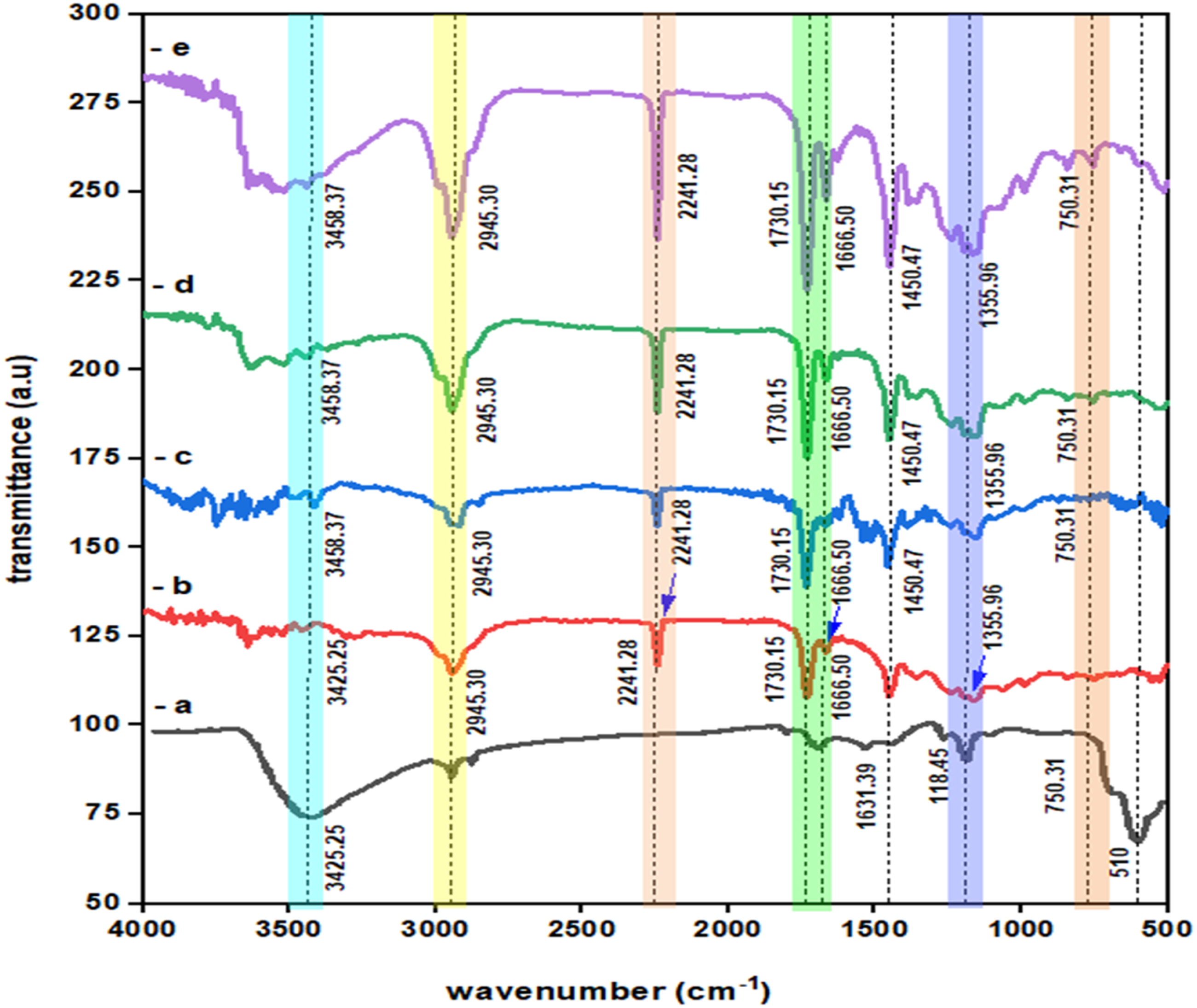
Moreover, an increase in the CuO amount (from 0 to 6.6 wt%) leads to a displacement of some characteristic peaks (with the maximal value of ∼10 cm−1) towards lower wavenumbers along with intensity changes. This indicates interaction between CuO nanoparticles and the PAN-PMMA matrix.
The above mentioned shifts could be explained by the presence of interaction between CuO nanoparticles and functional groups in the polymer matrix, such as nitrile (C≡N) in PAN and carbonyl (C = O) in PMMA. The formation of the latter can be caused by coordination or dipole-dipole interactions and does not involve the formation of new interaction. These results are consistent with.65,66 The lack of new peaks, coupled with peak shifting and intensity alteration, indicates the physical integration of CuO nanoparticles inside the polymer matrix with high interfacial compatibility. The results correlate with other studies on polymer-metal oxide nanocomposites, in which high dispersion and interfacial adhesion resulted in enhanced structural and functional properties. 67 Moreover, the reduction in the intensity of the C = O stretching vibration along with the shift towards lower wavenumber could imply partial interaction between CuO nanoparticles’ surface and carbonyl groups due to altered electronic environments inside the polymer chains. 68 Overall, FTIR spectroscopy reveals the effective addition of CuO nanoparticles inside the PAN-PMMA matrix, along with notable interfacial interactions.
XRD Analysis
The XRD patterns of synthesised nanometer-scaled CuO powder samples at pH 9, annealed at 500°C for 2.5 hours, are presented in Figure 5. The sample was scanned over the specified 2Ө range of 20 to 70° using CuKα1 radiation (λ = 1.5406 Å). The values of 2θ and the diffraction patterns for the four principal peaks were as follows: 32.535° (110), 35.452° (002), 38.748° (111), 48.705° (20-2); 32.509° (110), 35.438° (002), 38.731° (111), 48.743° (20-2); and 32.509° (110), 35.438° (002), 38.731° (111), 48.743° (20-2). These findings are in complete concordance with the values documented in reference code: 48-1548 and numerous scientific publications.69,70 The XRD data indicated that the growth of pure CuO was accurately indexed to the distinctive peaks of the monoclinic structure, with standard lattice parameters (a = 6.883 Å, b = 3.4229 Å, and c = 5.1319 Å). The interplanar distance (dhkl), microstrain (ε), and average crystallite size (D) were assessed using equations (2)–(4).
71
As inserted in Table1. Xrd diffractograms of CuO NPs. X-ray diffraction obtained results of CuO NPs.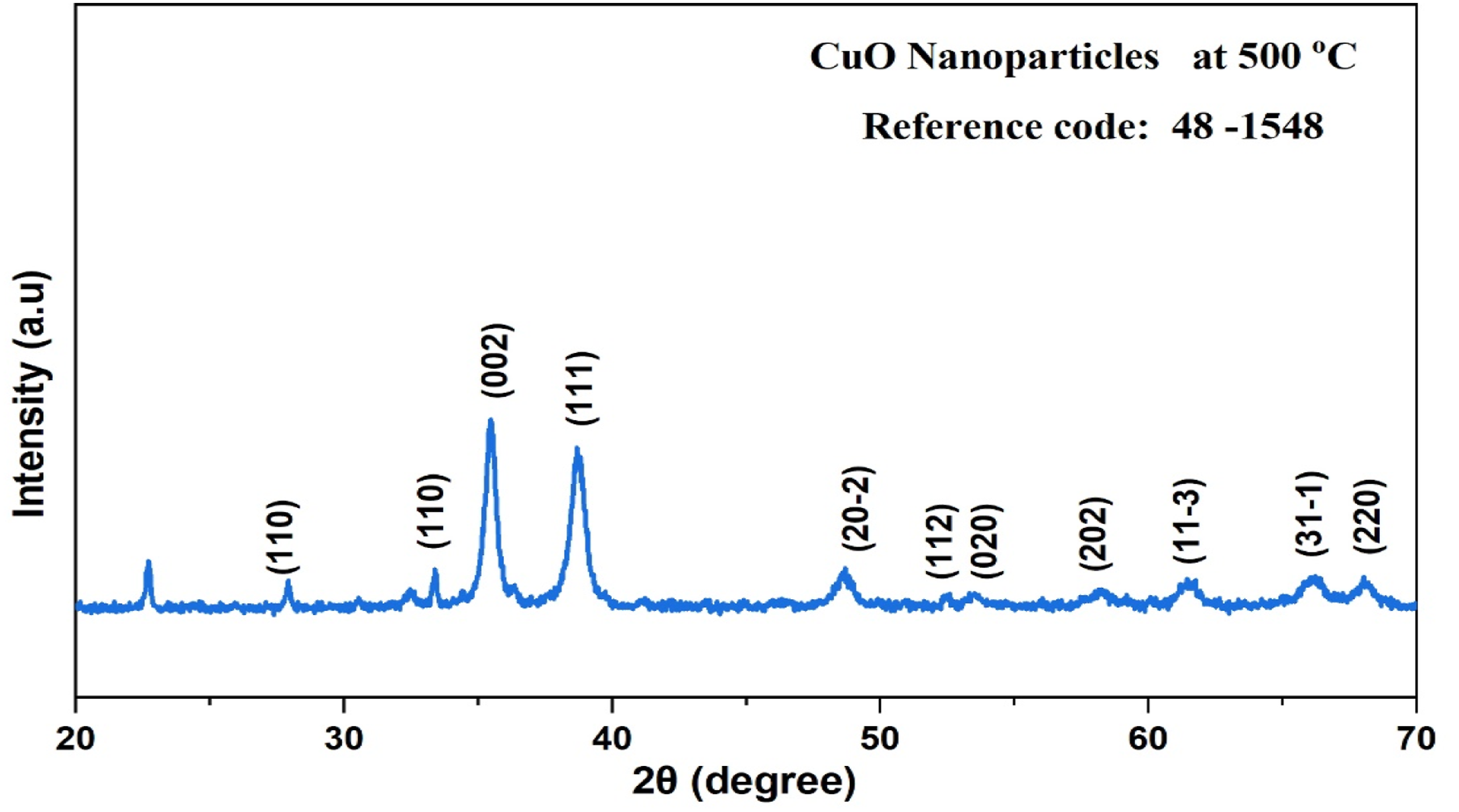
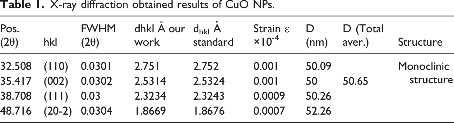


In this context, the symbol β represents the full width at half maximum (FWHM) of the peak, whereas θ symbolises Bragg’s angle. The data presented in Table 1 indicate that pH 9 considerably enhances the growth of NPs, as an adequate concentration of hydroxide ions (OH-) is available for the formation of CuO NPs. These findings align with the patterns recorded in prior scientific research. These findings align with the existing literature. 72
Morphological Analysis
FESEM and EDX of CuO NPs
Figure 6 illustrates the FESEM image and Gaussian distribution of synthesized CuO nanoparticles at pH 9. Synthesis of nanoparticles is strongly affected by various synthesis conditions such as stirring time and rate, reaction temperature, concentration of NaOH, and annealing conditions.
73
In the current study, such synthesis conditions were precisely regulated in order to obtain reproducible results and examine properties of the synthesized CuO nanoparticles. (a) FE-SEM picture and (b) average diameter with standard deviation of CuO NPs.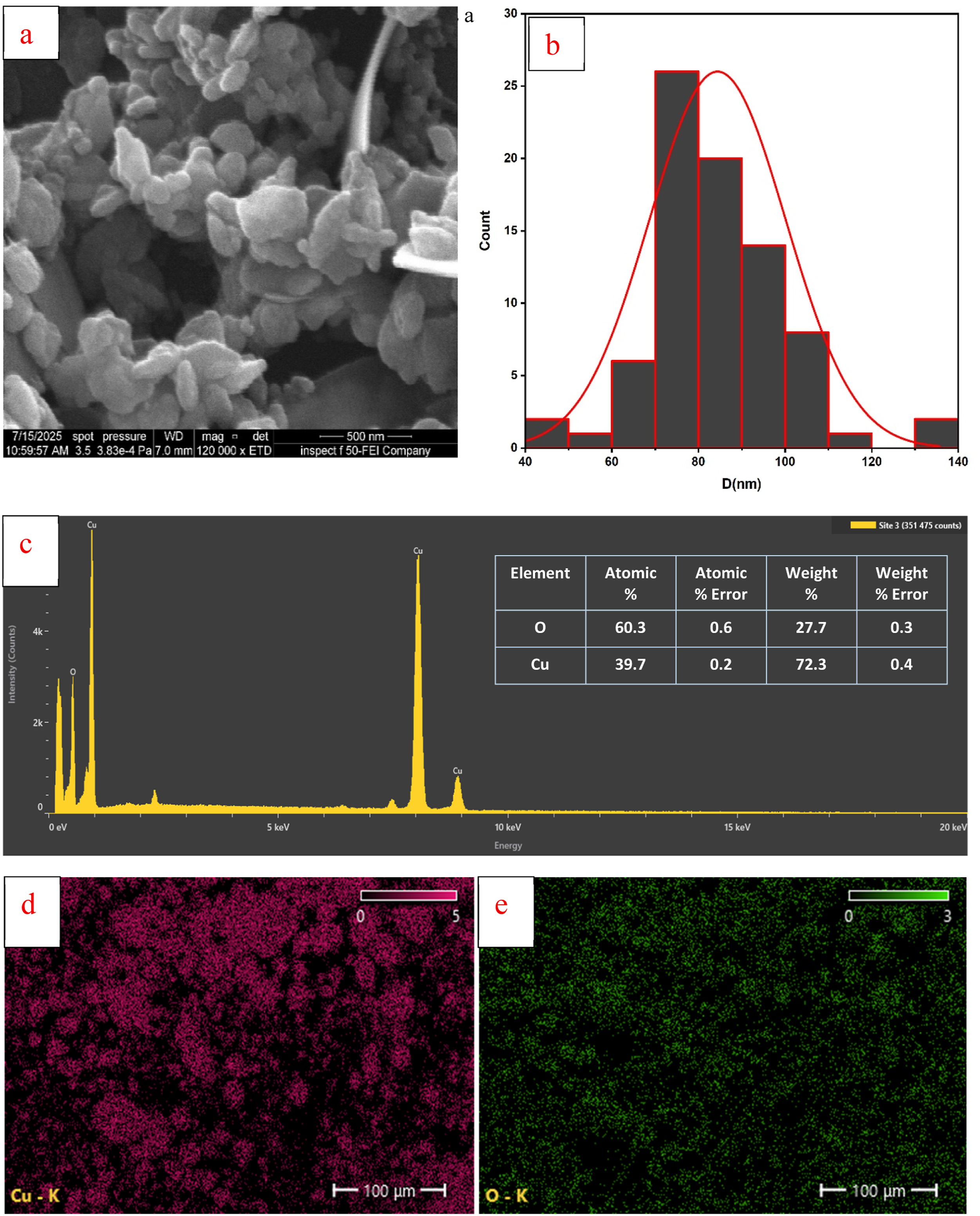
As can be seen in the FESEM images, morphology and average diameter of synthesized CuO nanoparticles are spherical to oval, with the average particle diameter of 84.41 nm the average particle diameter was calculated from FESEM images using ImageJ software through 100 measurements. The synthesized CuO nanoparticles are clustered on larger nanosheets. It is noteworthy that there is a certain level of nanoparticle agglomeration, as smaller particles tend to agglomerate on the surface of nanosheets, which will eventually lead to increased surface area and impact on functional characteristics of nanoparticles. Relative uniformity in the surface morphology implies efficient nucleation and particle formation, as previously discussed in literature. 74
The partial agglomeration observed with the CuO nanoparticles is typically linked with the presence of a high level of energy at the nanolevel, which increases the probability of interactions between the particles. Nonetheless, taking into consideration the general morphology of the material, it may be argued that the synthesis of the CuO nanoparticles at pH 9 represents a good compromise between growth and agglomeration. Similar results have been achieved before using hydrothermal synthesis of CuO nanoparticles.75,76
EDX spectra and the elemental distribution for CuO nanoparticles are presented in Figure. 6(c)–(e), and Figure 7(c)–(e). Elemental maps demonstrate the presence of copper and oxygen, denoted by various colours, and, thus, the successful preparation of CuO nanoparticles. The absence of any other peaks besides those for Cu and O in EDX spectra is consistent with earlier studies.77,78 According to the EDX analysis data, the concentration of copper is about 39.7 wt% and that of oxygen is about 60.3 wt%. Elemental mapping reveals that oxygen is more abundant than copper, implying that oxidation has been evenly achieved, and CuO nanoparticles have successfully been produced. (a)-(d) FE-SEM pictures of the PAN-PMMA mix and its NCFs containing 2.2, 4.4, and 6.6 wt% CuO, respectively.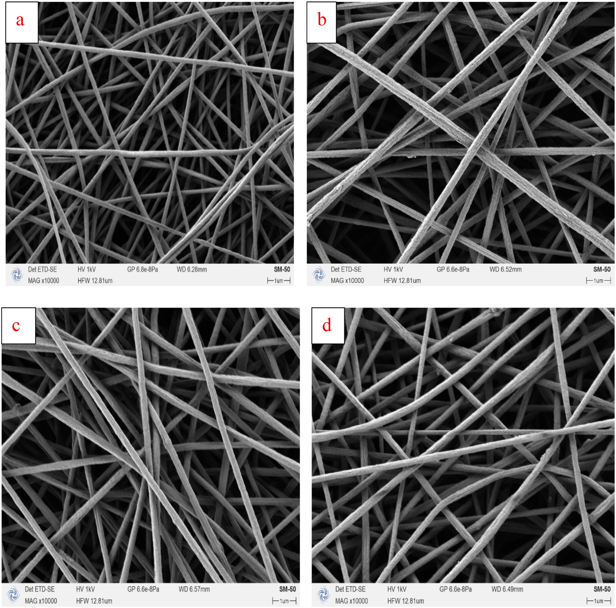
The lack of impurities in the EDX spectra, coupled with the relatively uniform distribution of the elements, suggests that the produced CuO nanoparticles are pure. Purity is crucial since it ensures efficient interaction between the CuO nanoparticles and the polymer matrix when producing nanofibers.
FESEM and EDX of PAN–PMMA/CuO nanofibers
Figures 7–9 show FESEM images, diameter distribution, and EDX analysis of PAN-PMMA nanofibers and PAN-PMMA/CuO NCFs, respectively. The pure PAN-PMMA nanofibers (Figure 7(a)) display a uniform and interconnected network of fibers, with the average diameter of ∼131.12 nm. The average diameter of the fibers was calculated from FESEM images using ImageJ software (n = 100), as shown in Figure 8. (a)-(d) Mean diameter with standard deviation of PAN-PMMA mix and its NCFs containing 2.2, 4.4, and 6.6 wt% CuO, respectively. (a)–(d) EDX analysis and average fiber diameter (±SD) of PAN–PMMA blend and PAN–PMMA/CuO NCFs with 2.2, 4.4, and 6.6 wt% CuO.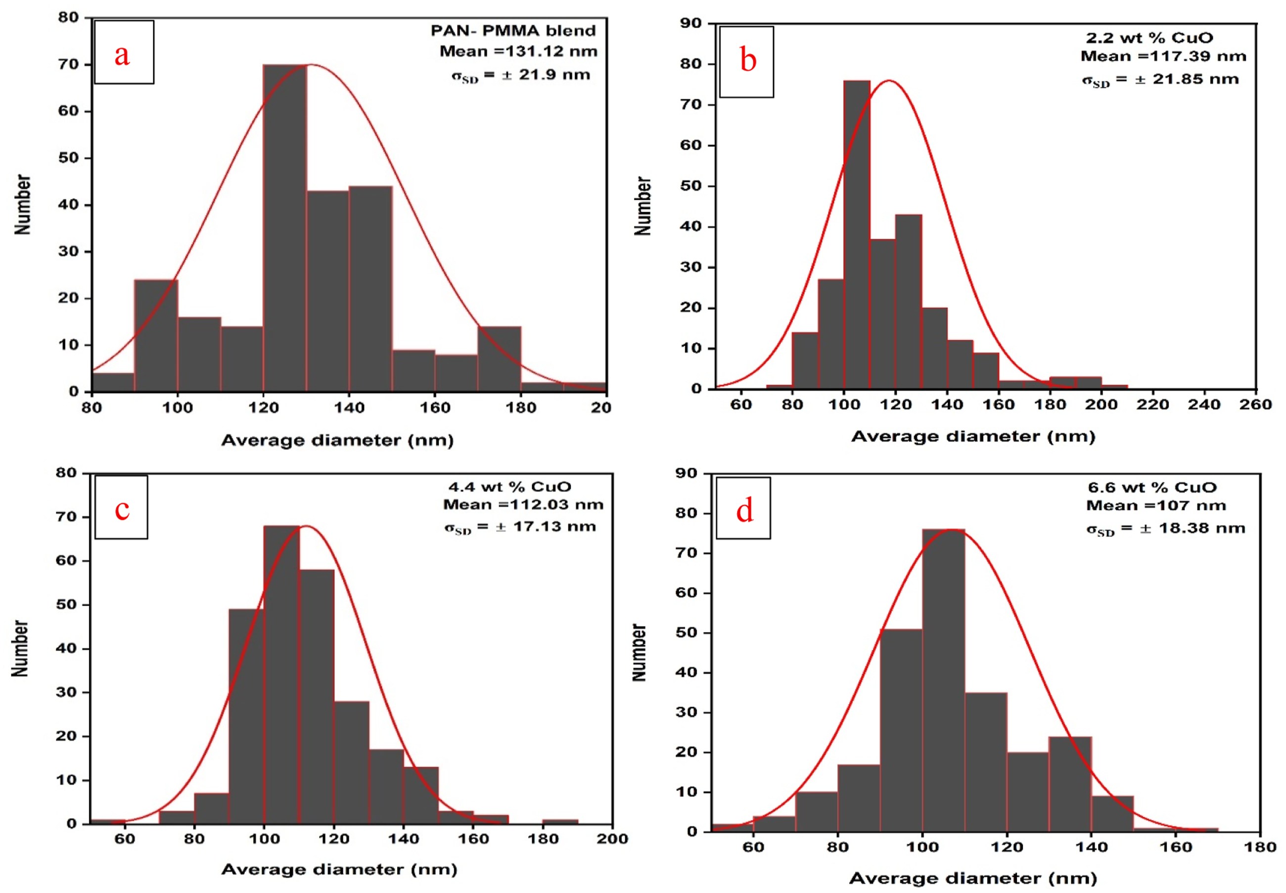
The inclusion of 2.2 wt% CuO (Figure 7(b)) caused the average fiber diameter to decrease to ∼117.4 nm. The average diameter further reduced to ∼112.03 nm with an increase in CuO concentration to 4.4 wt% (Figure 7(c)), with a more pronounced reduction in the diameter distribution range. In case of a maximum concentration of 6.6 wt% CuO (Figure 7(d)), the average diameter was found to decrease to ∼107 nm, as shown in Figure 8 however, some broadening of the diameter distribution was noted.
This reveals that the addition of CuO nanoparticles greatly affects the morphology and microstructure of the nanofibers, leading to a reduction in their diameter. This can be attributed to an increase in the conductivity of the solution and the stretching of the polymer jet during electrospinning, in accordance with previous reports. 79
It is further evident from the FESEM pictures that the dispersion of CuO nanoparticles in the PAN–PMMA nanofibers is quite good, especially at lower amounts of nanoparticles, due to the smoother and continuous nature of the nanofibers. In the case of high amounts of CuO, there might be slight differences in the appearance of the nanofibers as a result of possible agglomeration of nanoparticles; however, there seems to be no sign of agglomeration. It is clear from this that the PAN–PMMA mixture helps stabilize and disperse the CuO nanoparticles in the electrospinning process.
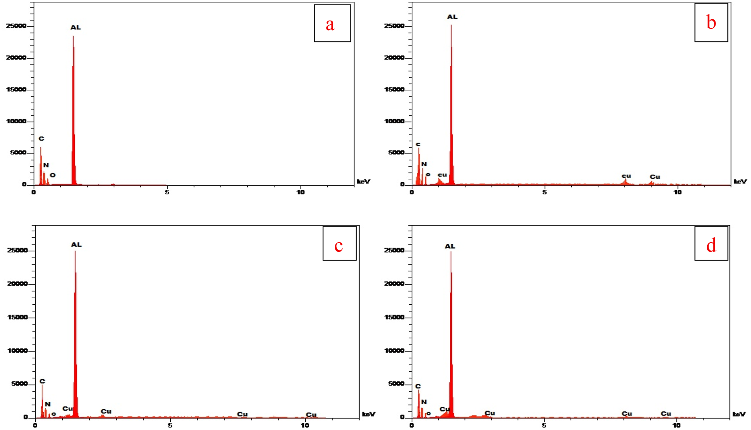
In addition, the EDX results provide proof that the CuO nanoparticles were indeed incorporated into the fibers successfully. It can be seen in all CuO-containing samples that there are peaks for copper and oxygen, proving the presence of CuO in the fibers. As depicted in Figure 9, Cu peaks increase with an increase in CuO nanoparticles in the samples, as would be expected. It must be mentioned that the EDX results represent relative element compositions, not amounts of CuO.
The even distribution pattern obtained from Cu and O elements in EDX mapping implies that the nanoparticles were efficiently dispersed within the nanofiber matrix, and the interface compatibility between the metal oxide nanoparticles and the polymer is thus very effective. Efficient nanoparticle dispersion in polymers–metal oxides nanofiber system was reported to have resulted from strong interfacial interaction between the two. 80
No other peaks besides the main ones in the EDX mapping imply that the synthesized fibers are highly pure. The presence of a peak associated with aluminum is due to the substrate utilized in sample preparation and does not influence the compositional analysis.
In general, all results show that CuO nanoparticles are efficiently incorporated into the nanofiber matrix. Such efficient incorporation and dispersion of the nanoparticles play an important role in the enhancement of their functionality, especially in the case of antimicrobial activity. These results agree with some previous reports.81,43
Antibacterial Assay
The outcomes showed that the generated nanofibers possess antibacterial activities on both bacteria types. Figure 10 depicts the antibacterial activity of PAN-PMMA/CuO nanofiber composites incorporated with different amounts (2.2, 4.4, and 6.6 wt%) of CuO nanoparticles on Gram-negative K. pneumoniae and Gram-positive S. aureus. The increase in the amount of CuO nanoparticles corresponded to an increase in the size of the inhibition zone, with radii of 0, 8, 16, and 18 mm for S. aureus and 0, 7, 12, and 18 mm for K. pneumoniae at 0, 2.2, 4.4, and 6.6 wt% of CuO, respectively. From the comparison of these results with those in previous investigations, it becomes clear that the effect of the PAN-PMMA blend matrix enhances the antibacterial capability, because it contributes to increasing the effective surface area and preventing the clumping of the nanomaterial. In contrast, the obtained inhibition zone diameter of 18 mm at 6.6 wt% CuO was significantly larger compared to previously reported values of the same amount of metal oxide-loaded PAN or PMMA nanofibers, which were in the range of 11-13.5 mm.
82
The digital photography and histogram for Staphylococcus aureus gram and Klebsiella aureus of PAN-PMMA/CuO Nanofiber.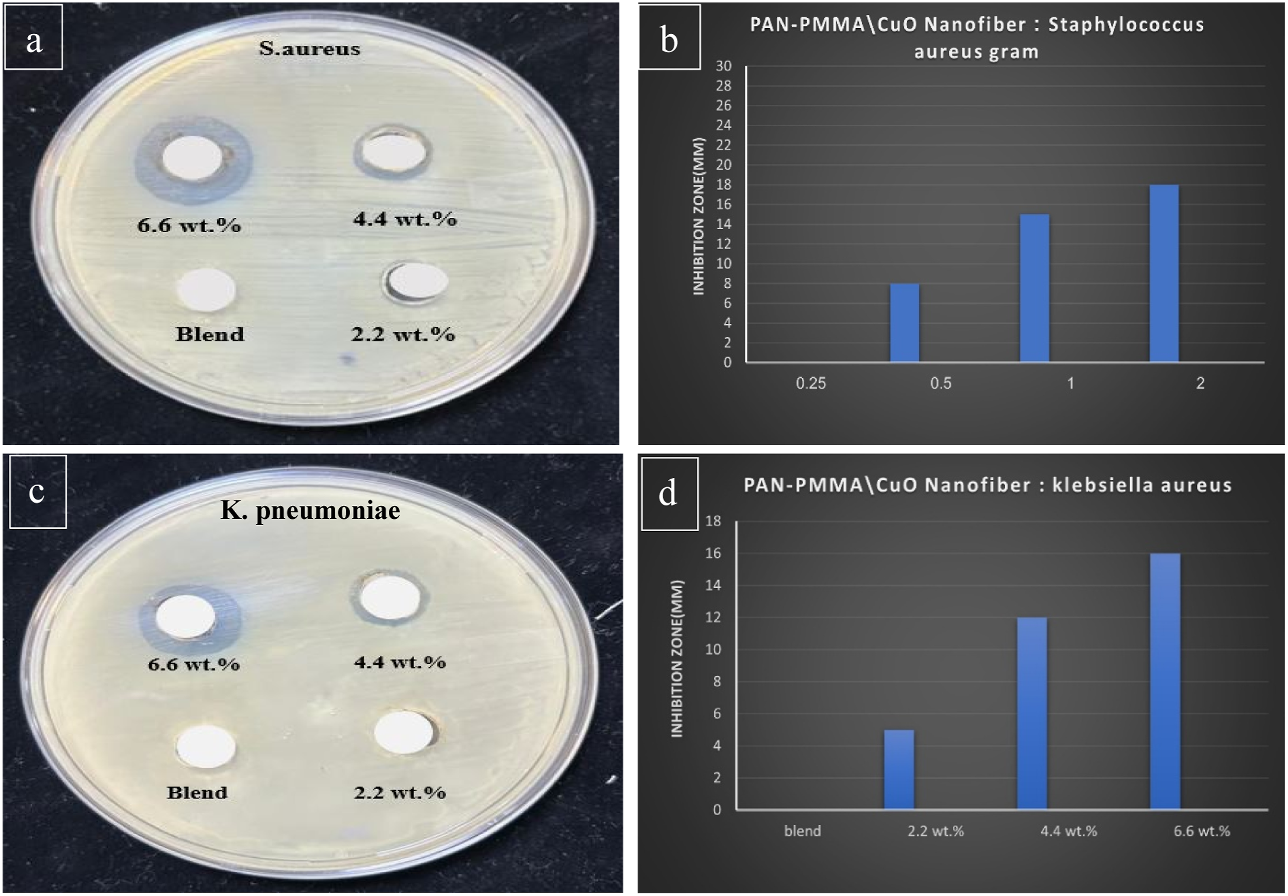
The observed effects have been explained by CuO nanoparticles’ attachment to the bacterial cell wall and their subsequent penetration into the cell membrane, thereby restricting growth and reproduction of the bacteria until their death. The findings are in agreement with.83,84 The variation in the reaction of antibacterial action on S. aureus (Gram-positive bacteria) and K. pneumoniae (Gram-negative bacteria) is due to the different structure of the bacteria’s cell walls. Gram-positive bacteria have a stronger cell wall composed of thick peptidoglycan, while Gram-negative bacteria not only have thick peptidoglycan but also an extra layer of membrane that may prevent penetration by nanoparticles. Metal oxides have limited solubility in water. In this case, it can be assumed that CuO nanocomposite immobilized on selected supports such as PAN-PMMA will lead to the creation of a highly stable and reusable material system. CuO NPs/PAN-PMMA showed improved antibacterial activity and effectively inhibited K. pneumoniae and S. aureus. This was mostly because the blend of PAN-PMMA was responsible for creating a strong scaffold that prevented aggregation of nanoparticles, thereby allowing a large effective surface for bacteria attachment. Thus, it appears that metal oxide (CuO) plays the major role of interaction in a hybrid composite with bacteria.85,86
Many factors contribute to making nanofibers effective anti-bacterial, one of which is the size of CuO NPs nanoparticles attached to the surface of the fibre. The small sizes of NPs help penetrate the cell wall, enabling the interactions between the particles and the bacteria through the creation of oxidative stress due to the generation of reactive oxygen species such as hydroxyl radical (OH•), singlet oxygen (1O2), superoxide hydroperoxide (OH−O2), and hydrogen peroxide (H2O2). Importantly, the particularity of hydrothermal method of nanoparticle synthesis at pH 9 is significant for obtaining higher crystal purity and optimal energy gaps between CuO NPs that help increase electron-hole pairs separation, thus generating ROS compared to non-tailored CuO particles or commercial. 87
The hydroxyl radical (OH•) is the most active oxygen radical which rapidly reacts with any type of molecule present in biological organisms. Under low concentrations, there are higher OH• levels with respect to CuO than Cu2O. Superoxide ions act on Cu(II) ions, reducing them to cuprous ions and generating hydrogen peroxide (H2O2). This hydrogen peroxide undergoes reactions like the Fenton reaction, which produces OH• radicals. Hydroxyl radicals (OH•) are produced by CuO NPs in much greater amounts even under low doses. These experiments suggest that free radicals, such as hydroxyl radicals, are produced in the redox cycles of copper ions. Reactive oxygen species (ROS).88,89 Can result in considerable harm to bacteria through disintegration of numerous organics including DNA, RNA, and proteins, when such compounds get attached to the membrane structure. Such conclusions were made based on results from previous research.
86
Nevertheless, it should be noted that this study did not include an analysis comparing performance of separate materials (i.e. PAN, PMMA, and CuO), which would be worth considering in future studies. However, it is important to mention that the assessment of the antibacterial activity of the materials used in this research was carried out on only two types of bacteria. It is suggested that future research be conducted using a wider variety of bacteria and quantitative assessments using MIC and MBC. Figure 11 illustrates the mechanism used for inhibiting bacterial activity in this study. Even though the antioxidant activity was not tested in the present study, the antibacterial activity has been explained by using the mechanism described in previous literature. The future studies will involve testing antioxidant activity to confirm the proposed mechanism. Mechanism of antibacterial action by PAN-PMMA/CuO nanofiber.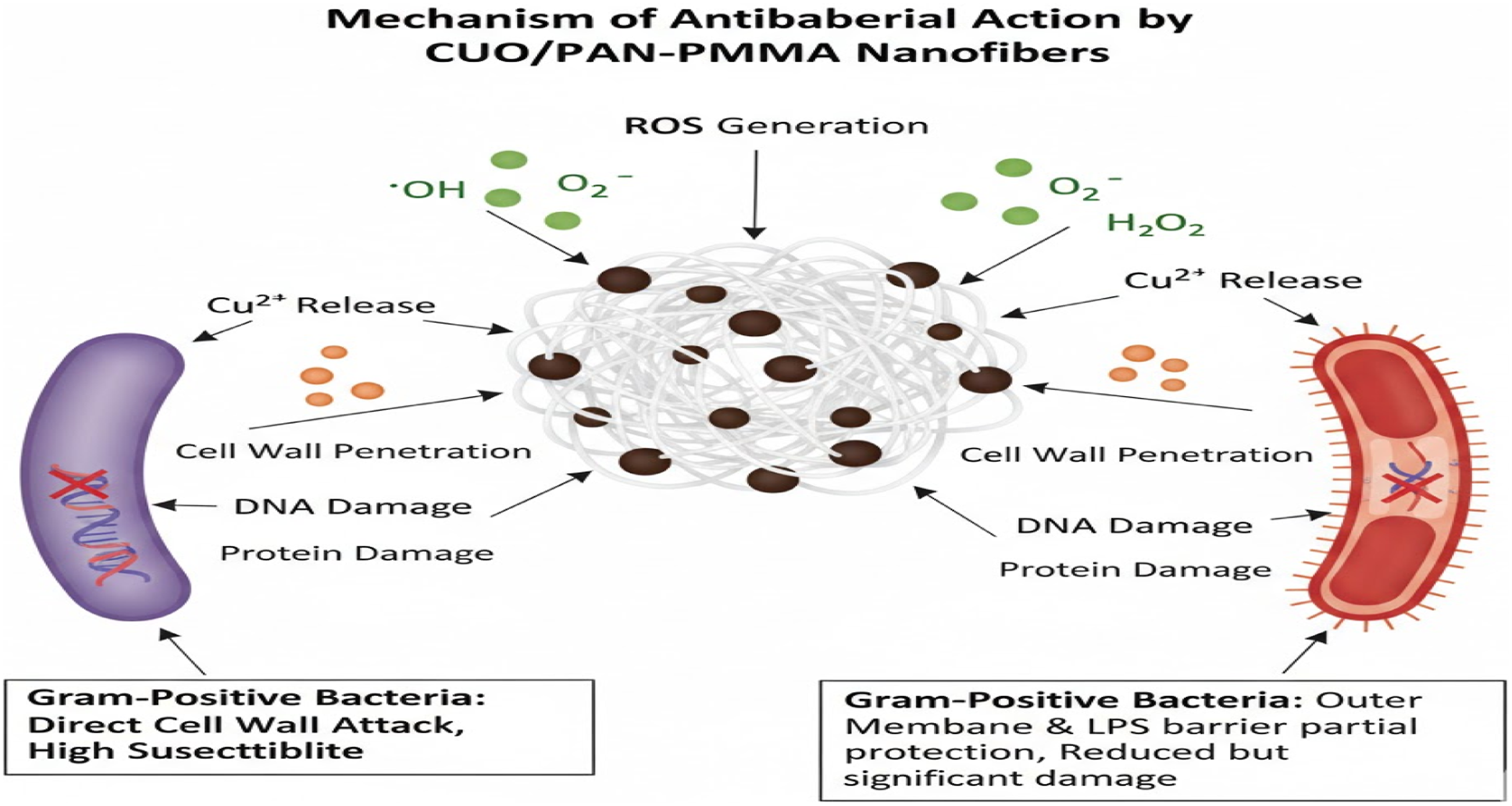
Limitations and Future Prospects
Although the current study offers useful information regarding the antibacterial activity of PAN-PMMA/CuO nanofibers, there may be some areas that can be explored in the future. First, the antibacterial testing involved only two model bacteria, offering preliminary results for material performance.
Furthermore, the discussed antibacterial effect mechanism, related to ROS production, was explained from the perspective of previous literature. Additional research could offer empirical evidence for this mechanism.
Moreover, further research could include a more extensive antibacterial assessment along with application-oriented and biocompatibility studies.
Conclusion
CuO NPs were effectively synthesised at pH 9 utilising highly pure aqueous copper sulphate (CuSO4·5H2O), sodium hydroxide (NaOH), and hydrochloric acid (HCl) at concentrations between 35% and 38% through hydrothermal technique. Nanofibers were synthesised from a PAN-PMMA mix and for this study reports PAN-PMMA/CuO NCFs via electrospinning. XRD examination indicated that the resultant material comprises pure CuO with a monoclinic structure. FTIR spectroscopy research shown that altering the CuO NPs content generates significant changes and strong interfacial interactions within the PAN-PMMA spectral range. FESEM images indicated that the size of the NPs is influenced by variations in sodium hydroxide (NaOH) concentration. The nanofiber pictures exhibited a pristine, linked network characterised by a homogenous, interwoven fibrous structure devoid of beads. EDX analyses validated the effective incorporation and distribution of CuO NPs inside the nanofiber matrix. The optical bandgap (Eg) for the direct allowed transition of CuO is 2 eV, but for the fibres, it diminishes from 3.3 eV to 2.8 eV with an increase in CuO content. These results suggest that the synthesised chemical is a viable material for several uses, including its antibacterial activity. When evaluated against two typical bacterial strains, Staphylococcus aureus S. aureus and K. pneumoniae, the antibacterial results consistently demonstrated superior bactericidal efficacy due to the immediate release of copper ions.
Footnotes
Acknowledgements
The authors express their gratitude for the assistance provided by the materials group College of Education for Pure Sciences, Department of Physics, University of Babylon, and the Nanomaterials Centre within the Ministry of Higher Education and Scientific Research, Iraq.
Author contributions
Mohammed A Kadhim, Fouad Sh. Hashim, and Shurooq S. Abed Al-Abbas conducted the experiments and evaluated the results. Mohammed A Kadhim authored the report, contributed to the optical aspects of the study, and analysed the data. Fouad Sh. Hashim and Shurooq S. Abed Al-Abbas were involved in the experimental design, enhanced the manuscript’s quality, and contributed to the FESEM, EDX, band gap, and photocatalytic action analyses. All writers reviewed and endorsed the final manuscript.
Declaration of conflicting interests
The authors declared no potential conflicts of interest with respect to the research, authorship, and/or publication of this article.
Funding
The authors received no financial support for the research, authorship, and/or publication of this article.
Author Information
Affiliations. University of Babylon, College of Education for Pure Sciences, Department of Physics, Iraq. Mohammed Abdul Kadhim. University of Babylon, College of Education for Pure Sciences, Department of Physics, Iraq. Fouad Sh. Hashim. University of Babylon, College of Education for Pure Sciences, Department of Physics, Iraq. Shurooq S. Abed Al-Abbas.
Ethical considerations
The research does not encompass investigations involving humans or their data.
Data Availability Statement
We affirm that all data and resources are genuine and accessible.