






Abstract
Veterinary dentistry has tended to follow the trends and advancements in the human dental field. As for any procedure performed on a patient, whether human or animal, an informed decision is based on the available evidence regarding treatment options. This is certainly true for the more involved treatments in veterinary dentistry which can include the disciplines of endodontics and dental implantology. A number of published case reports have been accepted by the specialty that endodontic therapy is indeed a predictable and a valuable service that can be offered to pet owners. Oral implantology has become an accepted and predictable procedure in the human dental field and can now be offered to pet owners if performed by suitable trained veterinary clinicians. The success rate for endosseous implant osseointegration is very high in humans. Success rates are also very high in animals used for implantology research. Canine studies have shown good bone-to-implant contact of around 73% in osseointegrated endosseous implants based on histological sections. Radiographic and histological findings demonstrate stable alveolar crestal bone levels after loading endosseous implants for up to 12 months in a dog model. This article discusses use of dental implants in the rostral mandible of a companion dog to replace periodontally diseased incisors, their restoration, to return the animal to full dental function and 3-year follow-up.
Introduction
The discipline of veterinary dentistry has followed the trends and advancements in the human dental field. This is the case for the area of dental implantology. 1 As for any procedure performed on a patient, the patient (in human dentistry) or the pet owner (in veterinary dentistry) makes an informed decision based on available evidence given by their clinician. The concept of informed consent is paramount in the relationship between clinician and patient/pet owner.
This is certainly true for the more involved treatments in veterinary dentistry which include the disciplines of endodontics and dental implantology. Years ago, in veterinary dentistry, there was very little evidence that root canal therapy was a viable and successful option for companion animals. However, over the years and with a number of published case reports, it has been accepted that endodontic therapy is a predictable, valuable service that can be offered to pet owners with confidence. 2 -6 Only time will judge whether oral implantology also becomes an accepted and predictable procedure that can be offered to pet owners. Human oral implantology research often uses other species such as the canine model. These results are then extrapolated back to treatment modalities in man.
The success rate for endosseous implant osseointegration 7 or functional ankylosis is very high in man. Success rates are also very high in animals used for implantology research. 8,9 Canine studies have shown good bone-to-implant contact of around 73% in osseointegrated endosseous implants based on histological sections. 10 Also, studies have shown through radiographic and histological findings that alveolar crestal bone levels are stable after loading endosseous implants for up to 12 months in a dog model. 11 The single implant crown survival rate in people has been documented to be 94.5% after 5 years, 12 although there is very little long-term follow-up of implant success in dogs. Crown design and type of retention (screw-retained versus cement-retained) did not influence the survival rate. 13 Other treatment planning decisions include whether to place the implant at the time of tooth extraction or at a time after tooth extraction.
Today, there is little debate over whether an implant can be placed at the time of extraction (immediate implant placement) when compared to the more conservative approach of placing the implant some weeks or months after extraction (delayed implant placement). Short-term survival rates and clinical outcomes of immediate and delayed implants were similar and comparable to those implants placed in healed alveolar ridges. 14 At present, to the authors’ knowledge, there are no long-term studies that support or contradict the use of immediate implants in extraction sockets.
At present, there are studies 15 -17 that have shown similar implant healing and survival rates after loading following immediate implant placement versus a delayed implant protocol. Most studies suggest that immediate implant placement is successful as long as implants are placed by experienced operators, not placed where acute infection may exist at the extraction site or where there is active periodontitis affecting the tooth to be extracted and replaced by an implant/crown. Other concerns that are raised with immediate implant placement include filling of the potential gap, if greater than 2 mm between the implant body and the alveolar wall. In these cases, the use of bone replacement materials with or without a barrier membrane may be required. 13 Also, soft tissue closure over the immediate implant may also be a problem, due to there often being a lack of gingiva for flap adaptation and to achieve primary healing. Inadequate primary stability may also be a concern when placing an immediate implant. Long-term stability of the soft tissues after immediate implant placement may also be less predictable than delayed placement. It has been shown that even with chronic apical infection affecting teeth to be extracted, an immediate implant can still be placed so long as the extraction socket is thoroughly curetted to remove chronic inflammatory/granulomatous tissue. 17
In veterinary dentistry, one of the more common reasons for tooth extraction is complicated crown and/or root fracture. 18,19 These teeth may not be suitable candidates for direct or indirect restorations or even endodontic treatment. One of the arguments for immediate implant placement after tooth extraction in veterinary dentistry would be reduction in anesthetic episodes for implant and implant crown placement. Also, the extraction socket can be used as a guide (to some extent) for placing the implant in a good restorative position. However, the caveats mentioned earlier need to be taken into account when treatment planning for implant placement.
Although it is assumed most animals can function and masticate reasonably well with missing teeth, no studies exist to support or refute this observation. There can be complications seen after tooth extraction, such as lip entrapment after the extraction of the maxillary canine tooth in cats and dogs. 20 There appear to be no published studies looking at implant and prosthetic crown survival rates in client-owned pets. This case report describes the replacement of 4 mandibular incisor teeth with 2 cantilevered implant bridges in a dog. Treatment options and discussion of surgical and postoperative complications should occur prior to obtaining informed consent from the pet owner. Owners should also be made aware that although forces that dogs can apply to implant crowns are similar to that of humans, forces applied to implant crowns from abrasive and hard objects such as bone chewing and unique duties of the teeth (such as bite work in police dogs) may adversely affect long-term survival when compared to human studies. These forces can lead to prosthetic failure. Therefore, as a part of management of any implant case, bone chewing and other potentially damaging habits are not recommended.
The follow-up for this case has been extensive, with regular 6-month reevaluations and reinforcement of oral hygiene practices to prevent mucositis and peri-implantitis. Mucositis and peri-implantitis are characterized by an inflammatory reaction in the tissue surrounding a dental implant that can lead to tissue destruction and implant failure. Peri-implantitis results in inflammation of tissue as well as bone loss. Risk factors include improper treatment planning, poor surgical and prosthetic execution, and improper maintenance. Proper maintenance includes regular tooth brushing by the owner, 21 routine appointments with the veterinarian either annually or semiannually, and various dental treats which are all helpful for regular plaque control. Implant/crown stability was assessed through periodontal probing depths, bleeding on probing, signs of pocket exudate and any crown mobility as well as crestal bone stability around the implant as seen on periapical radiographs.
Case Report
Phase 1: Extractions and Bone Grafting
A healthy 10-year-old, wheaten terrier presented for a scheduled dental cleaning and oral evaluation. During the initial examination, it was noted that a mild gingivitis calculus was present and both mandibular second incisors (302 and 402) were missing. Charting, probing, and dental radiographic examination were completed, and a diagnosis of stage 3/4 periodontal disease was made of the mandibular first incisor (301 and 401) teeth (Figure 1). Treatment options were discussed at length with the client, which included selective extractions, periodontal surgery, or extractions with bone grafting and dental implants to replace the 4 mandibular incisors in 6 to 8 months following site preparation. The owner elected to extract the affected teeth with socket preservation and place 2 endosseous implants at a later date. Prior to the surgical appointment, blood was drawn and a complete blood count and serum biochemistry profile were tested to check the patient’s general health. Atropinea and acepromazineb were administered via subcutaneous injection as a preanesthetic agent for sedation, to control salivation, and suppress bronchial secretions during the surgery. An intravenous (IV) catheter was placed and lactated Ringer’sc solution was started at a rate of 5 mL/kg/h. General anesthesia was induced with midazolam and ketamine via IV and maintained with sevoflurane.d Bupivacaine 0.5%e was administered via bilateral mental nerve blocks (0.25 mL [0.125 mg] per site), and butorphanolf was administered IV.
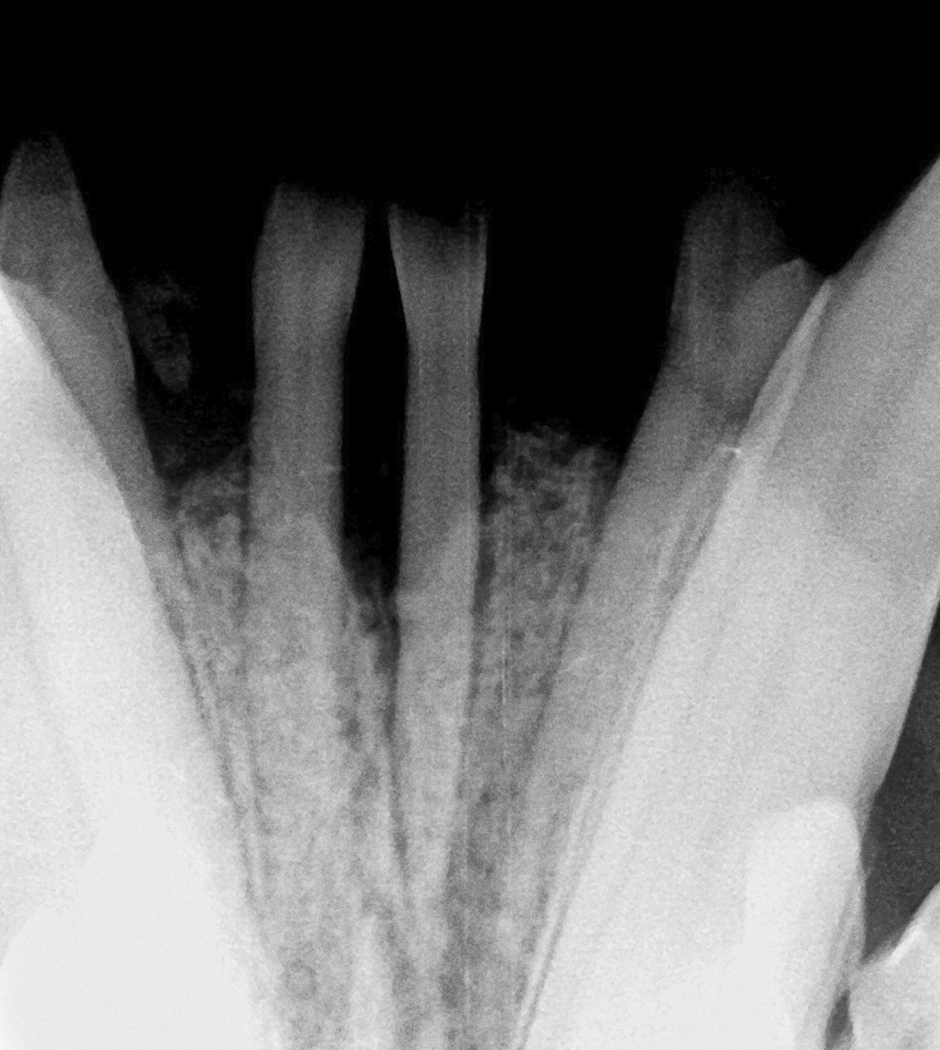
Radiograph of the rostral mandible demonstrating periodontal bone loss of the incisors.
A complete oral examination was performed on the patient to confirm the initial findings. The procedure was initiated by ultrasonic scaling to remove calculus supra- and subgingivally. A full thickness mucoperiosteal flap was reflected to gain access to the surgical site. Extraction of the periodontally affected teeth was accomplished using elevators and extraction forceps. The extraction sockets were debrided utilizing a periodontal curette and a football-shaped finishing bur (Figure 2). A demineralized freeze-fried particulate bone allograftg and a mini bone blockg (Figure 3) were utilized for guided bone regeneration (GBR) to fill the defects created by the periodontal bone loss and the extraction process. A resorbable collagen membraneh was placed over the ridge and tucked under the flap margins (Figure 4). Flaps were repositioned without tension, and the site was closed using a simple interrupted pattern using 4-0 absorbable suturei (Figure 5).
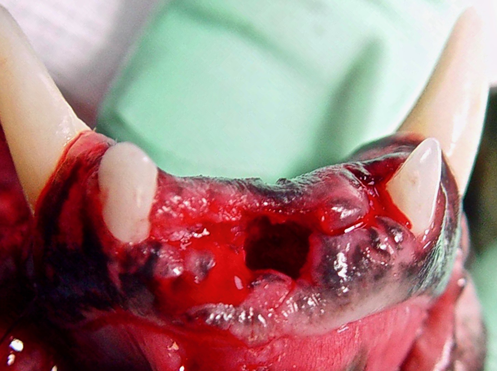
Mandible immediately after extraction of the incisors.
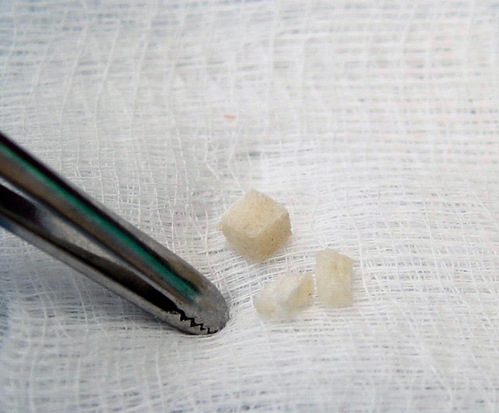
Bone blocks ready for placement into the surgical site.
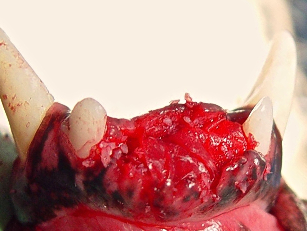
Extraction sites filled with graft material and resorbable collagen membrane prior to site closure, restoring the ridge for future implant placement.
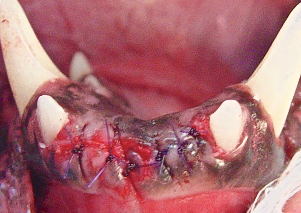
Extraction sites after closure.
Phase 2: Implant Placement
Eight months after extraction and GBR surgery, a radiograph was taken to evaluate the grafted site for possible implant placement (Figure 6). The patient was anesthetized following the same protocol utilized previously. A full-thickness envelope flap was elevated to expose the underlying bony ridge. Measurements were taken from the radiograph with regard to available width and length to assist in choosing the proper size implants for the sites (Figure 7). A sequential drilling protocol as recommended by the implant manufacturer was initiated to create osteotomies to accommodate 2 implants in the site available. Two endosseous hydroxyapatite-coated implantsj based on prior measurements (3.25 mm × 10 mm) were placed into the osteotomies prepared at 20 rpm and 40 N-cm (Figure 8). Stability was confirmed with a torque wrench with a final insertion torque measured at 50 N-cm for each implant (Figure 9), and a screw cover was hand tightened on the implants. Gaps at the alveolar margin were filled with a mixture of particulate bone allograftg and autologous leukocyte–platelet-rich fibrin (L-PRF) collected from the patient’s blood 22 (Figure 10) to maximize crestal height following healing and minimize future bone loss (Figure 11). The site was treated using a 2-stage surgical protocol. Implants with cover screws were submerged, and the flap was closed to achieve primary healing (Figure 12). A radiograph was taken to demonstrate implant placement and additional crestal grafting (Figure 13).
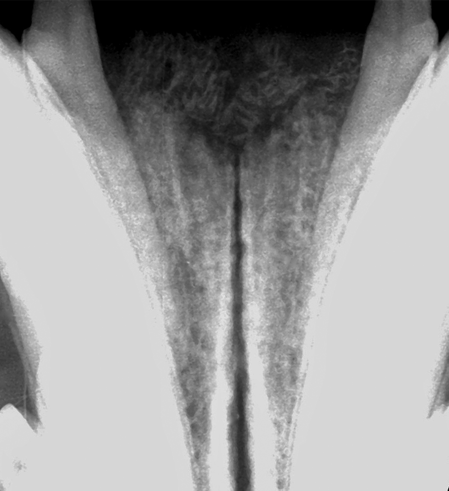
Radiograph demonstrating healing and integration of the osseous graft taken 8 months postoperatively.
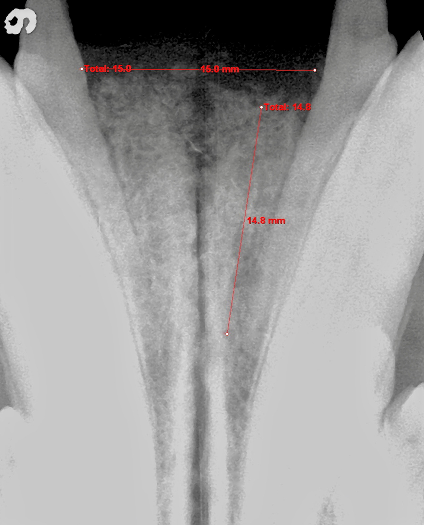
Measurements on the digital radiograph to determine available width and length for implant placement.
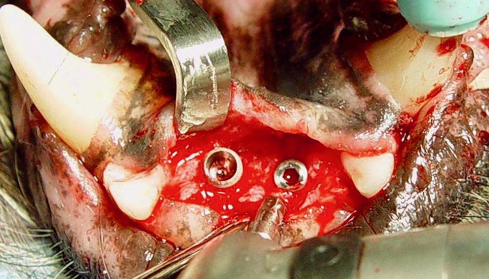
Endosseous implants have been placed into the mandible with a surgical handpiece to almost full depth with a setting of 40 N-cm.
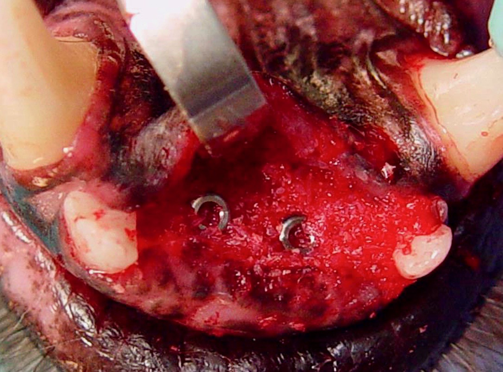
Final seating of the implants was accomplished with a torque wrench with final insertion torque measured at 50 N-cm.
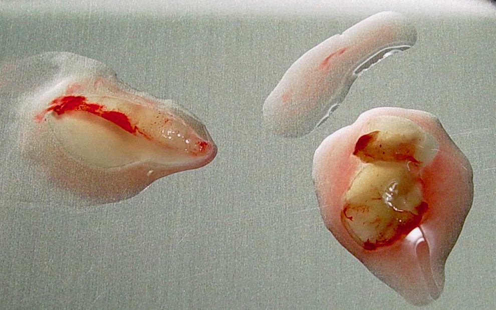
Autogenous platelet-rich fibrin was collected from the patient’s blood at the time of surgery by centrifugation.
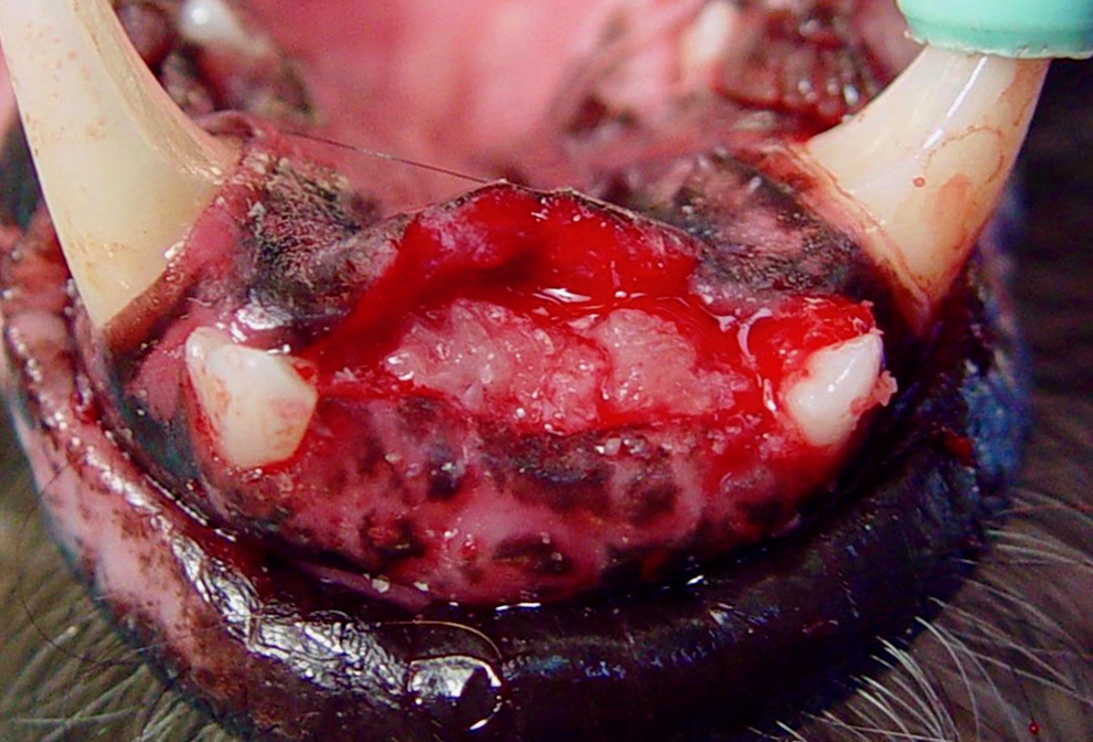
Surgical site immediately following placement of the osseous graft mixed with L-PRF, prior to flap closure.
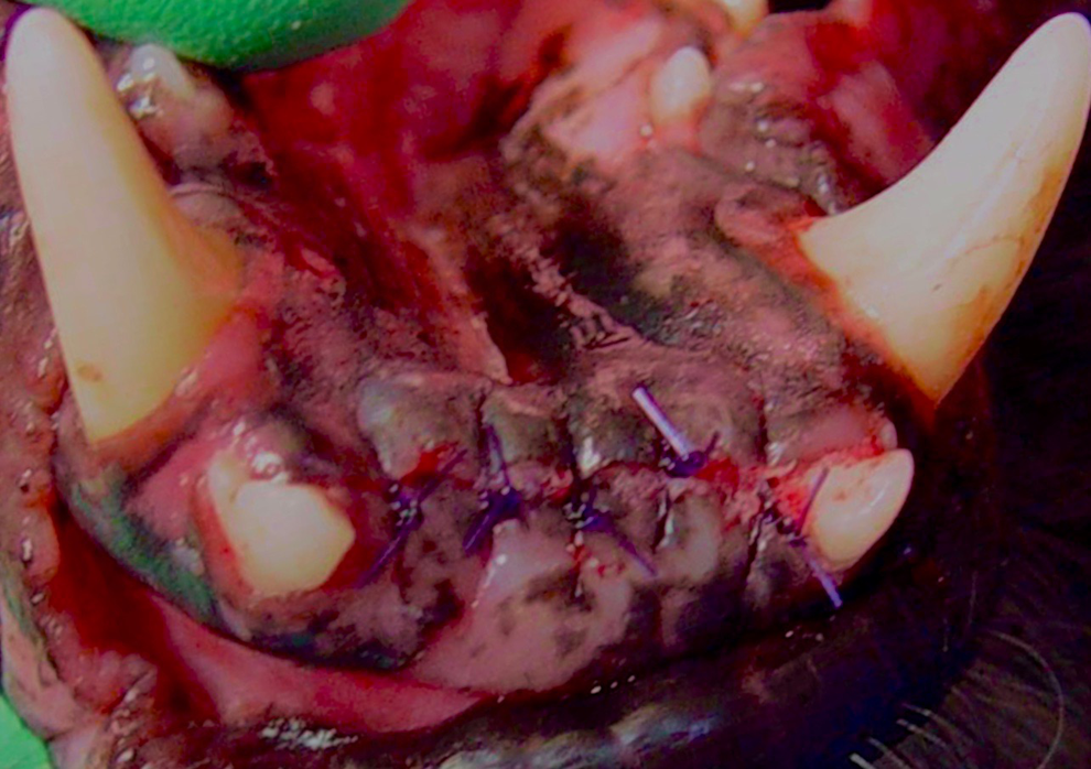
Surgical site was closed primarily after implant and graft placement.
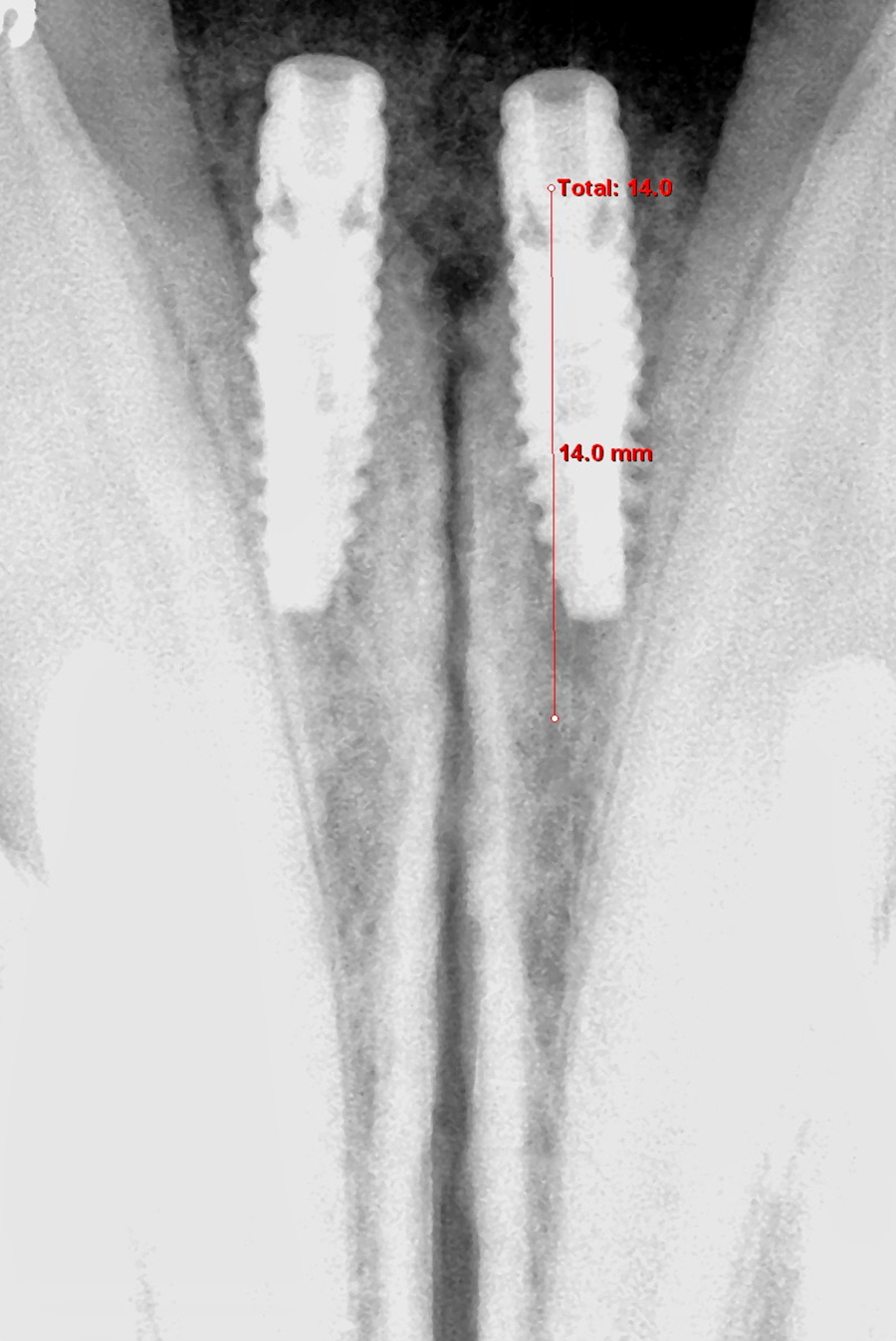
Radiograph following implant placement and additional crestal grafting demonstrating the site.
Restorative Phase
Four months after implant placement, the site was checked for tissue health, and radiographs were obtained to evaluate and verify implant integration. Implant cover screws were exposed with a CO2 laser (0.8 mm tip) to aid in exposure and emergence profile. Transmucosal healing abutments were placed before final tissue apposition (Figure 14), and the implants were determined to be stable and free of mobility. Cover screws were removed and implant-level headsj were placed and impressions were fabricated with vinylpolysiloxane (VPS) hand mix putty, and VPS detailed impression material, with custom formed trays.k Transmucosal healing abutmentsj were placed (Figure 15), and a radiograph was obtained to confirm seating of the healing abutments (Figure 16). An appositional flap was secured with sutures to encourage a proper emergence profile for future prosthetic components (Figure 17).
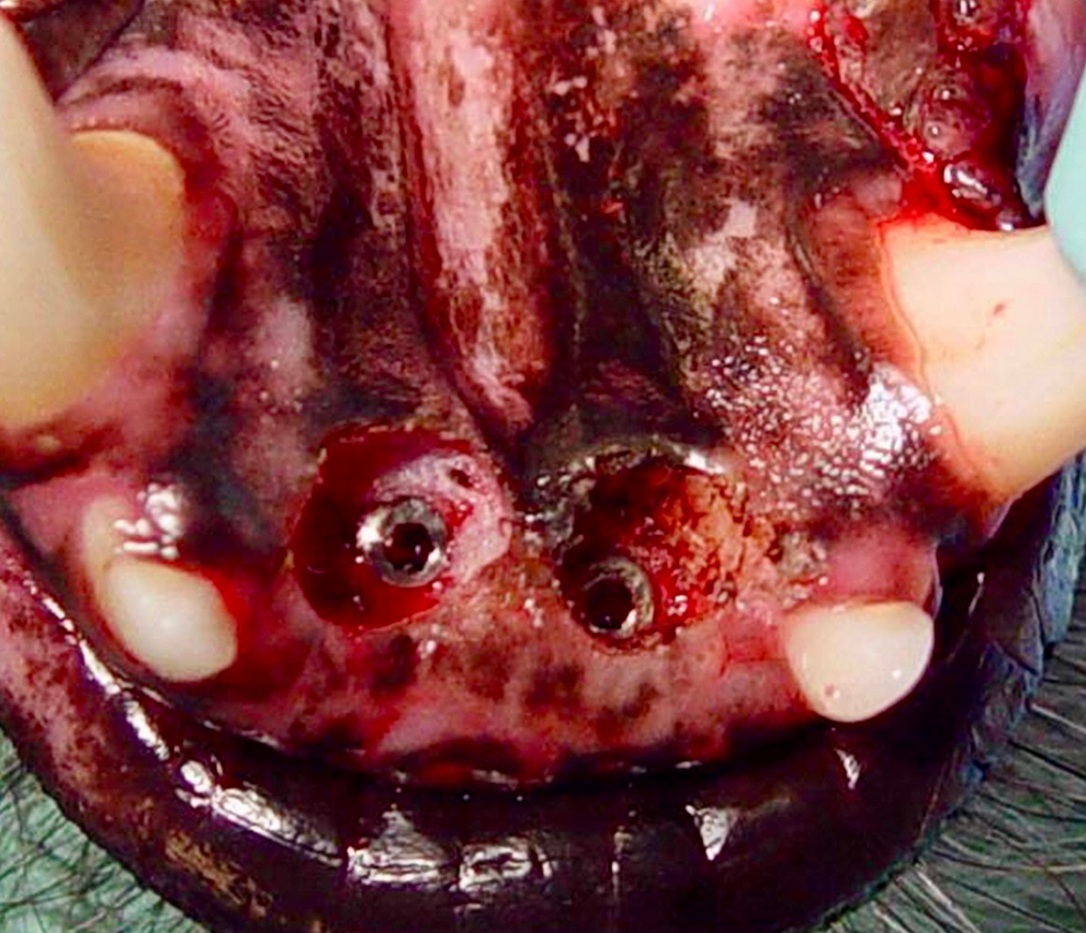
Implants were uncovered 4 months after placement.
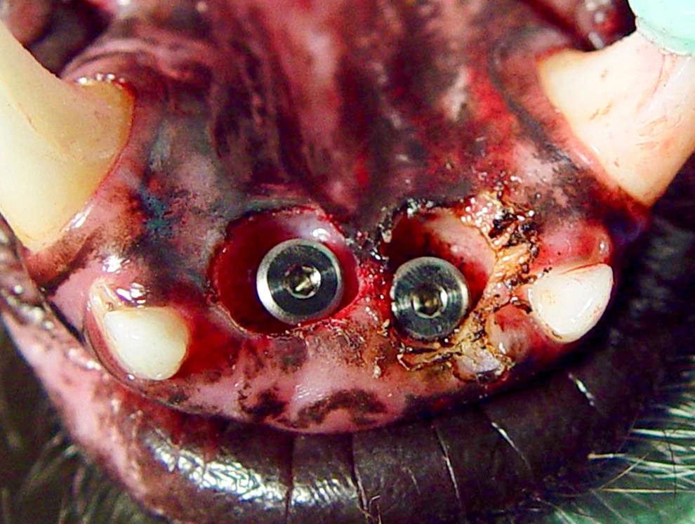
Healing abutments were placed in the integrated implants following implant exposure.
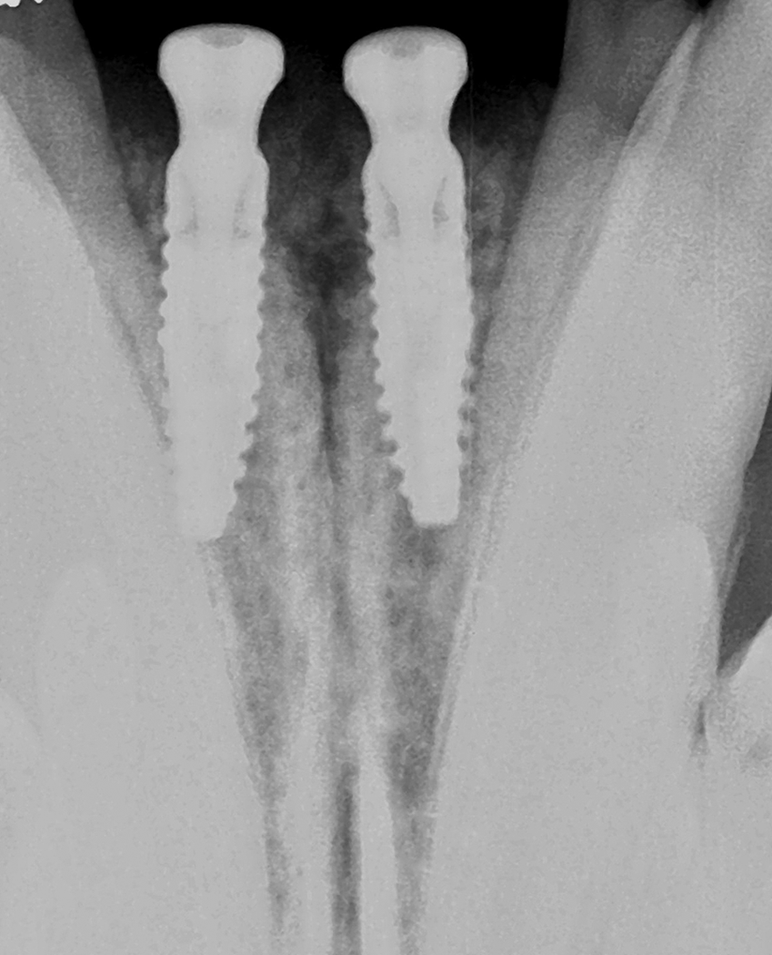
Radiographs were taken to verify seating of the healing abutments.
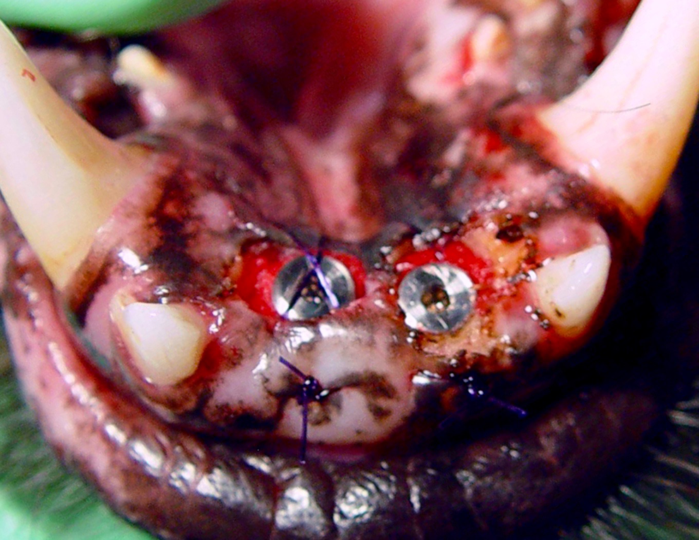
Soft tissue was opposed to surround as much of the healing abutments as possible.
The VPS impressions were sent to a dental laboratory,l and stone models were created with implant analogs embedded within the model. The maxillary and mandibular models were mounted in occlusion. The planned prosthetics were waxed up for the metal substructure of planned bilateral PFM cantilever crowns with a distal pontic at each site (Figure 18). The substructures were cast and porcelain was applied to complete the porcelain-fused-to-metal prosthetics (Figures 19 -21).
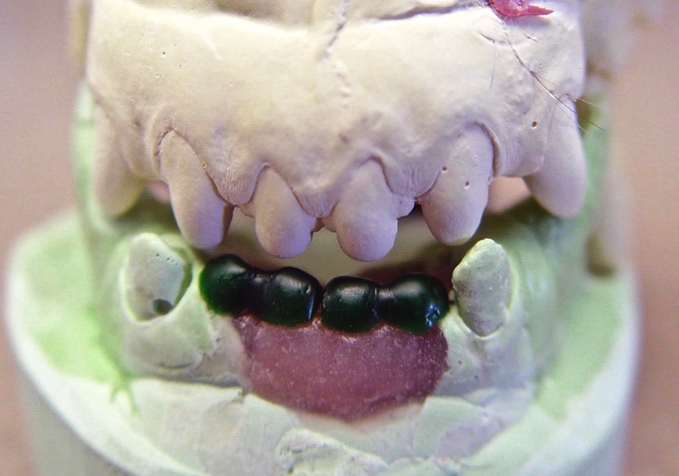
Prosthetic wax-up of the substructure for bilateral cantilever screw retained implant crowns.
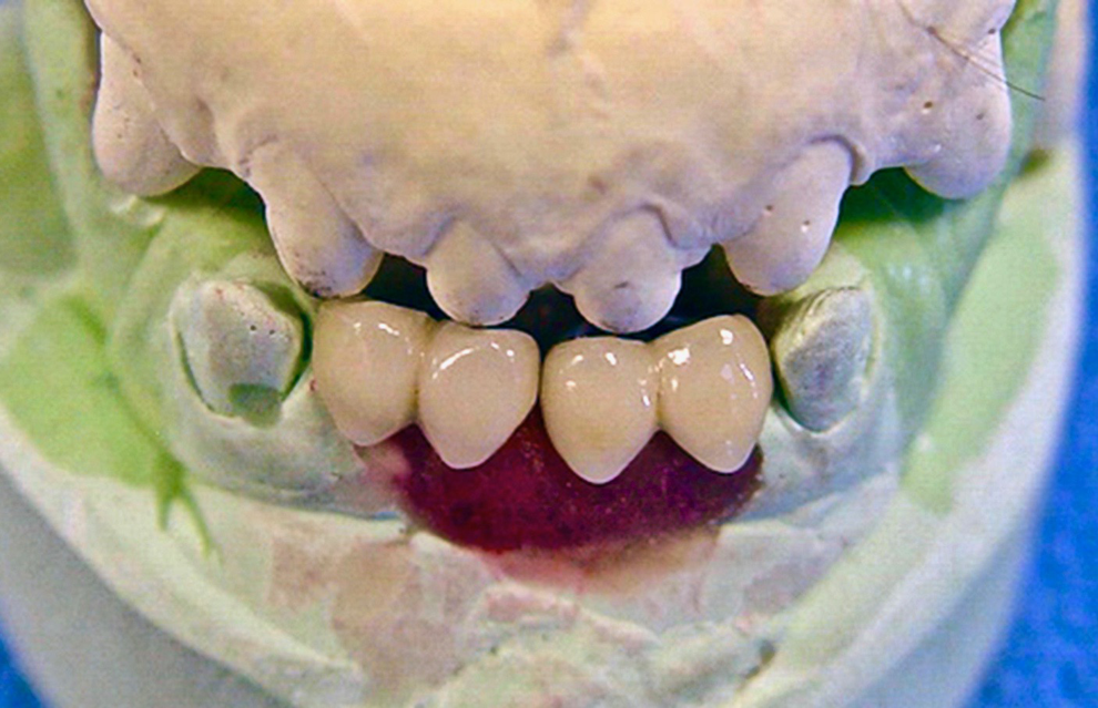
Ceramic was applied to the bilateral porcelain-fused-to-metal screw-retained cantilever prosthetics.
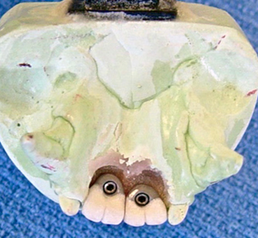
Lingual view of the screw-retained bilateral cantilever individual bridges demonstrating the access screw on the model.
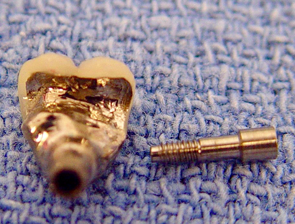
Individual screw-retained cantilever bridge with fixation screw.
One month later, the patient returned for placement of the prosthetic components. The patient was anesthetized following the same protocol utilized previously. The healing collars were removed to expose the healthy soft tissue collar and emergence profile. Emergence profile refers to the way the tooth emerges from the bone in relation to the gum tissue. It directly influences the surrounding tissue and adjacent teeth (Figure 22). 23,24 The prosthetics were tried in and fixation screws were hand-tightened and checked with a torque wrench (Figure 23). A radiograph was obtained to verify seating of the prosthetics (Figure 24). The prosthesis was completed.
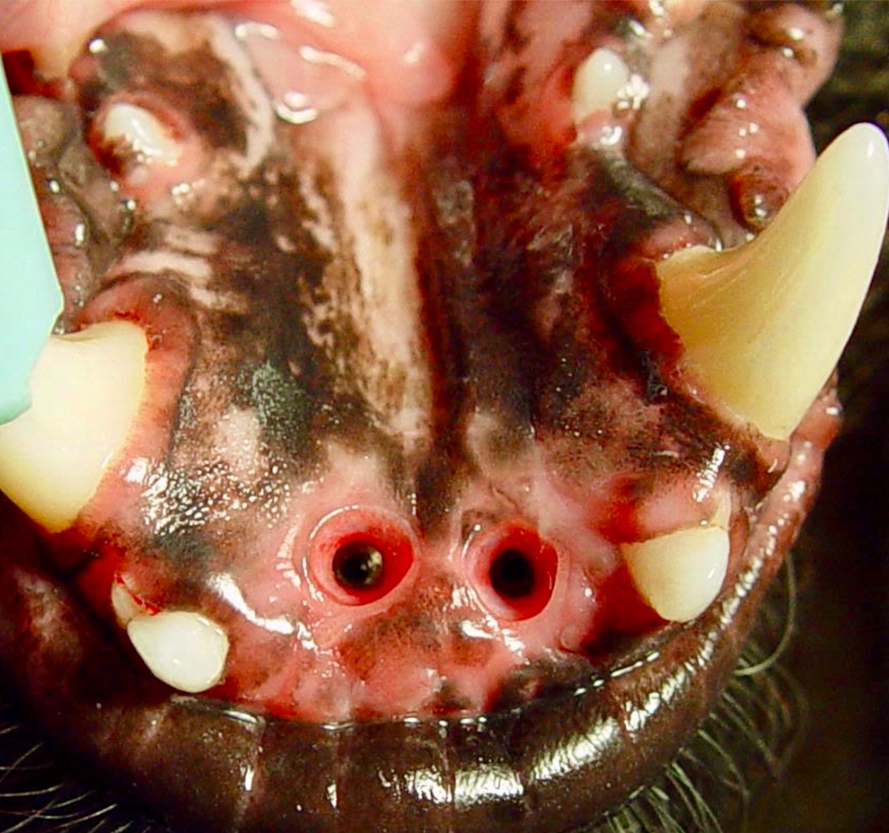
Healing abutments have been removed, demonstrating soft tissue healing and emergence profile for the prosthetics.
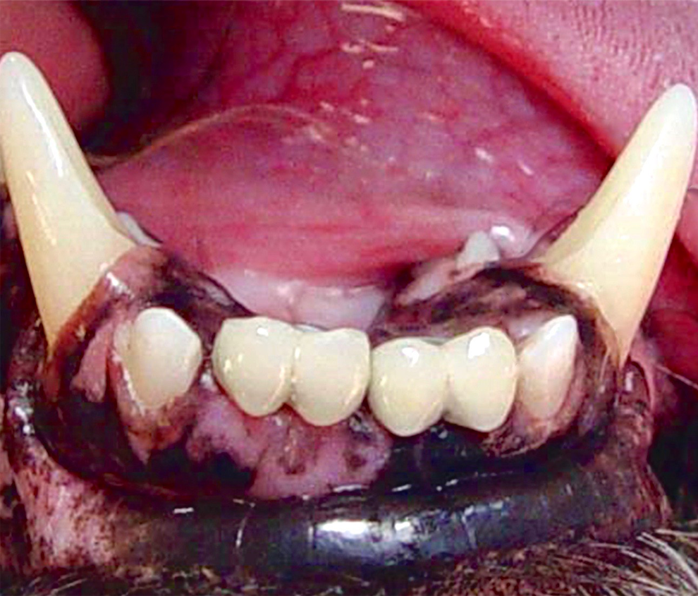
Facial view of the completed screw-retained prosthetics.
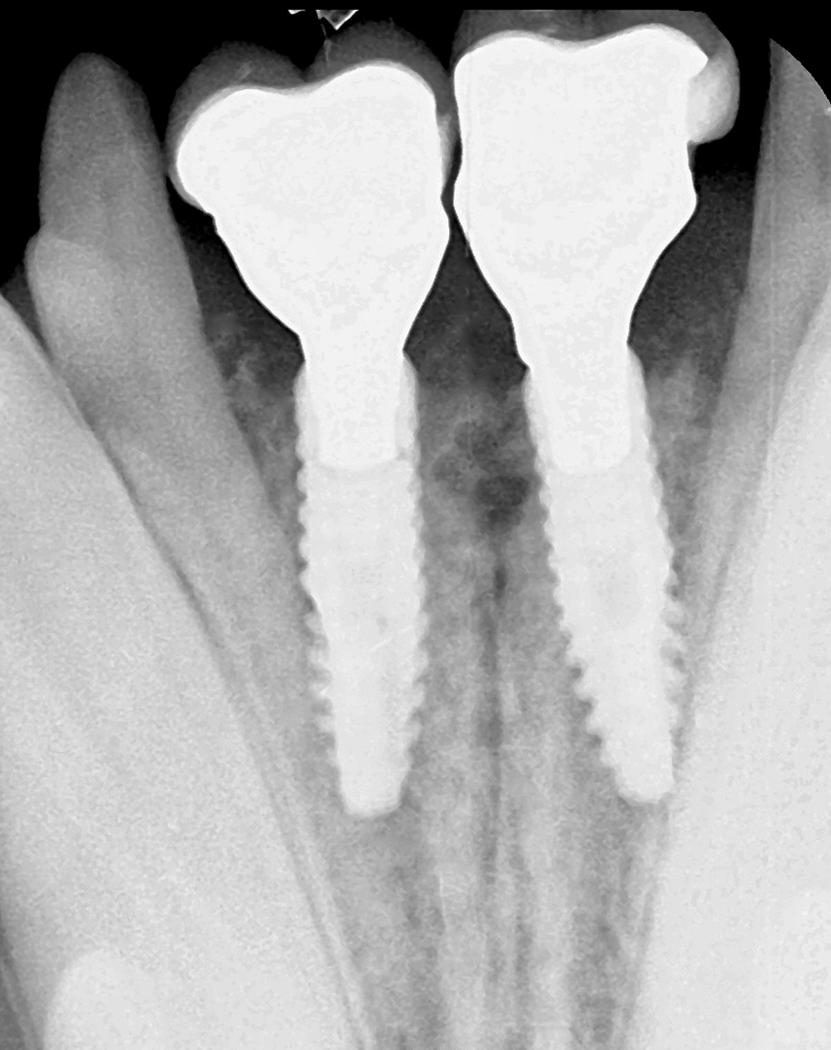
Radiograph to verify seating of the final crowns.
Follow-Up
The follow-up for this case was extensive, with regular 6-month reevaluations. At each visit, the importance was reinforced of oral hygiene practices to prevent mucositis and peri-implantitis. The patient was presented for a comprehensive oral examination and dental cleaning 3 years after dental implant restoration. Radiographs were obtained, and periodontal probing was performed to check pocket depths and general health of the implants (Figure 25). The surrounding tissue demonstrated with a healthy biotype and an absence of bleeding on probing with no evidence of peri-implantitis (Figure 26).
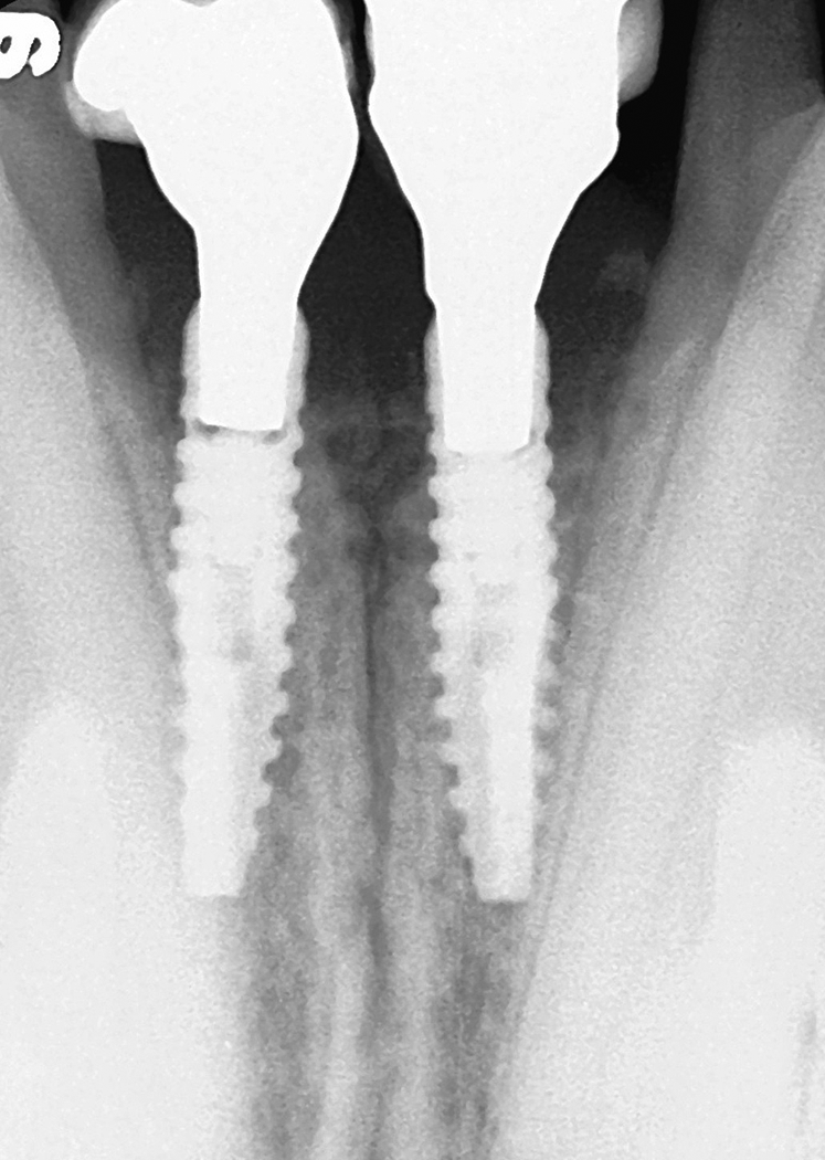
Radiograph taken 3 years after placement of the implants.
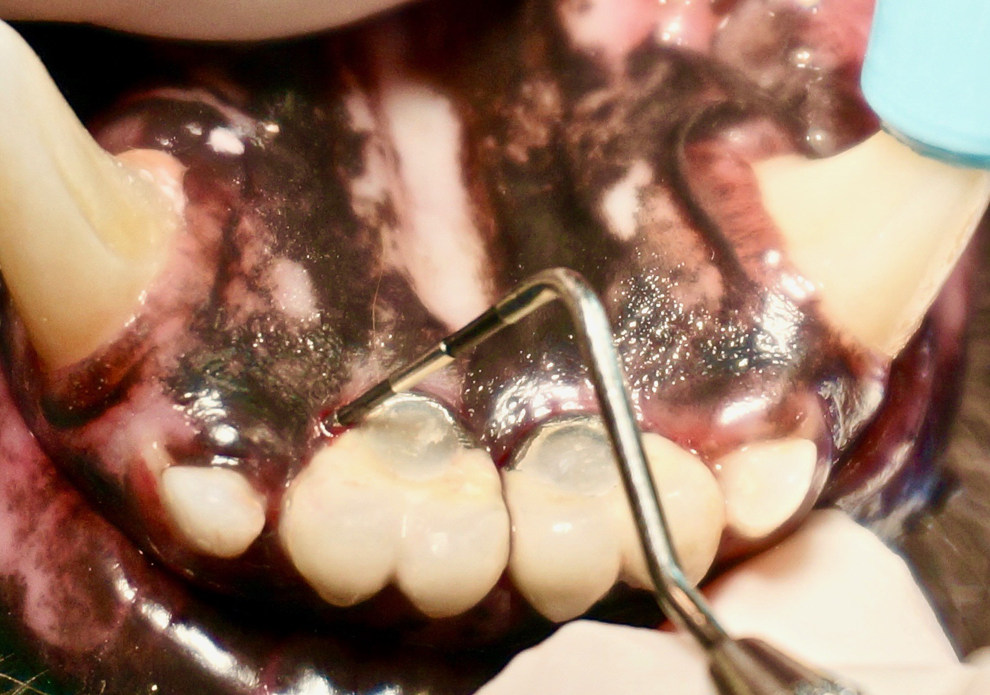
Periodontal probing 3 years after placement of the implants reveals deepest probing depth of 2 mm.
Discussion
Periodontal disease is the most common veterinary dental condition. Treatment options for periodontal disease include attempts to halt or reverse the disease, extraction without replacement of the missing dentition, or replacement with a dental implant followed by restoration with a fixed prosthesis. There are different schools of thought for each of these treatment options. Options should be presented to the pet owner with the advantages and disadvantages for each option so that the owner may make an informed decision.
It is widely accepted that domesticated animals benefit from some hard foods in their daily diet, as this aids in physical removal of plaque from dentition during chewing. When teeth are missing, the animal can be limited in what it can chew. An inability to chew hard foods with the resultant limitation of the diet could lead to further periodontal issues in the remaining dentition. As dogs derive enjoyment from chewing, replacement of missing teeth may influence animals’ mental well-being and help maintain physical health through a better ability to eat.
The patient discussed in this case was presented with stage 3 periodontal disease (bone loss and mobility) of the mandibular central incisors. Treatment options were discussed with the owner, which included extractions and no further treatment, extractions with periodontal surgery, grafting the sites with and without subsequent placement of implants following healing, and integration of the osseous graft. The cost/benefits were discussed, and the owner selected what he or she considered to be the best treatment available which was an implant fixed bridge treatment.
In human patients, when an immediate implant protocol is utilized, the patient is instructed to avoid chewing on the healing implants. In animals, it is impossible to have the patient avoid chewing on the implant restorations. This necessitates a need for a staged implant protocol, wherein the implants are placed in 2 stages and allowed to integrate for a set time. The implants are then uncovered, and the restorative phase can be accomplished without concern for early loading, which may lead to implant failure to integrate. Quality and quantity of the osseous bed determines whether the implants can be placed at the time of extraction. 25 A deficient site may need additional grafting to regenerate a new bony envelope that will accommodate future implants (2-stage procedure). In the case described here, the quality and quantity of the remaining mandibular bone would not allow initial stabilization of the fixtures that is required for immediate implant placement with an insertion torque of >30 N-cm. Therefore, a 2-stage approach was selected with extraction and site grafting followed by implant placement 5 months later.
Numerous grafting materials are available including autografts, allografts, xenografts, and synthetics. As the defect in this case was significant, it was decided to utilize a cancellous block and a particulate demineralized freeze dried bone allograft. Demineralizing of the osseous graft material exposes many of the known growth factors needed in bone production and remodeling. These types of materials are osteoconductive (lattice support) and osteoinductive (attracting immature cells and stimulating these cells to develop into preosteoblasts). This results in a faster maturation of host bone in the site, jump starting the bone production and yielding a denser bone at implant placement after healing. Autogenous grafting is more involved and causes increased morbidity, since 2 sites need to be managed (donor site and recipient site). It is the authors’ impression that use of allografts results in better outcomes than those obtained with synthetics for this particular purpose, although clinical veterinary studies are necessary to prove this.
Dental radiographs were utilized to estimate the size of implants used in this patient. Although radiographs showed a close approximation between the implant threads and the adjacent third incisor teeth, no radiographic or clinical evidence of problems were seen with teeth adjacent to the implants. Measurements taken from a dental radiograph are approximated unless the digital radiograph system is calibrated to known measurements prior to obtaining radiographs. More advanced diagnostic tests such as computed tomography (cone-beam or conventional) may provide further measurement capabilities.
Implants are available with various surface texture, thread spacing, depth of grooves, and angulation of threads. As smaller diameter implants are required in dogs or cats compared to those routinely used in humans, fewer brands are available for veterinary use. Implant designs with deeper threads provide increased bone-to-implant contact and are able to manage higher loads than those implants with shallower threads. 26 Implants with rougher surfaces also increase surface area, and bone is more easily adhered to the implant compared to smooth-surfaced implants. 27,28 With these considerations in mind, the authors selected a hydroxyapatite-coated titanium alloy fixture with deeper thread design. It is recommended that when using an hydroxyapatite-coated implant, the rough surface needs to be placed subcrestal to avoid plaque retention to the rough surface which can encourage periodontal disease. Titanium alloy is preferred to titanium (commercially pure or CP), as it has better physical properties. 29
The authors are unaware of any published reports with longer follow-up of implants in a privately owned companion animal. Animals have long been used as recipients of endosseous implants in research. This case demonstrates that implant/crown survivability and maintenance of the crestal bone levels around implants are indeed possible in dogs. Three years after treatment, alveolar bone has been maintained at the same level as when the implants were placed. Case selection, technical skills of the operator, limiting excessive forces to implant/crowns, and homecare maintenance are paramount to the success of the implant/crown.
Materials
Atropine sulfate injectable, MWI Veterinary Supply, Boise, Idaho.
Acepromazine maleate injectable, MWI Veterinary Supply, Boise, Idaho.
Baxter Healthcare, Deerfield, Illinois.
Abbott Animal Health, Abbott Park, Illinois.
Patterson Dental, Saint Paul, Minnesota.
Hospira World Wide, Lake Forest, Illinois.
Veterinary Transplant Services, Kent, Washington.
Neomem, Citagenix, Laval, Quebec, Canada.
Monocryl, Ethicon, Johnson & Johnson, New Brunswick, New Jersey.
Simpler Implants Inc, Vancouver, British Columbia, Canada.
Benco Dental Supply Co, Tucson, Arizona.
Dok’s Dental Lab, Tucson, Arizona.
Footnotes
Declaration of Conflicting Interests
The author(s) declared no potential conflicts of interest with respect to the research, authorship, and/or publication of this article.
Funding
The author(s) received no financial support for the research and/or authorship of this article.