






Abstract
Biomechanical studies of the elongated canine tooth of animals are few, and thus our understanding of mechanical and physical properties of animal teeth is limited. The objective of the present study was to evaluate the influence of force direction on fracture resistance and fracture pattern of canine teeth in an ex vivo dog cadaver model. Forty-five extracted canine teeth from laboratory beagle dogs were standardized by hard tissue volume and randomly distributed among 3 force direction groups. The teeth were secured within a universal testing machine and a load was applied at different directions based on testing group. The maximum force to fracture and the fracture pattern classification were recorded for each tooth. After correcting for hard tissue cross-sectional area in a multivariate analysis, no significant difference in the amount of force required for fracture was apparent between the different force direction groups. However, the influence of force direction on fracture pattern was significant. The results of this study may allow the clinician to educate clients on possible causal force directions in clinically fractured teeth and, thus, help prevent any contributing behavior in the future.
Introduction
The study of tooth fracture biomechanics evaluates how material properties and tooth morphology relate to fracture patterns and critical stress loads. Currently, this field is in its infancy, and the majority of available studies are focused on vertical (longitudinal) and chip fractures in bunodont molars. 1 -7 However, there are a few articles that describe the biomechanics of fractures in elongated canine teeth of carnivores and compare the defined fracture patterns among different tooth morphologies. 4,8 -13
The 3 basic fracture types that are often described in the veterinary and human literature when discussing fracture biomechanics include vertical, chip, and transverse fractures. Vertical fractures are defined as cracks that extend vertically through the enamel from the occlusal cusp (radial cracks) or the base of the tooth (marginal cracks). These fractures generally occur with axial (ie, occlusal/compressive) loading of the tooth. Chip fractures are defined as scallop-shaped segments coming off the side of the enamel caused by off-axial loading. Lastly, there are transverse fractures, which are defined as fractures that course through the transverse plane of the entire tooth and generally occur under lateral loads. 1,4 -6,8,10,11 Although oblique fractures are described clinically in both human and veterinary literature, to the authors knowledge, no study has evaluated the association between specific force directions and oblique fracture patterns.
Tooth elongation confers a protective factor to the tooth preventing vertical fractures when exposed to axial loads. 8 Finite element analysis (FEA) of teeth has shown that when the height to diameter ratio of the crown base (ie, tooth crown excluding the cusp) exceeds 1, vertical crack propagation will arrest approximately one-third of the way down the cylindrical base of the tooth, even with increasing load. 8 Thus, increasing height is protective against crack propagation to failure when a longitudinal load is placed on the cusp tip. Furthermore, it has also been demonstrated that the small rounded cusp of canine teeth protects the tooth from chip fractures when exposed to off-axial loading. 4,10 However, despite the decreased susceptibility to chip and vertical fractures, canine teeth are more vulnerable to other fracture types, namely, transverse fractures. 4,10,11 Thus, transverse fractures are the most common fracture type in canine teeth. 4,8,10,11 Studies that evaluate fracture types in canine teeth and describe how canine tooth morphology affects fracture susceptibility are limited.
The structural strength was studied of teeth that received a lateral load delivered at 70% of the crown height (0.7 H) using both mounted cadaver models and FEA to calculate the base bending stress of the tooth material. 10 This calculation allowed an allometric scale of structural tooth strength to be constructed based on the fact that transverse fractures will occur when the maximum tensile stress exceeds the strength of the material. However, a number of assumptions and simplifications were made in the calculations of the maximum tensile stress in bending. The most notable drawback was that the dentin and enamel were considered as a homogenous unit, thus disregarding the fact that fractures can stall at the dentinoenamel junction and that crack propagation is an important concept of ultimate tooth failure. 10,11
Another study focused on the evaluation of crack propagation in a stepwise fashion leading to transverse fractures of canine teeth. 11 Again, the critical load required to cause transverse fractures with a lateral load applied at 0.7 H was evaluated utilizing finite element modeling. The authors considered the canine tooth crown morphology to be a tapered coaxial beam of elliptical cross section with a uniformly thin enamel coat. This study culminated in an equation for the critical load needed to cause transverse fracture at 0.7 H based on differing tooth morphologies. The equation demonstrated that greater loads are required with increasing base radius and dental toughness. Furthermore, the study also described an inverse relationship with height, demonstrating that lower loads are required to fracture teeth with longer crowns. The authors of the present study have also confirmed this relationship in ex vivo dog canine tooth studies. 12
Finite element models of canine teeth are inherently based upon some assumptions that potentially lower their clinical applicability. Among these assumptions are that enamel and dentin thickness and material properties are homogenous throughout the entirety of the tooth. In reality enamel and dentin are anisotropic, meaning these tissues are not identical in thickness and material properties throughout the entire tooth. Additionally, the material properties of teeth, such as modulus of elasticity and dentinal toughness, are often based on known values for human teeth as no published studies have evaluated these properties in dogs. Lastly, the influence of the unique morphological features of the canine tooth, such as the dynamic distal curvature of the canine tooth, has not been adequately taken into account.
The effect of off-axis loading (45° to the longitudinal axis of the crown) at the distoclusal line angle of mounted cadaver canine teeth has been evaluated by our group. 12 This force direction was chosen to simulate the load typically encountered in canine teeth during biting and pulling behavior. The study found that decreasing the height of the canine tooth may confer protective benefits to the future integrity of the tooth, which confirmed previous finite element modeling findings. 9 -11 By performing biomechanical analysis on cadaver teeth, the previous assumptions made by FEA, including material properties, shape, and isotropic enamel/dentin, were to a large degree, negated. However, the results of our previous study also suggested that decreasing the height of a single canine tooth of a clinical patient may also increase the fracture susceptibility of the remaining canine teeth. 12
From the limited biomechanical studies on canine teeth, we appreciate that increased crown height makes a tooth more susceptible to bending stress and thus more susceptible to fracture at lower loads. We also know that increased base radius and increased dentinal toughness confer protection from transverse fractures when laterally loaded. However, our understanding of the influence of force direction on fracture resistance and fracture pattern of canine teeth is limited. The objective of this study was to evaluate this influence in an ex vivo canine cadaver model.
Materials and Methods
Teeth
Mandibular and maxillary canine teeth were surgically extracted from fresh or fresh-frozen canine cadavers that had been euthanized for reasons unrelated to the study. In order to minimize the variability in the size of the teeth included in this study, the estimated hard tissue volume was calculated for each tooth as previously described. 12 Briefly, the crown height as well as the major (mesial to distal) and minor (facial to lingual) base diameters were measured at the level of the cementoenamel junction (CEJ). The estimated total crown volume was then calculated using the formula for a right elliptical cone. Lateral radiographs were obtained to evaluate the pulp chamber diameter and, utilizing this information, the estimated pulp chamber volume was calculated based on the formula for a right circular cone. Finally, the hard tissue volume for each tooth was calculated by subtracting the pulp chamber volume from total crown volume. 12
A convenience sample of 45 teeth were randomized into 3 groups of 15 (groups A, B, and C). After randomization the hard tissue cross-sectional area (CSA) was calculated for each tooth based on the following equation 1, where D1 is the base diameter of the crown measured between the mesial and distal axial walls at the CEJ and D2 is the pulp diameter measured with intraoral radiography at the same location.

The CSA was used for further evaluation of the influence of the geometry of the tooth on fracture biomechanics. The teeth were stored in 10% formalin 14 at room temperature until prepared for biomechanical testing. Prior to biomechanical testing, the crowns of the teeth were visually examined for evidence of preexisting crown trauma or defects. All crown trauma and/or defects were noted in a spreadsheet. Teeth with significant crown trauma or defects (eg, abrasion/attrition, vertical or transverse fractures, evidence of developmental enamel defects) were excluded from the study. Teeth with minor enamel fractures on the distal surface of the crown were included and noted prior to testing. One tooth from group A, 3 teeth from group B, and 3 teeth from group C were excluded from the study due to preexisting vertical crown fractures.
Force to Fracture Testing
The teeth were thoroughly rinsed and dried before being potted in clear autopolymerizing orthodontic acrylic.a The roots of the teeth were potted such that the acrylic remained 1 to 2 mm below the CEJ. The acrylic was allowed to cure for a minimum of 48 hours in a fume hood before biomechanical testing.
Each tooth was secured with a custom-made fixture within a universal materials testing machine. A displacement with a crosshead speed of 1 mm/min was applied at 3 different force directions based on group assignment. For group A, the load was applied to the disto-occlusal line angle in a distal to mesial direction at an angle of 45° to the long axis of the crown (Figure 1). For group B, the load was applied to the labio-occlusal line angle in a labial to lingual direction at an angle of 45° to the long axis of the crown (Figure 2). For group C, the load was applied to the mesio-occlusal line angle in a mesial to distal direction at an angle of 45° to the long axis of the crown (Figure 3). A 0.25 mm thick piece of aluminum foil was placed between the tooth and the load applicator in order to prevent chipping of the tooth at the point of contact.
11
The maximum measured force (F) represented the force required to fracture the tooth. The maximum transverse shear stress (τmax) at the base of each tooth was estimated using Equation 2, where F/√2 represents the lateral component of the force to fracture.
15

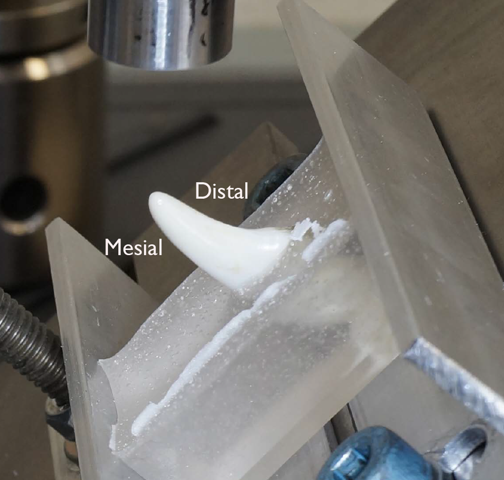
Photograph depicting a tooth from group A with the load being delivered in the distal–mesial direction 45° to the long axis of the tooth.

Photograph depicting a tooth from group B with the load being delivered in the labial–lingual direction 45° to the long axis of the tooth.

Photograph depicting a tooth from group C with the load being delivered in the mesial–distal direction 45° to the long axis of the tooth.
All tooth fragments were collected and saved for fracture pattern analysis. Fracture patterns were reviewed and categorized into 4 general fracture classes based on fracture orientation and fracture propagation direction (Tables 1 -4). Classes 1 to 4 represented transverse fracture, oblique fracture with propagation in distal to mesial direction, oblique fracture with propagation in mesial to distal direction, and oblique fracture with propagation in labial to lingual direction, respectively. Transverse fractures were further categorized into 3 subclasses: (a) crown only, (b) root only, and (c) any combination of subclass a or b and a vertical fracture. Groups 2, 3, and 4 were each further categorized into 4 subclasses: (a) crown only, (b) crown–root into cervical 1/3 of root, (c) crown–root into middle 1/3 of root, (d) crown–root into apical 1/3 of root, and (e) any combination of subclass a, b, c, or d and a vertical fracture. Additional characterization of fracture patterns included notation of the presence of an enamel–dentin fracture at the distal ridge of the tooth (if there was no preexisting enamel distal ridge abrasion), the fracture initiation point (defined as the distance between the load application point to the point of fracture initiation) and classification as either noncatastrophic or catastrophic based on the ability to clinically restore or not restore the crown, respectively.
Fracture Class 1: Transverse Fractures of the Crown or Root.

Abbreviations: M, mesial; D, distal.
Fracture Class 2: Oblique Crown Fracture With Propagation in the Distal–Mesial Direction.

Abbreviations: M, mesial; D, distal.
Fracture Class 3: Oblique Crown Fracture With Propagation in the Mesial–Distal Direction.

Abbreviations: M, mesial; D, distal.
Fracture Class 4: Oblique Crown Fracture With Propagation in the Lingual–Lateral Direction.

Abbreviations: Li, lingual; La, labial.
Statistical Analysis
Data were summarized by group with mean (95% confidence interval) or frequency (%). For numeric outcomes, group comparisons were made with univariate and multivariate analysis of variance (ANOVA) controlling for CSA. Two-way comparisons between groups were made with Tukey family-wise adjusted t tests. For categorical outcomes, group comparisons were made with Fisher exact tests. Two-way comparisons between groups were made with Holm adjusted Fisher exact tests. Due to a lack of occurrence of all fracture patterns (1a, 1b,…, etc), analysis was conducted based on class of fracture (1, 2, 3, 4) regardless of fracture subclass/characteristic (a, b, c, d, e) and then separately for fracture subclass/characteristic regardless of fracture class. For fracture class 1, the subclassification c (additional vertical fracture) was recategorized as subclassification e (additional vertical fractures in classes 2-4) to allow accurate comparison across the different fracture classes. All tests were conducted with a .05 significance level and were completed with an R version 3.0, utilizing lsmeans package.b
Results
Table 5 summarizes tooth hard tissue volume, CSA, force to fracture, maximum transverse shear stress, and initiation point between the 3 force direction groups. Univariately, there were significant differences in volume (ANOVA P = .009), CSA (ANOVA P = .011), and force to fracture (ANOVA P = .035), based on force direction group. Specifically, the hard tissue volume in group A was significantly larger than group B (Tukey P = .018) and group C (Tukey P = .026). The CSA was significantly larger in group A compared to group B (Tukey P = .010), and marginally larger than group C (Tukey P = .091). The force needed to fracture the teeth was significantly greater in group A compared to group B (Tukey P = .027). However, the significant difference in force to fracture was no longer significant when controlling for CSA as a covariate (multivariate analysis).
Summary of Tooth Hard Tissue Volume, CSA, Force to Fracture, Maximum Transverse Shear Stress, and Initiation Point Between the 3 Force Direction Groups.d

Abbreviation: CI, confidence interval.
aTukey family-wise adjusted P values comparing 2 groups at a time.
bUnivariate ANOVA P value.
cMultivariate ANOVA P value controlling for CSA.
dGroup A indicates distal to mesial force direction; group B is labial to lingual force direction; and group C is mesial to distal force direction.
Figure 4 summarizes tooth fracture class (1-4) by force direction groups. Loads applied in the distal–mesial direction (group A) resulted in a class 2 fracture pattern prevalence of 85.7%, and a class 1 fracture pattern prevalence of 14.3%. When the load was applied in the labial–lingual direction (group B), 41.7% of fractures were class 1 and 58.3% were class 4. When the load was applied in the mesial–distal direction (group C), 25% of fractures were class 1, 66.7% were class 3, and 8.3% were class 4. There was a statistically significant association between force direction group and fracture class (Fisher P < .001). Furthermore, each group had a different distribution of fracture class than any other group (Holm P values <.01).
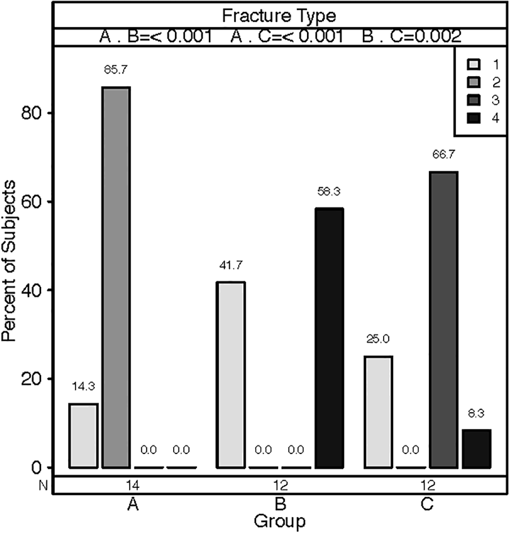
Bar graph depicting the influence of force direction on the fracture type classifications (1-4). 1 = transverse fractures; 2= oblique crown fracture with propagation in the distal–mesial direction; 3 = oblique crown fracture with propagation in the mesial–distal direction; 4 = oblique crown fracture with propagation in the lingual–lateral direction. Group A = distal to mesial force direction; B = labial to lingual force direction; C = mesial to distal force direction.
Figure 5 summarizes tooth fracture subclasses (a-e) by force direction groups. It was found that there was no significant association between group and fracture subclass (Fisher P = .216).
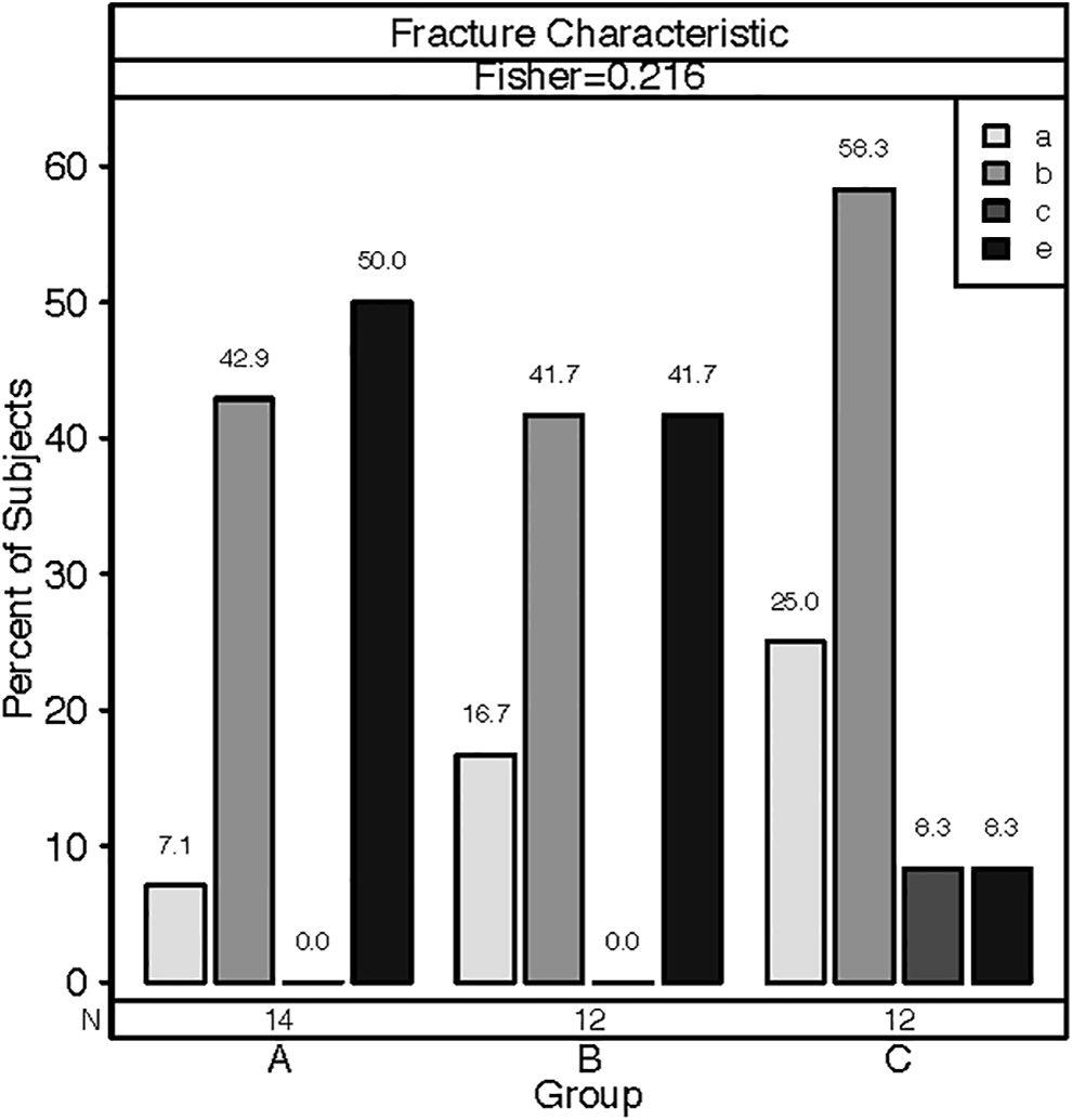
Bar graph depicting the influence of force direction on the fracture subclassification (a-e). a: Fracture propagated through the crown only; b: fracture propagated into the cervical one-third of the root; c: fracture propagated into the middle one-third of the root; d: fracture propagated into the apical one-third of the root; and e: any fracture subclassification (a-d) along with a vertical fracture. Group A = distal to mesial force direction; B = labial to lingual force direction; C = mesial to distal force direction.
Lastly, there was also no significant association between force direction group and the presence of enamel–dentin distal ridge fracture (Fisher P = .052) or the presence of catastrophic fractures (Fisher P = .420; Figures 6 and 7).
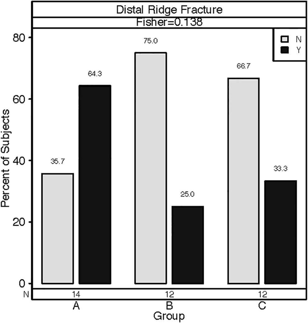
Bar graph depicting the influence of force direction group on the presence or absence of a distal ridge fracture. Y = yes; N = no. Group A = distal to mesial force direction; B = labial to lingual force direction; C = mesial to distal force direction.
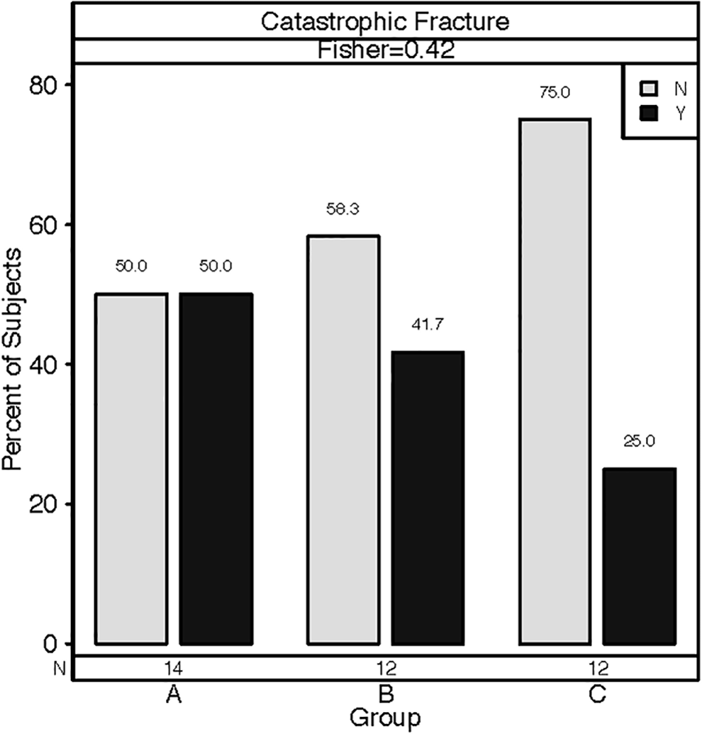
Bar graph depicting the influence of force direction group on the severity (catastrophic vs noncatastrophic) of the fracture. Y = yes; N = no. Group A = distal to mesial force direction; B = labial to lingual force direction; C = mesial to distal force direction.
Discussion
The present study found a trend toward a lower force to fracture requirement when load was delivered in a labial–lingual direction (group B), and a significant difference in mean force to fracture was observed when directly comparing this direction to the distal–mesial direction (group A). However, it is evident from the statistical analysis that there was a significant difference in both the hard tissue volume and the hard tissue CSA between these 2 groups (B < A). It is known from previous studies that the amount of dentin is one of the key factors dictating ultimate force to fracture values. 10,16 -18 It has been previously shown that tooth strength at the crown base was reduced by 20% in young coyotes due to the influence of the larger root canal/pulp chamber. 10 This finding has also been documented in humans with both ex vivo studies and FEA, which indicate that the more hard tissue that is removed with cavity and root canal preparation, the greater the risk of fracture. 16 -18 Furthermore, teeth with a smaller base radius have an increased susceptibility to transverse fractures. 11 Although the hard tissue CSA of a tooth cannot be calculated without knowledge of the pulp diameter, the assumption can be made that for 2 similarly aged animals with equal crown height, the canine tooth with the smaller base radius will have the lower hard tissue CSA. Thus, the critical load equations described by Lawn et al indirectly reiterates the importance of hard tissue thickness in fracture susceptibility. 11 In the present study, hard tissue CSA was significantly correlated with force to fracture, highlighting the importance of this tooth characteristic in evaluation of required fracture loads. Thus, hard tissue CSA was corrected for in the multivariate statistical analysis, which resulted in lack of a statistical difference for force to fracture between groups A and B. The results of the comparison of maximum transverse shear stress between the different force directions reiterated this finding.
Interestingly, in the present study there was a high degree of variability in the load required to cause fracture (71.7-811 N) and the maximum transverse shear stress at fracture (2.52-23.4 MPa). To the authors’ knowledge, there are only 2 studies that have fracture data from canid cadaver elongate canine teeth that are available for comparison to the data from the present study. 12,19 Corrêa et al evaluated the force to fracture in a fragment reattachment study, in which the load was applied perpendicular to the tooth at 4 mm from the CEJ versus the present study where the load was applied at the cusp tip. 19 When load is applied at the base of the tooth, the moment arm is decreased resulting in decreased bending stress on the tooth and increased resistance to fracture. 10 As expected, the mean force to fracture (942.12 ± 271.04 N) observed in the control teeth in Corrêa’s study was much greater than the values within the present study. Conversely, the overall mean force to fracture values (501 ± 154 N, group A; 289 ± 192 N, group B; 404 ± 247 N, group C) are similar to what was reported for the unaltered teeth by Soukup et al (494 ± 125 N). 12 However, the minimum force to fracture detected in the present study was substantially lower than what has been historically reported. 12,19 This is most likely due to the different force directions evaluated, as the minimum loads required for tooth fracture were found specifically in group B (labial–lingual). Soukup et al evaluated the influence of crown height to diameter ratio on force to fracture when a distal–mesial force direction was applied 45° to the long axis of the crown. The study found only 1 tooth that failed at less than 200 N. 12 In the present study, 0 of 14 teeth fractured at forces less than 200 N when the load was applied in the distal–mesial direction (group A), while half of the teeth (6 of 12) within group B fractured at less than 200 N. This suggests an increased susceptibility for fracture at lower loads with a labial–lingual direction. Lastly, 2 of 12 teeth fractured at loads less than 200 N when load was applied in the mesial–distal direction (group C). The natural shape of the canine tooth has evolved for resisting force in the biting pulling motion (distal–mesial force direction). The shape of the dog’s canine tooth, similar to that of a crane hook, may be optimized as a short, curved beam under the bending moment caused by a force in the distal–mesial direction. Thus, the force to failure in the labial–lingual direction, and to a lesser extent the mesial–distal direction, would be expected to fail at lower loads. No statistically significant difference was detected when mean force to fracture was compared between groups in the multivariate analysis. Similarly, there was no statistical difference in the mean maximum transverse shear stress between groups (11.66 ± 3.04 MPa, group A; 8.56 ± 6.00 MPa, group B; 11.08 ± 6.58 MPa, group C); however, the small sample size and large variance may have precluded significance.
There were significantly different fracture patterns observed between the 3 groups. Specifically, there was a significant difference in fracture direction propagation (fracture classifications 1-4) based on the force direction. It was concluded that the most common fracture propagation pattern (ie, fracture class) for each group was propagation in the same direction as the load direction. An explanation for this finding is that the shape of the canine tooth is a short, curved, hollow beam; and therefore, although under the combined loads of axial compression, bending, and transverse shear, the failure mode is dominated by the transverse shear force. As a brittle material, the tooth fails under maximum normal stress conditions. Given the orientation of the test setup, assuming pure shear, the maximum normal stress may be estimated as equal in magnitude to the maximum transverse shear stress acting at 45° to the long axis of the crown. Failure due to maximum normal stress is most clearly reflected in the observed fracture classes 2a, 2b, 2c, 3a, 4a, and 4e. The shear stress across the CSA of a beam is maximum at its neutral axis, which is most clearly reflected in the crack propagation at the center of the tooth, as seen in fracture classes 1c, 2d, 2e, 3c, 3d, 3e, 4c, 4d, and 4e. The shear stress in a beam is maximum at its neutral axis, which is most clearly reflected in the crack propagation at the center of the tooth, as seen in fracture classes 1c, 2d, 2e, 3c, 3d, 3e, 4c, 4d, and 4e. Interestingly, transverse fractures can be clinically observed with any of the evaluated force directions. However, when a transverse fracture is seen, the most common causal direction is the labial–lingual direction. Thus, the fracture propagation direction observed clinically (classes 2-4) may be used to determine the causal force direction with reasonable confidence. However, it is important to note that due to the small sample size the fracture patterns had to be analyzed based on groupings of class or subclass, rather than as each individual fracture pattern, which was not the original intention of the study and may have biased the results. Thus, despite the significance found between the overall fracture classes and force direction, this finding may best be considered observational.
Although fracture propagation is correlated with force direction, no significant association was seen between force direction and the degree of root involvement (ie, subclasses a-e). Similarly, force direction was not a significant predictor of catastrophic or noncatastrophic fractures. Again, sample size may have precluded the determination of any significance between force direction and root involvement or fracture severity.
Interestingly, a feature of a number of the observed fractures was the development of enamel–dentin fractures at the distal ridge after fracture loading (Figure 8). There was a trend to see more enamel–dentin fractures with the distal–mesial direction compared to the other force directions, and this finding neared significance. However, this fracture pattern was observed with all force directions. The clinical importance of this finding is that it brings into question whether these lesions, which have typically been considered “abrasion” injuries, are, in actuality, acute traumatic dentoalveolar injuries that may require a different therapeutic and preventative approach. Further evaluation of this finding is warranted to discern if this pattern is truly indicative of an acute traumatic distal–mesial direction injury.
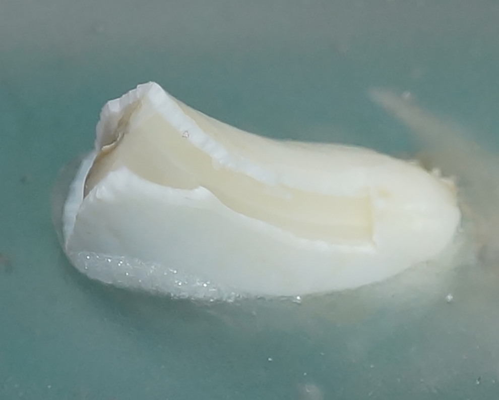
Photograph depicting an enamel–dentin fracture on the distal ridge of a canine tooth.
Excluding characterization systems that define tooth fractures by the dental tissue that is affected (enamel, enamel–dentin, enamel–dentin–pulp, etc), 20 -23 there is a paucity of both human and veterinary literature that characterize dental fracture patterns. As described in the introduction, the most common method to classify fractures when discussing tooth fracture biomechanics is to define fractures as vertical, chip, or transverse fractures. However, this classification system is not specific enough for clinical implementation and was not applicable to the goals of the present study. The classification system utilized for the present study is a modification of a system published by Loomba et al in 2010. 24 Modifications were necessary to account for the unique morphology of the dog canine tooth as well as to fit the goals of the present study for the classification system to apply to only 1 tooth rather than the entire mouth. Modifications of Loomba classification system included combining type 1 and type 2 fractures to accommodate for only the canine tooth being evaluated (became class 1 fractures in present study). Class 1 (transverse fractures) was then subclassified as crown fractures or root fractures. Loomba classification system did not adequately describe oblique fractures, which were the most common fracture pattern noted in the present study. Thus, the 3 fracture classes (2-4) in our modified Loomba classification represent the different oblique fracture patterns that were observed after loading: oblique fractures with mesiodistal propagation, labial–lingual propagation, and distal–mesial propagation, respectively. These fracture classes were then subdivided based on the degree of root involvement as proposed historically in Loomba classification. Crown involvement was not subdivided because this rarely impacts treatment in the veterinary patient. Root involvement, on the other hand, can significantly affect the ability to preserve the tooth, thus subdivision of root involvement was included within the modification. Lastly, we excluded fractures that occurred exclusively in the longitudinal plane (vertical fractures) from the 4 basic classifications because they were not anticipated nor observed as the primary fracture pattern in the present study. However, vertical fractures were observed in addition to the primary oblique fractures in the present study (Figure 9). Thus, vertical fractures were incorporated as a subclass of the fracture classes (1c, 2-4e). Additional fracture classes, such as a class specifically for isolated vertical fractures, may be necessary for future studies.
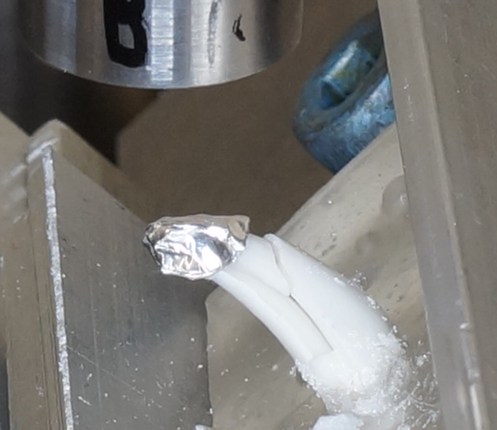
Photograph depicting a fracture class 4e with the presence of a vertical fracture concomitant with an oblique crown fracture.
The authors acknowledge limitations in the present study, the most notable being the small sample size. The limited amount of cadaver teeth may have led to the analysis being underpowered in some of the statistical tests. With the small sample size in each group, the present study is to be considered exploratory and further study on a larger number of teeth may be warranted to confirm the findings of the present study. Furthermore, the teeth were all from 1 breed and population of dogs, potentially limiting the clinical applicability of the data. Study design, and thus data, may not accurately represent true clinical scenarios. First, teeth were stored in formalin before testing. Sterilization of biological specimens is a critical step in the provision of safety for researchers whom interact with biological tissues. In order to achieve sterilization of teeth, the Center for Disease Control and Prevention has established guidelines that require teeth to be either heat sterilized or stored in 10% formalin. 25 In order to prevent desiccation, the teeth in the present study were stored in 10% formalin prior to mechanical testing, which may have been influential on the outcome of the present study. However, a study comparing the effect of storage in deionized water, saline, and 10% formalin on the mechanical properties of human dentin revealed no significant difference between the 3 storage media on the strength or toughness of dentin and concluded that all teeth should be stored in 10% formalin prior to mechanical testing. 14 Second, the teeth were not tested in a moist environment and humidity was not controlled during testing. Third, 2 groups of teeth (B and C) were tested after a longer drying time, due to equipment technical issues, which may have altered the material properties. Lastly, methyl methacrylate may not adequately represent the biomechanics of the periodontal ligament space and alveolar bone and may have led to a stall in fracture propagation at the methyl methacrylate–tooth interface, potentially limiting the ability to fully evaluate root fractures. To the authors’ knowledge, there are no studies that evaluate how methyl methacrylate affects fracture propagation. However, it has been discussed previously that this stiff material most likely does not compromise results of fracture pattern analysis as clinically relevant fractures continue to be observed despite the absence of a synthetic periodontal ligament. 12
The directions chosen for the applied force may not necessarily represent the applied force directions that the canine tooth would encounter in the natural environment. However, when considering the force the canine tooth encounters during bite work, the distal–mesial direction accurately represents the biting pulling motion at the tip of the tooth when pulling back on the sleeve. The labial–lingual direction represents the direction the tooth encounters when dogs swing their heads to the side while actively biting on the sleeve. The mesial–distal direction, on the other hand, may only be encountered by blunt force trauma to the maxillofacial region, which would more likely be delivered perpendicular (90°) to the long axis of the tooth rather than at 45°. The natural morphology of the dog’s canine tooth precluded true perpendicular delivery of the load. The curvature of the tooth near the cusp results in slipping of the load point when delivered at 90° to the long axis of the tooth and would have provided unreliable force to fracture data. In addition, the teeth were loaded at the cusp tip, which may have falsely led to a decreased load required for fracture than what would be observed clinically. When the tooth is loaded at the most coronal aspect, the moment arm is at its longest, and thus the bending stresses are increased lowering the load required to cause failure. 10 It is presumed that significant load would be carried at the cusp of the tooth during normal behavior. However, no data exists regarding the natural point at which the load would be applied. A previous study suggested application of force at a point 70% of the height of the tooth to mimic where the canine tooth would extend into prey. 10 This height may be an appropriate load point when delivering a perpendicular load. However, delivering the load at 45° to the long axis of the tooth to accurately model the forces during biting and pulling is impossible at 70% height. Force delivered at 45° represents a combination of both compressive (ie, axial) and transverse (ie, off-axial) loads that arise when a dog bites (compressive) and pulls (transverse) on an object, and this load can only be delivered at the tip of the tooth. Thus, load application at the tooth cusp may be a more appropriate predictor of force to fracture seen in nature versus application of a perpendicular force at 70% height. Lastly, the equation used to calculate the maximum shear stress for each tooth assumes a hollow, circular, thin-walled cylindrical geometry. The cross-sectional geometry of the teeth was more reflective of an ellipse than a circle. Use of a circular cross-section likely resulted in an underestimation of the true maximum shear stress. However, no statistically significant differences were found between the ellipse morphology of the teeth in each group. Therefore, the relative magnitude in maximum shear stresses between groups would remain the same for circular or elliptical cross-sectional geometries.
In conclusion, the results of this study support the hypothesis that different force directions lead to significantly different fracture patterns. Thus, the clinician may be able to inform the client of potential force directions based on the observed fracture pattern as classified by the suggested classification system above. The clinician may also be able to draw conclusions as to what type of behavior may have contributed to the fracture and make recommendations to avoid similar injuries in the future. Furthermore, although a trend for different force to fracture with different force directions was noted, no significance was found between the 3 groups leading to the observation that regardless of force direction a similar force to fracture is required for clinical failure. Future studies with a larger sample size are recommended to confirm the findings of this study regarding force direction and resultant fracture resistance and fracture pattern.
Materials
Hygenic orthodontic resin, Coltene/Whaledent, Inc, Cuyahoga Falls, Ohio, USA. R open access statistical software, version 3.0, The R Project for Statistical Computing, www.r-project.org. R Core Team (2013). R: A language and environment for statistical computing. R Foundation for statistical Computing, Vienna, Austria. URL http://www.%20R-project.org/.
Footnotes
Declaration of Conflicting Interests
The author(s) declared no potential conflicts of interest with respect to the research, authorship, and/or publication of this article.
Funding
The author(s) received no financial support for the research, authorship, and/or publication of this article.