
Abstract
During daily routine oral examinations in a research colony of nonhuman primates (NHPs, Macaca fascicularis), a variety of oral–dental lesions were identified. A dental care program was established based on these findings. Based on the presence of dental clinical signs and their severity, 31 animals were triaged to be examined and treated by a veterinarian. Clinical examination consisted of visual inspection using a periodontal probe/explorer and full or partial mouth dental radiographs. Treatment was performed during the same procedure. Some animals had a follow-up examination including radiographs months later. Four common dental diseases were diagnosed: periodontal disease, caries, tooth fracture, and tooth attrition. Less frequent were dental abscess, enamel hypomineralization, gingival hyperplasia, hypercementosis, tooth luxation, tooth dysplasia, root resorption, abrasion. Less severe periodontal disease was treated conservatively. If severely affected, teeth were extracted. Well-circumscribed caries without endodontic involvement were treated by composite restoration. Teeth with extensive caries and pulp involvement were extracted. Teeth with exposed pulp were treated via extraction or orthograde root canal treatment. In this case series, 27 (87%) of 31 NHPs exhibited at least 1 moderate to severe dental lesion that required treatment. The presumable improvement in welfare and weight of oral/dental lesions for the overall health status in research NHPs encourages us to continue this program prospectively.
Keywords
Introduction
During routine examinations in a research colony of nonhuman primates (NHPs, Macaca fascicularis), a variety of oral–dental lesions such as broken tooth, caries, or gingivitis were identified. Dental lesion may lead to local or distant infection, pain, and distress, which may hinder the well-being of animals. Furthermore, preexisting dental disease may confound experimental outcomes due to pain and infection unrelated to the actual studies. 1,2
Therefore, a dental care program was deemed necessary to reliably diagnose dental lesion. By improving the knowledge of dental health status, this would increase the well-being of NHPs and the reproducibility of experimental results. This program practically addresses the third principle of Russell and Burch’s “Refinement”, defined as the use of methods that alleviate or minimize potential pain, suffering, or distress and enhance animal welfare for the animals used. 3
The purpose of this clinical study was to describe dental lesion of NHPs and to outline the steps of an established dental care program. Possible etiology and correlation with age and diet are discussed.
Material and Methods
The animals of this case series were part of a research colony of purpose-bred Chinese, Philippian, and Mauritian M fascicularis, males and castrated males. They were 9 to 22 years old, weighing between 5.6 and 12.2 kg (median 9.4 kg; Table 1).
Listing of Diagnoses and Treatment.
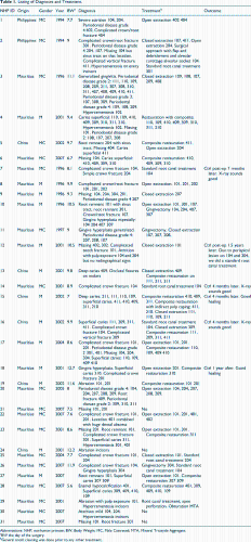
Abbreviations: NHP, nonhuman primate; BW, Body Weight; MC, Male Castreted; MTA, Mineral Trioxyde Aggregate.
aBW the day of the surgery.
bGeneral tooth cleaning was done prior to any other treatment.
The NHPs lived in highly hierarchized social groups of 3 to 8 individuals. They were housed exclusively indoor in big pens of concrete and metal in 1 air-conditioned barrier unit. Room temperature was kept constant between 22.5°C and 22.8°C and relative humidity between 50% and 60% (range: 40%-80%), with artificial and natural light. They were fed a laboratory monkey food (70-100 g/day/animal) a and since 2011 received a predominantly vegetable-based diet (daily rotation of fresh tomato, potatoes, salad, cucumber, fennel, chicory). Fresh fruits (apple, banana) were given occasionally as reward. Before 2011, fruits and vegetables were offered in equal proportions. The fresh vegetables are placed to enhance foraging and active search. Popcorn and alternatively a mix of cereals, soy, and sunflower seeds were given freely on the ground, once weekly as part of the olfactory enrichment program. Raspberry syrup in water and dry raisins, peanuts, and jelly sweets (Haribos, Bonn, Germany) were also given weekly during their training sessions as positive reinforcement. Nonchlorinated drinking water b was available ad libitum.
Serological testing for tuberculosis, measles, cercopithecine herpes I virus, and simian virus (SIV [simian immunodeficiency virus], SRV [simian retrovirus], and STLV [simian T-cell lymphotropic virus]) was performed annually, while complete blood counts and clinical chemistries, diabetic markers, and bacteriological and parasitological fecal examination were performed quarterly as part of clinical health checks.
A structured enrichment program was available with structural elements always present in the pen (ladders, barrels, sand box) and hanging or free items (kongs, nut cages, puzzle feeders, paint and paper rolls, PET [polyethylene terephthalate] bottles, surprise boxes) as auditive (TV) and olfactory (popcorn) elements on a rotational weekly base. The structural elements are used also as visual barriers additional to colored divisions. The facility is Association for Assessment and Accreditation of Laboratory Animal Care (AAALAC) accredited.
For the purpose of this program, the NHPs were arbitrarily divided into 3 groups based on the year of birth: group 1 from 1994 to 1997, group 2 from 2001 to 2002, and group 3 from 2004 to 2007.
A dedicated team consisting of a specialized veterinary dentist (diplomate of the European Veterinary Dental College), an anesthesia technician, and a veterinary technician cared for the animals.
Scaling with powered equipment should be considered a contaminated procedure as it creates aerosolized bacteria (and possibly viruses) which can travel several meters in the air. In NHPs, there is always a potential concern for herpes B virus transmission. 4 To decrease exposure risk, all people present in the surgical room wore a powered air-purifying respirator. c Additionally, gloves, Tyvek sleeves, and bonnets were worn as personal protective equipment (Figure 1).
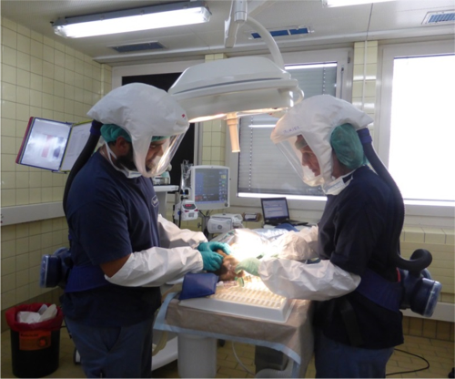
Mandatory personal protective equipment including a powered air-purifying respirator for all people present in the surgical suite to decrease the risk of exposure to potential zoonotic pathogens (herpes B, aerosolized bacteria, etc).
Equipment
A dental unit, d an aspiration pump, e and a portable X-ray unit f with direct g or indirect h digital radiography systems were used. Various instruments and materials were available to perform soft tissue surgery, extractions, endodontic treatment, and restorations.
The animals were screened and selected by the veterinary technician based on the detection of obvious or suspicious oral–dental lesion after clinical examination on conscious or sedated animals during daily general health controls and routines. The clinical signs taken into account were calculus accumulation (light, moderate, severe), gingival condition (healthy or inflamed), tooth position, missing teeth, fractured teeth, and worn teeth.
A priority list was compiled based on the presence of the above findings and their severity; the animals with fractured teeth, caries, severe soft tissue inflammation, marked and generalized calculus accumulation, and other oral or dental lesions were scheduled to be examined by the veterinarian dentist at quarterly intervals.
Every patient underwent general anesthesia with a protocol according to their American Society Anesthesiologists (ASA) status. Animals classified ASA status 1: medetomidine i (0.05 mg/kg)/ketamine j (5 mg/kg) IM (intramuscular) followed by sevoflurane 2% k to prolong anesthesia as needed. Animals classified ASA status 2 and 3: ketamine (6 mg/kg) IM/alfaxalone l induction followed by 2 mg/kg IV (intravenous) bolus and 8 to 4 mg/kg/h CRI (Constant Rate Infusion). 5 All animals underwent endotracheal intubation (cuffed ET [Endotracheal] tube, 4 mm ID [Internal Diameter]), received 100% oxygen via a pediatric rebreathing circle system, and were fully monitored. m Normothermia was achieved with a warmed air blanket. n All animals received preemptively 20 mg/kg IV cefazolin o as antibiotic coverage, 10 mL/kg/h IV lactated Ringer’s solution until recovery, and vasopressors and/or inotropes as needed to maintain a mean arterial blood pressure above 60 mm Hg. For intraoperative analgesia, a locoregional nerve block with 2 mg/kg bupivacaine p was performed for teeth extractions and 10 μg/kg SC buprenorphine was injected at recovery. Postoperatively, animals received 0.1 mg/kg meloxicam SC (subcutaneous) followed by 0.1 mg/kg meloxicam q orally after dental treatments (root canal) for 3 to 5 days. In case of dental extractions, 10 μg/kg SC buprenorphine r every 8 hours for 3 days was added.
A thorough clinical examination was done using a periodontal probe/explorer. Full-mouth or partial mouth radiographs were obtained. Some dental abnormalities were also photographed. The signs were registered on an adapted chart using the Triadan teeth numbering system (Figure 2). 6 The dental chart is a useful tool to document dental lesion and its treatment. Based on the diagnosis, a treatment plan was decided and performed immediately during the same procedure. Some animals had follow-up examinations under anesthesia including dental radiographs several months later.
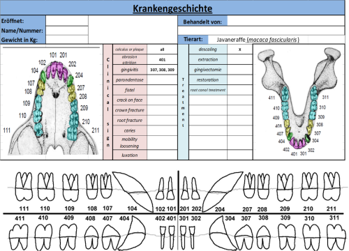
Dental chart using the Triadan numbering system.
Results
Diagnostic Findings
During the first 2 years of the program (from 2013 to 2015), 31 animals were examined and treated under general anesthesia. Based on the clinical and radiographic examination of the dentition, 4 common dental diseases were diagnosed: periodontal disease, caries, tooth fracture, and tooth attrition. Some less frequent lesions were found such as dental abscess, dental cyst, enamel hypomineralization, gingival hyperplasia/enlargement, hypercementosis, tooth luxation, tooth dysplasia, root resorption, and abrasion (Table 1 and Figure 3).
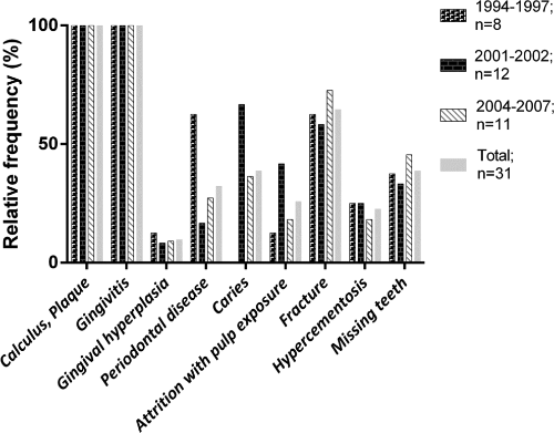
Relative frequency (%) of pathologies by age-group and for all examined nonhuman primates (NHPs).
Four (13%) NHPs of 31 had no serious dental lesion except calculus, plaque, mild gingivitis, slight attrition and/or abrasion, or missing teeth. Scaling and polishing was the only treatment needed in these cases.
Periodontal disease was the most commonly encountered disease. Calculus and plaque were present in every animal combined with mild to moderate gingivitis in 21 NHPs. The 10 (32%) other NHPs had stage 2 periodontal disease or higher, with a total number of 46 teeth affected. Periodontal disease was more common and more severe on molar (20 teeth) and premolar (20 teeth) than on canine (3 teeth) and incisor (3 teeth; Figure 4).
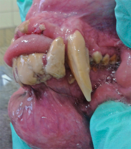
Calculus and severe generalized periodontal disease.
Caries was observed in 12 (38%) of 31 NHPs. Forty-four caries in total were observed mainly on molar (43 teeth), rarely on premolar (1 tooth), and not seen on incisor and canine teeth. Caries were most commonly located on the occlusal surface and were identified as small black spot with a softer hard tissue compared with the neighboring healthy hard tissue. Caries extend rarely below the enamel–dentin junction. One animal had several teeth affected with severe, destructive, deep caries (Figure 5).
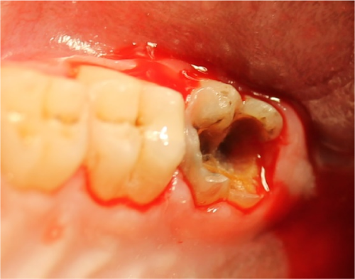
Deep caries of the left maxillary third molar tooth.
Tooth fracture was a very common occurrence. A total of 30 teeth were fractured in 20 (64%) animals. Most fractures were complicated crown fractures or complicated crown–root fractures, but root fractures and root remnants were also seen. Incisor teeth (19 teeth) were most commonly affected by fracture, followed by canine teeth (8) and also molar teeth (3; Figure 6).
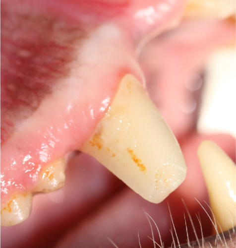
Complicated crown fracture of tooth 104.
Twenty-two teeth with attrition and/or abrasion (Figure 7) including pulp exposure were found in 8 (26%) NHPs and affected mainly the canine (8), but also some incisor (2). Every NHP had at least a minor degree of attrition and/or abrasion on all 4 canines and some incisors presented a minor degree of attrition and/or abrasion without pulp exposure. We hardly found signs of attrition on the mandibular third premolar, despite the fact that its mesial coronal surface is responsible for attrition of the distal surface of the maxillary canine. From the 8 maxillary canines with pulp exposure, the affected area was always on the distolingual surface of the maxillary canine. In all cases, the pulp was already necrotic and periapical radiolucency was detectable.
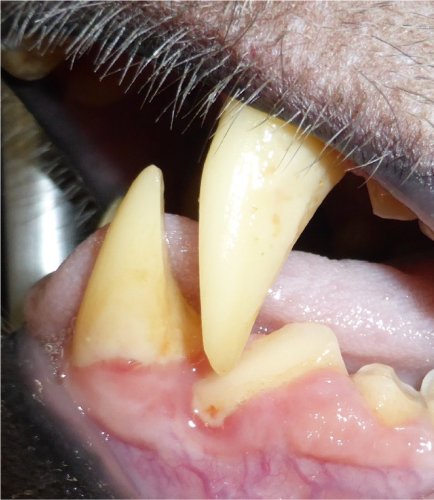
Honing mechanism illustrated by the attritional signs on the contact faces of the lower canine, upper canine, and lower third premolar.
In this case series, 7 of 31 (22%) NHPs showed one or several incisors with radiographic evidence of hypercementosis (Figure 8). No other tooth type was affected by hypercementosis. Twelve (39%) of 31 NHPs had incomplete dentition and a total of 22 teeth were missing: incisor (8 teeth), canine (9 teeth), premolar (1 tooth), and molar (4 teeth).
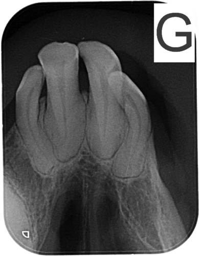
Image showing enlargement of the apices of the 4 maxillary incisors suggestive of hypercementosis.
Treatment
Patients with stage 1 and 2 periodontal disease were usually treated conservatively. Teeth were thoroughly rinsed with a disinfectant solution containing chlorhexidine 0.12%. Calculus and plaque were removed using an ultrasonic scaler. Exposed root surfaces were planed using a Gracey curette when needed. Finally, the crowns were polished using a prophy cup with polishing paste.
Teeth affected by some stage 2 and all stage 3 and 4 periodontal disease were extracted, generally using a closed extraction technique. Well-circumscribed caries without endodontic involvement were treated using a classical restoration technique with flowable composite. Teeth with deeper, extensive caries with pulp involvement were extracted.
Teeth with complicated crown or crown/root fracture or teeth with exposed pulp due to wear were treated by either extraction or orthograde root canal treatment. Root canal treatment was chosen whenever possible for affected canine teeth and sometimes for incisors. Access was determined depending on the fracture line level and the root curvature. For the majority of affected canines, we made an access hole 3 to 4 mm above the gum line on the mesial aspect of the crown, in the deep mesial groove. Preparation and disinfection of the canal was done using veterinary 60-mm-long reamers and Hedstrom files and a chelator and disinfection gel. s For the observation, we used 60-mm gutta-percha combined with zinc oxide–eugenol cement. t Restoration was finalized with an intermediate layer of glass ionomer u followed by composite. Each step of the procedure was assessed radiographically (Figure 9A and B).
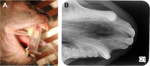
A, Debridement of the pulp chamber and root canal of tooth 104 using a 60-mm endodontic file. B, Radiograph of orthograde root canal therapy and restoration of tooth 104.
Three NHPs with gingival hyperplasia were treated with gingivoplasty using a conical medium diamond bur (Figure 10). Dental radiography revealed a big radiolucency in 1 NHP that didn’t show external sign of disease. It was a large cystic lesion of the right mandible extending from the right mandibular canine to the right first incisor (Figure 11A-G). The lesion was about 3 cm in diameter, both lower right incisors were displaced. After incision, a white-yellow, creamy material was extracted and aspirated out of the lesional cavity. The lesion was managed by raising a mucoperiosteal flap, extracting both incisors, debriding the defect using a bone curette, and abundant rinsing with a physiological solution. The exposed root surface of the mandibular right canine was planned using a Gracey curette, and the gingiva was finally closed using simple notch closure.
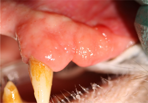
Gingival enlargement over teeth 104, 107, 108. The crowns of 107 and 108 are completely covered.
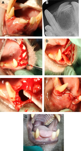
A, Swelling of the rostral right mandible causing displacement of teeth 401 and 402. B, Radiograph of the right mandible showing a large radiolucency and displaced incisors. C, White, creamy material coming out of the wound after incision of the gingiva. D, Extraction of the displaced incisors. E, Image showing the large cystic structure. F, Closure of surgical site. G, Appearance of the site 4 months later.
Discussion
Macaca fascicularis, the long-tailed macaque, is a small cercopithecine monkey, weighing between 3 and 8 kg, with a natural distribution that stretches from Burma to the Philippines. It lives in large multi-male groups of between 15 and 30 individuals and is almost exclusively arboreal in rain forest. They are widely used in research mostly for safety testing of pharmaceutical products and devices. They are opportunistic omnivores with fruits and seeds making up 60% to 90% of their diet in the wild, but also eat leaves, flowers, roots, bark, vertebrates, invertebrates, and birds. This is reflected in their dentition which is very similar to human beings. Macaca fascicularis also have 32 teeth, composed of 2 incisors, 1 canine, 2 premolars, and 3 molars on each quadrant. The tooth morphology and the number of roots are similar to humans with some exception, the most obvious one is the very large and sharp canines. Also the mandibular third premolar (which is the next tooth distal to the mandibular canine) has a triangular shape and is in occlusion with the maxillary canine. The mandibular third molar is longer than all other molars with a distal extension, the hypoconulid. Finally, a wide diastema is present between the maxillary canine and maxillary second incisor to enable occlusion of the mandibular canine when the mouth is closed. 7
In this case series, 27 (87%) NHPs examined of 31 suffered at least 1 moderate to severe dental lesion that needed a treatment more involved than tooth cleaning and polishing. The high prevalence of dental lesion was likely affected by the fact that these 31 NHPs were preselected after the first visual examination for dental signs of lesion. The real incidence of dental disease in this research colony will be available when all animals have been screened. In a previous study on Macaca mulatta, free-ranging population partially fed with commercial food, and the percentage of animals with dental lesion was only 7.4% overall. 8 This low percentage can be explained by a broad spectrum of individuals compared to the exclusively adult male colony in our case series. Young animals or females have naturally less dental lesions compared to adult males, as they have fewer tendencies for aggressive behavior that can lead to teeth trauma. In the same study, only 1 NHP had caries, 8 NHPs had fractured teeth, and no periodontal disease was described. This is surprisingly very low compared to the present case series. This difference in the incidence of dental lesion can be explained by 2 factors: environment and diagnostic mean. Environment certainly plays a role as here the NHPs are living in captivity in small groups composed of adult males housed in big pens of concrete and metal and were fed a mixed diet of natural and artificial food. These conditions may increase the risk of fight, trauma, and abnormal behavior such as cage biting syndrome, and the diet may foster the risk of periodontal disease and caries. The second point is the diagnostic mean: In this program, the examination was done by a veterinarian specifically trained and accustomed to diagnosing dental lesions. Moreover, the systematic use of intraoral radiography enabled to detect root and bone modifications. Past studies in dogs and cats have shown the importance of intraoral radiography, not only to improve the information about dental lesion but also to increase the number of dental lesion diagnosed. 9
Periodontal disease is a major problem in dogs and cats and also in humans; NHPs have been used as a model of periodontal disease for years with Macaca species, demonstrating naturally occurring periodontal disease that increases with age. 10,11 –14 It has also demonstrated familial susceptibility or resistance to periodontitis in Rhesus macaque. 15 Periodontal disease may show 4 stages. Stage 1 is gingivitis without alveolar bone involvement. Stages 2, 3, and 4 represent increasing loss of alveolar bone attachment. Unfortunately, even with good oral hygiene, bone loss can’t regenerate spontaneously, so stages 2, 3, and 4 are not easily reversible. The prevalence of periodontal disease stage 2 and over is high in dogs and humans. 16,17
Dental caries, also known as tooth decay, is a breakdown of teeth due to activities of bacteria. Caries forms through a complex interaction over time between acid-producing bacteria and fermentable carbohydrates and many host factors including teeth and saliva. 18 The etiology includes elevated colonization levels of Streptococci mutans, high-frequency sugar consumption, and developmental defects on primary teeth. 19 The impact of sugar-containing food in the monkey diet and also the use of sweets as reward can explain the high prevalence of caries detected compared to studies with animals living in the nature. 8 Past studies have shown the weight of cariogenic high-sucrose diet. 20 Dental caries were seen mainly on molar teeth and develop especially on natural fissure of the occlusal surface. This fissure, corresponding to developmental grooves, may favor food debris impaction. Moreover, the thin or even absent enamel layer at the fusion point of developmental lobes may have contributed to the ease of caries initiation. 21 This is similar to observations in human clinical studies. 22 Caries is not a new finding in NHP and is induced by the same factors as in humans. 20
Attrition is the wear off of dental hard tissue due to tooth-on-tooth contact. The intentional attrition used for sharpening of canine teeth in monkey is called the honing mechanism. Honing is a natural behavior of male cercopithecidae to improve their capacity in defense of individual or troop against predators, in fighting among individual males within or between troops and as part of “threat” gestures. 23 There are actually 3 wear surfaces: the mesial face of the maxillary canine wearing against the distal face of the mandibular canine, the cingulum and adjacent superior distal surface of the mandibular canine wearing against the cusp tip of the maxillary canine, and the distolingual surface of the maxillary canine wearing against the mesiobuccal surface of the mandibular third premolar. The maxillary canine attrition is faster and deeper compared to the mandibular canine and above all the mandibular third premolar. This difference can be explained by the much thicker enamel covering the mandibular premolar compared to those of the canine. 23 Attrition is generally a slow mechanism that enables the formation of reparative dentin to protect the pulp cavity against pulp exposure. However, if the attrition is faster, this self-protection mechanism may not be sufficient. Abrasion against hard toy, metal, or concrete can surely be an accelerating factor. Considering the severe biting lesion due to male behavior and the risk of pulp exposure due to overstimulated attrition/abrasion, a preventive crown reduction would be of great benefit if done properly with respect to vital pulp tissue using good veterinary practice.
Tooth fracture, especially complicated crown fracture of the canine and incisor teeth, is a common finding in NHPs. Fights, hits against walls or any other hard material, and mastication on hard substance (corn, nuts, wood, stone, etc) are the most common causal factors. In a previous study, the main cause of canine size reduction at Gombe, Tanzania, was breakage in fights. 24
One animal showed a vertical fracture on a molar tooth. This rare fracture is most likely due to mastication of something hard from the food intake. The lesion was severe enough to justify extraction. Another rare fracture was a radicular fracture without endodontic lesion. The fracture line was well identified, some signs of reparation were visible, and no periapical lesion seen on radiographs. This tooth necessitated no treatment. All other fractures were typical crown or crown–root fracture with pulp exposure. The decision to treat conservatively or to extract the tooth was taken depending on the severity of the clinical and radiographic signs. As often as possible, a standard root canal treatment was used. Extraction was selected in the following cases: severity of the periapical lesion, difficulty to perform a good root canal treatment, pathological apical opening, and root resorption. To perform a root canal treatment, occasionally an access hole 2 to 3 mm supragingival to the gingival margin in the mesial groove was performed, especially when the remnant crown was too long.
Hypercementosis is a nonneoplastic condition in which excessive cementum is deposited in continuation with the normal radicular cementum. Apart from its idiopathic nature, this condition is associated with several local and systemic factors. 25 In humans, hypercementosis is rare and when it occurs, it affects premolar, molar more frequently than incisors. 26 In horses, however, hypercementosis is more frequent in incisors and associated with aging. 12 Without further study, it is difficult to speculate about the causative factors. The most potential factors are genetic, periodontal disease, occlusal forces, and endodontic stimulation.
Gingival hyperplasia, also described as gingival enlargement in the absence of histological confirmation, is an abnormal, excessive growth of the periodontal tissues. The etiology in humans is multifactorial and includes age, genetic predisposition, and induction due to drugs (like cyclosporine) or plaque. In the veterinary field, this is a common condition in boxer, French bulldog, and giant-breed dogs where hyperplastic tissues are sometimes so overgrown that they can completely cover the teeth. 27 The 3 NHPs in this study with generalized gingival hyperplasia were treated by gingivectomy and professional dental cleaning. No biopsies were taken to differentiate gingival overgrowth of inflammatory origin from benign or malignant tumor by histopathology.
The wide radiolucent lesion found in 1 NHP can be related to abscess, tumors, and cysts. Abscesses often develop periapically, associated with broken teeth or deep periodontal disease. Tumors may be associated sometimes with cystic lesions or with soft or hard tissue overgrowth. One case of an ameloblastic fibroma in Macaca mulatta has been published, showing a cyst-like structure on radiographs. 28 Dentigerous cysts originate from the dental follicle and are associated with unerupted teeth. Odontogenic keratocyst are sometimes very aggressive and often multilobulated. 29 Some produces orthokeratin, a foul-smelling cheese-like material that is not pus but rather collected degenerating keratin. In the present case, the lesion was more likely to be of cystic nature. Unfortunately, no biopsy was obtained.
Missing teeth may be missing for various reasons: inheritance, congenital, extraction, or avulsion. Occasionally, the crown may be absent but a root remnant is visible on radiographs. In this colony, no history is available to confirm or exclude congenital or inherited missing teeth. The most likely causes here were fracture or prior extraction by a veterinarian.
Considering the pathological lesions and their relation to aging, it can be concluded that periodontal disease is age related. The older the NHP, the more severe and extended the periodontal disease level. Other lesions appear not to be age related.
Conclusion
The dental care program (specialized diagnosis and treatment) performed in the research colony of NHPs proved necessary and valid as all NHPs were diagnosed with a number of periodontal and dental lesions of varying severity.
These painful conditions are often associated with local or systemic infection or inflammation and need to be addressed and treated. As far as possible, a conservative treatment as root canal treatment or restoration was attempted with great success. In some circumstances, the severity of the dental lesions and the difficulty to maintain good oral hygiene with uncooperative animals resulted in a more radical treatment. This case series emphasizes the importance of using radiographs in diagnosis of lesions, treatment, and follow-up of procedures.
The implementation of treatment enhances the well-being of the NHP colony by causally eliminating pain. Furthermore, treatment may prevent bacteremia, pain, and secondary lesions (cardiac, hepatic, renal), which can bias scientific studies. Healthy animals are a prerequisite to provide researchers with sound and robust animal models. This encourages us to continue this program prospectively with regular examinations and, if necessary, treatment of dental disease. The strategic design and implementation of this dental program upholds the principle of Russell and Burch’s “refinement” concept. 3
Footnotes
Acknowledgments
The authors thank Dr Guido Steiner for advising with presentation of the data.
Declaration of Conflicting Interests
The author(s) declared no potential conflicts of interest with respect to the research, authorship, and/or publication of this article.
Funding
The author(s) received no financial support for the research, authorship, and/or publication of this article.